DOI: 10.1534/genetics.109.110171
A Genomewide RNAi Screen for Genes That Affect the Stability, Distribution
and Function of P Granules in
Caenorhabditis elegans
Dustin L. Updike and Susan Strome
1Department of Molecular Cell and Developmental Biology, University of California, Santa Cruz, California 95064 Manuscript received September 22, 2009
Accepted for publication September 28, 2009
ABSTRACT
P granules are non-membrane-bound organelles found in the germ-line cytoplasm throughout
Caenorhabditis elegansdevelopment. Like their ‘‘germ granule’’ counterparts in other animals, P granules
are thought to act as determinants of the identity and special properties of germ cells, properties that include the unique ability to give rise to all tissues of future generations of an organism. Therefore, understanding how P granules work is critical to understanding how cellular immortality and totipotency are retained, gained, and lost. Here we report on a genomewide RNAi screen in C. elegans, which identified 173 genes that affect the stability, localization, and function of P granules. Many of these genes fall into specific classes with shared P-granule phenotypes, allowing us to better understand how cellular processes such as protein degradation, translation, splicing, nuclear transport, and mRNA homeostasis converge on P-granule assembly and function. One of the more striking phenotypes is caused by the depletion ofCSR-1, an Argonaute associated with an endogenous siRNA pathway that functions in the germ line. We show thatCSR-1and two other endo-siRNA pathway members, the RNA-dependent RNA polymerase EGO-1and the helicase DRH-3, act to antagonize RNA and P-granule accumulation in the germ line. Our findings strengthen the emerging view that germ granules are involved in numerous aspects of RNA metabolism, including an endo-siRNA pathway in germ cells.
G
ERM granules are large, non-membrane-bound, ribonucleoprotein (RNP) organelles found in the germ-line cytoplasm of most, if not all, animals (Eddy 1975; Saffman and Lasko 1999). The term ‘‘germ granule’’ encompasses what are known as P granules in Caenorhabditis elegans, polar granules in Drosophila melanogaster, germinal granules in Xenopus laevis, and the perinuclear nuage in mouse and human germ cells. These large RNP complexes contain a heterogeneous mixture of RNAs and proteins. To date, most of the known germ granule proteins across species, and all of the known P-granule components in C. elegans, are associated with RNA metabolism, which suggests that a main function of germ granules is post-transcriptional regulation (Strome2005; Seydoux and Braun2006). Germ cells are unique in their ability to give rise to all tissues of future generations of an organism. Consequently, germ cells are considered to be both totipotent and immortal. The widespread presence of germ granules in germ cells across species and the ability of germ granule transplantation to induce functional germ cells suggest that germ granules arekey determinants of the identity and special properties of germ cells (Smith1966; Illmenseeand Mahowald 1974; Ephrussiand Lehmann1992). As more compo-nents and regulators of germ granules are identified, we will better understand their role in conferring germ cell identity and properties.
One of the earliest identified constitutive compo-nents ofC. elegansP granules isPGL-1(Kawasakiet al. 1998). Identification of genes whose loss alters the level and/or distribution of PGL-1 offers an avenue to identify other P-granule components and components that regulate P-granule assembly and stability. For example, theC. elegansVASA homologGLH-1, another constitutive P-granule component, acts ‘‘upstream’’ of PGL-1; in glh-1 loss-of-function mutants,PGL-1 is not properly localized to P granules and instead a significant portion ofPGL-1is diffusely distributed in the germ-line cytoplasm (Kawasaki et al. 1998; Spike et al. 2008a). More recently another constitutive P-granule compo-nent,DEPS-1, was identified in a forward genetic screen for mutants that displayPGL-1localization defects similar to those seen in glh-1(lf) mutants (Spike et al. 2008b). Targeted studies have demonstrated that mutation or RNAi of other genes also disruptsPGL-1in the germ line. These include genes encoding the nuclear transportins IMB-2,IMB-3, IMB-5, andIMA-3( J. Ahringer, personal communication; Gelesand Adam2001); the maternal zinc finger proteinMEX-1 (Mello et al.1992; Guedes Supporting information is available online athttp://www.genetics.org/
cgi/content/full/genetics.109.110171/DC1.
1Corresponding author:Room 329, Sinsheimer Labs, Molecular, Cell and Developmental Biology, 1156 High St., Santa Cruz, CA 95064.
E-mail: [email protected]
and Priess1997); many of the Sm spliceosome compo-nents (Barbee et al. 2002); the eIF-5A homolog IFF-1 (Hanazawaet al.2004); the germ-line enriched protein MEG-1 (Leacock and Reinke 2008); and the oocyte maturation factorsOMA-1and OMA-2(Shimadaet al. 2006). Of these, MEX-1, MEG-1, OMA-1, and the Sm proteins are also P-granule components. This led us to predict that additional genes that affect the composition and behavior of P granules could be identified through a genomewide RNAi screen forPGL-1accumulation and localization defects. Unlike the forward mutagenesis that identified deps-1, an RNAi screen would not require mutants to be viable and fertile. In addition, the variable expressivity of RNAi phenotypes could be used to discover P-granule defects accompanying the incomplete loss of essential genes.
In this article we report the identification of 173 genes required for the normal assembly and localization of PGL-1. Many of these components fall into specific gene classes with shared P-granule phenotypes, allowing us to better understand how cellular processes converge on P-granule assembly and function. We looked closely at the striking phenotype, accumulation of enlarged P gran-ules, caused by RNAi depletion ofCSR-1, an Argonaute that was previously shown to target its mRNA slicing activity through secondary siRNAs (Yigit et al. 2006; Aokiet al.2007). Loss of other predicted members of a secondary siRNA pathway, the RNA-dependent RNA polymerase EGO-1 and the DExH helicase DRH-3, causes a similar phenotype. We propose that CSR-1(1), EGO-1(1), and DRH-3(1) antagonize the growth and accumulation of P granules through an endogenous siRNA pathway that functions in the germ line.
MATERIALS AND METHODS
Strains and culture: C. elegans strains were maintained as described by Brenner (1974). Strains used for this study include N2(Bristol) as wild type (WT), SS747
bnIs1(pie-1TGFPTPGL-1; unc-119(1)) (Cheeks et al. 2004), ZT3
csr-1(fj54)IV/nT1[qIs51](IV:V), FX892 csr-1(tm892)/unc-24 IV,
NL2098 rrf-1(pk1417)I, NL2099 rrf-3(pk1426)II, EL500
ego-1(om84)I/hT2G(I:III), and SS889rde-3(r459)I/hT2G(I:III).
GFPTPGL-1 RNAi screen: SS747 worms carrying a
GFPTPGL-1transgene inserted into LGI were maintained at
24°. The following screen protocol was followed for 10 weeks: Day 1. Worms were washed off of 8 recently starved plates into 100 ml of S medium with 2 g ofHT115bacteria and grown at 24° with shaking (150 rpm) (Lewis and Fleming 1995). Three liters of NGM medium160mg/ml Carbenicillin1
1 mmIPTG (Kamath et al. 2001) were poured into 80 24-well plates (1 ml/well).
Day 2. Five 384-well plates from the Ahringer RNAi library (Kamath et al. 2003) were replicated onto Nunc plates containing LB agar150mg/ml tetracycline150mg/ml ampicillin and incubated overnight at 37°.
Day 3. The 5 spotted Nunc plates were used to inoculate LB medium1 60mg/ml Carbenicillin cultures in 5 384-well plates (50ml/well), which were grown overnight at 37°.glh-1
RNAi feeding bacteria were inoculated into 4 empty wells of the 384-well plates each week as a positive control.
Day 4. Worms were collected from the liquid culture (started on day 1) by centrifugation. Embryos, prepared by bleach treatment of adults, were transferred to 3 NGM plates without food and allowed to hatch overnight at 24°. RNAi bacteria from the 50-ml cultures were seeded onto the 80 24-well plates and incubated overnight at 37°.
Day 5. The 80 seeded 24-well plates were placed at room temperature. Hatched L1s were washed off the unseeded plates into 100 ml of S media12 g of bacteria and incubated at 24°with shaking.
Day 6. After 30–33 hr of growth, the synchronized worms (mainly L3/L4 larvae) were washed and distributed onto the 80 24-well plates using a COPAS Biosort (Union Biometrica) to deliver 12–15 worms/well. Worms were incubated on RNAi plates at 24°.
Day 7. Approximately 28 hr after sorting, worms were placed at 15°to slow growth. All observable P0adults and F1embryos
in each well were screened for GFPTPGL-1 phenotypes
using a Zeiss SV11 fluorescence dissecting microscope fitted with a stereo and 103compound objective. Secondary and tertiary screens were performed using a Leica MZ16F fluorescence dissecting microscope fitted with a stereo and 53compound objective.
Immunocytochemistry: Embryos and worms were fixed using methanol/acetone (Strome and Wood 1983). Anti-body dilutions were 1:30,000 rabbit anti-PGL-1 (Kawasaki
et al. 1998), 1:5000 mouse anti-NPC MAb414 (Covance)
(Blobel 1985), 1:5000 rat anti-PGL-3 (Kawasaki et al. 2004), 1:1000 rabbit anti-GLH-1(Gruidlet al.1996; Kawasaki
et al.2004), 1:400 Alexa Fluor 488 goat anti-rabbit IgG, 1:400
Alexa Fluor 594 goat anti-mouse IgG, and 1:400 Alexa Fluor 594 goat anti-rat IgG (Molecular Probes, Eugene, OR). Images were acquired with a Volocity spinning disk confocal system (Perkin-Elmer/Improvision, Norwalk, CT) fitted on a Nikon Eclipse TE2000-E inverted microscope.
Quantitative RT–PCR: Four biological replicates of N2 worms fed empty vector or csr-1 RNAi bacteria at 24° for 30 hr were washed and frozen prior to RNA isolation using Trizol (Invitrogen, Carlsbad, CA). cDNA was synthesized using Superscript III First Strand synthesis (Invitrogen). Quantita-tive PCR was performed using a 23SYBR green mix (Roche, Indianapolis) on a Roche LightCycler 480.pgl-1,pgl-3, and glh-1amplicons were normalized toact-2. Primers were as follows:
pgl-1, f-tgttgttggagtcgcgaag; pgl-1, r-tccgcaatggctcgtctt; pgl-3,
f-ctcgagcagtgcttttctca; pgl-3, r-tttcgttgttcaactcgcttt; glh-1, f-actctggttttggggaagga; glh-1, r-gtcactcgatcgatgtcctg; act-2, f-cgtcatcaaggagtcatggtc; andact-2, r-catgtcgtcccagttggtaa.
SYTO14 staining of RNA: Gonads fromN2worms 3 days posthatching were dissected in 118 mmNaCl, 48 mmKCl, and 5 umSYTO14 (Schisaet al.2001), incubated for 10 min, and then imaged with a confocal microscope.
targetscct-8, JA:Y65B4B_13.b targetsimb-5, JA:ZK1058.2targets
rpn-1, JA:Y47D9A.d targetsrps-3, JA:C24H12.6targets an
AT-rich area with multiple targets, JA:T05H10.3targetsF15E11.5, and JA:ZK675.1targetsF59E12.11.
Western blot analysis: L3 larvae were fed control or csr-1(RNAi) bacteria for 30 hr. Forty adult worms boiled in SDS– PAGE sample buffer were loaded in each lane. Blots were probed with 1:50,000 rabbit anti-PGL-1and 1:1000 mouse anti-tubulin [Sigma (St. Louis) DM 1a] for a loading control. Normalized ratios of PGL-1 were compared between six samples of control andcsr-1(RNAi) lysates.
RESULTS
Multiple components are required for the proper organization and localization of PGL-1: To identify effectors of P-granule assembly, stability, and function,
we used the Ahringer RNAi feeding library (Kamath et al. 2003), which targets 16,757 genes (87% of theC. elegansgenome), to screen for abnormal accumulation and/or localization of GFP-taggedPGL-1in adult germ lines and their embryonic progeny (Figure 1A). Histor-ically,PGL-1antibody and GFPTPGL-1have been used
to identify and characterize P granules, so often the terms P granules and PGL-1 granules are used inter-changeably; however, the phenotypes observed in our screen rely solely onPGL-1localization unless otherwise stated. Our visual GFP screen allowed us to observe patterns that deviated from normal GFPTPGL-1
pat-terns, including differences in GFP intensity, subcellu-lar and organismal distribution, and granule shape throughout adult germ-line and embryonic develop-ment (Table 1; Figure 1, B and C). We initially identified Figure1.—Genomewide GFPTPGL-1 screen. (A) GFPTPGL-1 screen strategy. L3/L4 stage worms were placed on bacteria from the Ahringer RNAi feeding library for 30 hr at 24°. GFPTPGL-1 phenotypes were examined in the adult (P0) germ line
of living animals and their embryonic progeny (F1) under a fluorescence dissecting microscope. (B) RNAi depletion of 173
dif-ferent genes caused GFPTPGL-1 phenotypes in F1embryos and/or phenotypes in P0germ lines. The range of phenotypes
in-cluded diffuse, high, low, and undetectable (und.) GFPTPGL-1 accumulation. ‘‘Diffuse’’ GFPTPGL-1 included both dispersed
small granules and more homogeneously distributed GFPTPGL-1. Diffuse GFPTPGL-1 in embryos was either throughout the
embryo (see glh-1) or restricted to germ-line progenitor (P) cells. (C) GFPTPGL-1 accumulation in the P0 germ line (top)
and F1embryos (bottom) of wild-type,glh-1, andcsr-1RNAi worms showing wild-type, diffuse, and high-accumulation phenotypes,
respectively. Wild-type and control embryos display small GFPTPGL-1 granules in somatic cells from the24- to200-cell stage.
Small somatic GFPTPGL-1 granules are much more prevalent inglh-1andcsr-1RNAi embryos. csr-1RNAi embryos also often
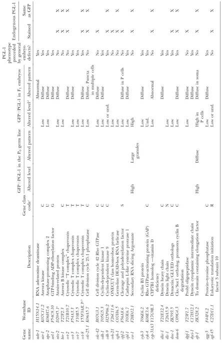
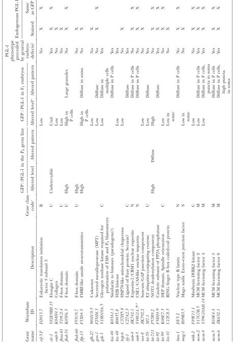
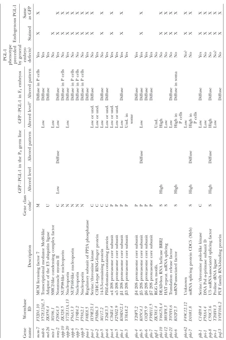
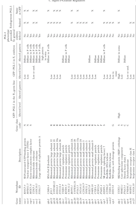
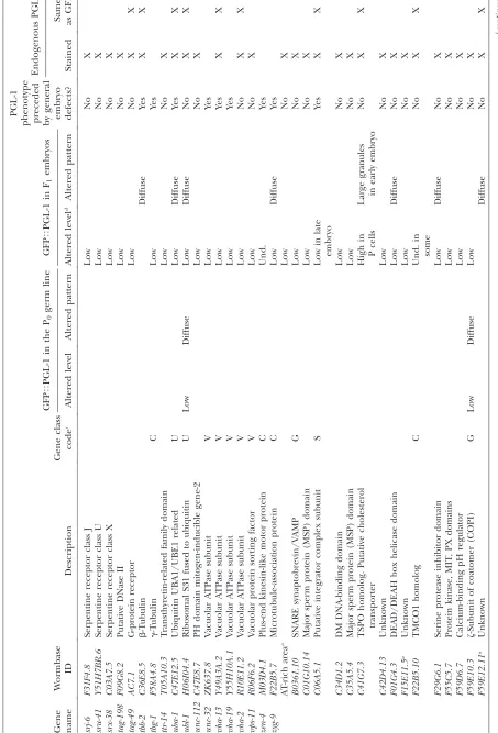
T ABLE 1 GFP T PGL-1 screen positives GFP T PGL-1 in the P0 germ line GFP T PGL-1 in F1 embryos PGL-1
phenotype preceded by
general embryo defects? Endogenous PGL-1 Gene name W ormbase ID Description Gene class code c Altered level Altered pattern Altered level d Altered pattern Stained
Same asGFP
T ABLE 1 (Continued) GFP T PGL-1 in the P0 germ line GFP T PGL-1 in F1 embryos PGL-1
phenotype preceded by
general embryo defects? Endogenous PGL-1 Gene name W ormbase ID Description Gene class code c Altered level Altered pattern Altered level d Altered pattern Stained
Same asGFP
T ABLE 1 (Continued) GFP T PGL-1 in the P0 germ line GFP T PGL-1 in F1 embryos PGL-1
phenotype preceded by
general embryo defects? Endogenous PGL-1 Gene name W ormbase ID Description Gene class code c Altered level Altered pattern Altered level d Altered pattern Stained
Same asGFP
T ABLE 1 (Continued) GFP T PGL-1 in the P0 germ line GFP T PGL-1 in F1 embryos PGL-1
phenotype preceded by
general embryo defects? Endogenous PGL-1 Gene name W ormbase ID Description Gene class code c Altered level Altered pattern Altered level d Altered pattern Stained
Same asGFP
T ABLE 1 (Continued) GFP T PGL-1 in the P0 germ line GFP T PGL-1 in F1 embryos PGL-1
phenotype preceded by
gen eral embryo defects? Endogenous PGL-1 Gene name W ormbase ID Description Gene class code c Altered level Altered pattern Altered level d Altered pattern Stained
Same asGFP
234 genes whose depletion by RNAi caused GFPTPGL-1
phenotypes. These initial positives were taken through two additional rounds of screening, which narrowed our list of positives to 173 genes whose depletion consis-tently caused aberrant GFPTPGL-1 phenotypes (Table
1). GFPTPGL-1phenotypes observed under the
fluores-cence dissecting scope were placed in broad classes of high, low, or undetectable (und.) GFPTPGL-1
accumu-lation (Figure 1B, Table 1). GFPTPGL-1dispersed in the
cytoplasm instead of in perinuclear aggregates was classified as ‘‘diffuse’’; this category included dispersed small granules and PGL-1 signal homogeneously dis-tributed in the cytoplasm. Altered GFP distributions were scored as throughout the embryo or restricted to the germ-line blastomeres. Examples of wild-type GFPTPGL-1 accumulation, diffuse GFPTPGL-1 in
glh-1(RNAi), and high GFPTPGL-1 accumulation in
csr-1(RNAi)are shown in Figure 1C.
We wanted to know if GFPTPGL-1 defects reflect
defects associated with endogenous PGL-1 or instead are specific to the GFP-tagged transgenic protein. The GFPTPGL-1transgene was integrated into the genome
through particle bombardment (Cheeks et al. 2004) and as a consequence should be present at low copy number and shielded from transgene silencing effects. However, since the reporter uses the germ-line-specific pie-1 promoter (Cheeks et al.2004), it is possible that some RNAi depletions specifically affect expression of the transgene and not endogenous PGL-1. It is also possible that the post-transcriptional processing, ex-pression, or stability of transgenicPGL-1is affected by the GFP tag. Therefore, we repeated RNAi against 126 screen positives in wild-type (not transgenic) worms, stained with antibody against PGL-1, and examined the P0germ line and F1embryos using both widefield fluorescence and spinning disk confocal microscopy (Table 1). In the majority of RNAi depletions that we tested (76 of 126), endogenousPGL-1recapitulated the GFPTPGL-1 phenotypes. Furthermore, the improved
resolution of fixed and stained samples allowed us to examine thePGL-1phenotypes in greater detail. How-ever, subtle differences inPGL-1 levels were harder to see in fixed and stained embryos than by GFP imaging. Fifty of 126 RNAi depletions that caused reduced levels of GFPTPGL-1did not cause noticeably reduced levels
of endogenous PGL-1. This set of 50 genes may specifically affect expression of the GFP transgene or may result in changes inPGL-1protein levels or distribu-tion that are more easily seen by GFP than by antibody staining.
As expected, the list of screen positives is significantly enriched for genes expressed in the germ line. Seventy-four of the 173 genes identified are in the set of 3144 genes with germ-line-enriched expression defined by Reinkeet al.(2004), which is a 2.7-fold enrichment over the number expected by chance. One hundred twenty-seven of the 173 genes identified are in the set of 4699
T
ABLE
1
(Continued)
GFP
T
PGL-1
in
the
P0
germ
line
GFP
T
PGL-1
in
F1
embryos
PGL-1
phenotype preceded by
general
embryo defects?
Endogenous
PGL-1
Gene name
W
ormbase ID
Description
Gene
class
code
c
Altered
level
Altered
pattern
Altered
level
d
Altered
pattern
Stained
Same asGFP
K06H7.1
Serine/threonine
protein
kinase
Low
Diffuse
Yes
K12H4.3
rRNA
maturation
of
60S
ribosome
Low
No
X
T03G6.3
Phosphodiesterase
Low
No
X
T04A8.7
Glucosidase
acid
b
-orthologs
Low
or
und.
No
X
X
T06E6.1
Possible
60S
ribosomal
Nop7
factor
Low
No
X
T12D8.6
Calmodulin
containing
Low
or
und.
Yes
T19C3.6
Unknown
Low
No
X
Y47D3A.29
DNA
polymerase
primase
Low
Diffuse
in
P
cells
Yes
ZC477.4
Unknown
Low
No
X
a T
argeted
gene
originally
misannotated
in
Ahringer
RNAi
Library
.
Correct
sequenced
target
is
shown.
b Gene
obtained
more
than
once
in
the
screen.
c Gene
class
letter
code
(see
T
able
2).
d Und.,
genes expressed in dissected gonads as detected using SAGE (Wanget al.2009), which is a 3.5-fold enrichment over the number expected by chance.
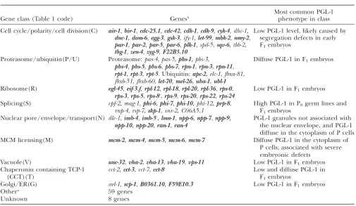
The majority of screen positives were grouped into large gene classes that display common phenotypes (Table 2). These classes are discussed below, followed by more in-depth analysis of an argonaute gene identified in our screen.
Cell cycle/polarity/cell division:The largest group of screen positives can be classified by their role in the cell cycle, establishing cell polarity, and the segregation of cellular components during early cell divisions. This group includes 28 genes of the 173 total screen positives (Table 2). In wild-type animals, P granules are evenly distributed in oocytes and newly fertilized zygotes, but prior to each P-cell division the granules are segregated to the cytoplasm destined for the next P-cell daughter (Strome and Wood1982). P granules left behind in somatic daughters are disassembled or degraded (Hird et al.1996; Saffman and Lasko 1999; DeRenzoet al. 2003; Spike and Strome 2003). RNAi depletions that cause early cell cycle arrest or altered cell division patterns and eventually embryonic lethality may lead to a diffuse or ‘‘missegregated’’ P-granule pattern. In fact, P granules have been used as a marker to study cell polarity for.20 years, and some examples of P-granule
missegregation have been intensively studied (e.g., Gonczyand Rose2005; Cowanand Hyman2007). In our secondary and tertiary screens, we more closely examined whether a defective GFPTPGL-1 phenotype
appeared to be correlated with abnormal nuclear morphology, embryo patterning defects, and/or early arrest (Table 1).PGL-1patterning defects appeared to precede general embryo defects for only 8 of the 28 genes in this class (bir-1, cdc-25.1, cdc-42, let-99, mbk-2, par-1,par-5, andF22B5.10).
As shown in Figure 2, RNAi depletion of genes in the cell cycle/polarity/cell division class tended to show two types of phenotypes. As exemplified bypar-1and cdk-9, RNAi depletion causedPGL-1to be present in all cells of early embryos and reduced or absent from all cells of later embryos. This pattern likely reflects both missegregation of P granules and failure to properly separate germ-line and somatic lineages, with their different abilities to retain (germ-line)vs.disassemble (somatic) P granules (Kemphues et al. 1988). As exemplified by let-99, par-5, and cdc-25.1, RNAi de-pletion causedPGL-1to be present in all cells of early embryos and to persist in numerous cells of later embryos (Rose and Kemphues 1998; Morton et al. 2002). We suspect that the extraPGL-1-containing cells in later embryos are in most cases a consequence of the TABLE 2
Most prominent PGL-1 phenotypes associated with each gene class
Gene class (Table 1 code) Genesb
Most common PGL-1 phenotype in class
Cell cycle/polarity/cell division(C) air-1,bir-1,cdc-25.1,cdc-42,cdk-1,cdk-9,cyk-4,dhc-1,
dnc-1,dom-6,egg-3,gsk-3,ify-1,let-99,mbk-2,nmy-2,
par-1,par-2,par-5,par-6,plk-1,spd-5,sqv-6,tbb-2,
tbg-1,zen-4,zyg-9,F22B5.10
Low PGL-1 level, likely caused by segregation defects in early F1embryos
Proteasome/ubiquitin(P/U) Proteasome:pas-4,pas-5,pbs-1,pbs-3,
pbs-4,pbs-5,pbs-6,pbs-7,rpn-1,rpn-3,rpn-11,
rpt-1,rpt-3,rpt-5. Ubiquitin:apc-2,elc-1,fbxa-81,
fbxb-51,fbxb-60,let-70,mel-26,uba-1,ubl-1
Diffuse PGL-1 in F1embryos
Ribosome(R) egl-45,eif-3.f,rpl-12,rpl-18,rpl-20,rpl-36,rps-0,
rps-3,rps-5,rps-8c,rps-9,rps-20,rps-22,rps-24
Low PGL-1 in F1embryos
Splicing(S) cpf-2,mag-1,phi-6,phi-7,phi-10,phi-12,prp-8,
rnp-4,rsp-7,skp-1,snr-2,C06A5.1
High PGL-1 in P0germ lines and
F1embryos
Nuclear pore/envelope/transport(N) dlc-1,imb-4,imb-5c,lmn-1,npp-6,npp-7,npp-9,
npp-10,npp-20,ran-1,ran-4
PGL-1 granules not associated with the nuclear envelope, and PGL-1 diffuse in the cytoplasm of P cells MCM licensing(M) mcm-2,mcm-4,mcm-5,mcm-6,mcm-7 Diffuse PGL-1 in the cytoplasm of
P cells; associated with severe embryonic defects
Vacuole(V) unc-32,vha-2,vha-13,vha-19,vps-11 Low PGL-1 in F1embryos
Chaperonin containing TCP-1 (CCT)(T)
cct-2,cct-3,cct-7,cct-8c Low and diffuse PGL-1 in
F1embryos
Golgi/ER(G) arl-1,scp-1,B0361.10,F59E10.3 Low PGL-1 in F1embryos
Othera 59 genes
Unknown 8 genes
a
Groups with fewer than three genes not shown.
b
Genes in boldface type exhibit the most common PGL-1 phenotype in their class (described in right column).
c
earlier segregation defects, but another interesting possibility is that they arise by extra divisions of the P-cell lineage.
Proteasome/ubiquitin: The next largest group of screen positives contains 23 protein degradation com-ponents from the proteasome and ubiquitin pathways (Table 2). RNAi depletion of the majority of genes in this class resulted in a characteristic GFPTPGL-1
phe-notype: the uterus was filled with early embryos that exhibit a diffuse PGL-1 distributed throughout the embryo (Table 1, Figure 3A). Often the embryonic phenotype was accompanied by diffusePGL-1 distribu-tion in the P0 germ line. The PGL-1 phenotype displayed by this class is likely a result of compromised protein degradation mechanisms, which may stabilize PGL-1, and by extension P-granule proteins, in both the P0germ line and in embryos (DeRenzoet al.2003; Spike and Strome 2003). This phenotype is probably com-pounded by the early embryonic arrest associated with
the depletion of all but 4 of the 23 genes. These 4 include ubl-1(RNAi), whose RNAi depletion caused PGL-1 granules to persist in multiple cells prior to embryonic arrest, and the F-box-containing genes fbxa-81,fbxb-51, andfbxb-60, whose depletion did not cause embryonic lethality under our experimental condi-tions. This class emphasizes the importance of protein degradation in regulating P-granule patterns in germ lines and embryos.
Ribosome: We obtained 14 genes that encode ribo-somal components in our screen (Table 2). In contrast to the groups mentioned above, most PGL-1 pheno-types in this class (11/14) preceded apparent cell/ embryo death (egl-45,eif-3.f, rpl-12, rpl-20, rpl-36,rps-0, rps-3, rps-5, rps-9, rps-22, and rps-24) (Table 1). RNAi depletion of all 14 of these genes resulted in reduced PGL-1 signal and smallPGL-1 aggregates (Figure 3B). While compromised translation of GFPTPGL-1 or
is also possible that the accumulation of P granules is dependent upon active translation. In comparison to somatic cell differentiation, which relies heavily on transcriptional regulation, this ‘‘ribosome’’ class empha-sizes the crucial role of post-transcriptional regulation in the identity and development of the germ line. Indeed, recent transcript profiling from dissected germ lines revealed that a relatively large percentage of germ-line expressed mRNAs are involved in trans-lation and ribosome structure and biogenesis (Wang et al.2009). P-granule integrity and distribution may be sensitive to the translational status of germ cells and early embryos.
Splicing: Our screen identified 12 genes that encode known or putative splicing factors (Table 2). ThePGL-1 phenotypes of 9 of these genes appeared to precede cell/embryo death (cpf-2,mag-1,phi-6,phi-7,phi-10, phi-12, prp-8, rsp-7, and skp-1) (Table 1). Several PGL-1 phenotypes were shared by multiple members of the splicing class of genes (Figure 4). RNAi depletion of 5 genes (phi-6,phi-7,phi-10, prp-8, and skp-1) resulted in increasedPGL-1signal in the germ line of P0adults and in F1embryos. RNAi depletion of 9 genes (cpf-2,phi-6, phi-7,phi-10,phi-12,prp-8,rnp-4,rsp-7, andskp-1) resulted in some diffuse PGL-1 in the embryonic germ-line
blastomeres and PGL-1 granules that are dissociated from the nuclear periphery. In wild-type embryos, between the 4- and 8-cell stages, P granules start to become perinuclear in the germ-line blastomere, while embryos depleted for splicing factors often contained PGL-1 granules dispersed in the cytoplasm, which persisted until the embryos arrested around the 100-cell stage. This arrest was accompanied by failure of the primordial germ cell (P4) to migrate internally.
Sm components of the splicesome have been shown to localize to germ plasm in multiple species, and previous RNAi depletion of theC. elegans Sm proteins disrupted P-granule patterns in embryos (Moussaet al. 1994; Barbeeet al. 2002; Chuma et al. 2003; Bilinski et al.2004; Barbeeand Evans2006). However, previous RNAi depletion of other splicing components did not disrupt P-granule patterns in embryos (Barbee et al. 2002), suggesting that P-granule integrity and perinu-clear localization in embryonic cells do not depend on pre-mRNA splicing. Our screen demonstrated that at least some pre-mRNA splicing factors are necessary for proper PGL-1 localization to granules at the nuclear periphery in embryos. Since the embryonic germ-line blastomeres are not actively engaged in transcription (Seydoux et al. 1996), the requirement for proper Figure3.—Proteasome/ ubiquitin and ribo-some classes. GFPTPGL-1
(green) in live worms (A) and PGL-1 (green), nu-clear pores (red), and DNA (blue) in fixed em-bryos (B) are shown. Dashed boxes indicate the location of zoomed images on the right. (A) Protea-some/ubiquitin class. De-pletion of proteasome
(e.g., pbs-5) and ubiquitin
components often causes early embryonic arrest with diffuse PGL-1 throughout the embryo. Arrested em-bryos accumulate in the uterus (bracketed bar) af-ter proteasome or ubiqui-tin depletion, which creates a distinctive GFPTPGL-1 phenotype.
(B) Ribosome class. Four-cell embryos are shown. Depletion of core ribo-somal components such as
rps-0, rpl-20, and rpl-36
splicing regulation likely reflects that maternally gener-ated mRNAs must be properly spliced for P granules to localize normally in embryos. Whether properly spliced mRNAs or the process of splicing affect P-granule pattern-ing directly or indirectly remains to be determined.
We noted that several splicing factors obtained in our screen represent one subunit of multisubunit splicing complexes. This finding raised two possibilities: the other subunits in the complex do not participate in regulating P-granule distributions or the other subunits were missed in our single-pass RNAi screen. To distin-guish between these possibilities, we tested all subunits of the SF3b complex, which is absolutely essential for pre-mRNA splicing (Will et al. 1999). This complex contains seven proteins, of which six have clear homo-logs in worms, encoded by the genesW03F9.10,phf-5,
C46F11.4,sap-49,phi-11, andphi-6(supporting informa-tion,Table S1). Onlyphi-6was identified in our screen. Of the six worm genes, only phi-6,phi-11,sap-49, and C46F11.4are targeted by the Ahringer RNAi library. We depleted these four genes by RNAi and found that in addition tophi-6, loss of bothphi-11andsap-49resulted in diffuse PGL-1 in the embryonic germ-line blasto-meres, PGL-1 granules that are dissociated from the nuclear periphery, and failure of P4to migrate internally (Figure 4B,Table S1). Late embryonic arrest was used as a measure of RNAi effectiveness, and unlike phi-6, phi-11, andsap-49, depletion ofC46F11.4did not result in late embryonic arrest, diffusePGL-1, or a P4migration defect. Interestingly, the Ahringer RNAi clone targeting C46F11.4 contains a short sequence that is present in numerous copies throughout the genome, potentially Figure4.—Splicing class. Antibody-stained PGL-1 (green), nuclear pores (red), and DAPI-stained DNA (blue) in fixed embryos are shown. Dashed boxes indicate the location of zoomed images on the right. (A) Approximately 16-cell (left) and150-cell (right) embryos. In wild-type embryos, PGL-1 granules are attached to the periphery of the nucleus in the primordial germ cell P4(left embryo,16 cells) and its daughters Z2and Z3(right embryo,150 cells). P4, Z2, and Z3nuclei are marked with asterisks.
phi-12,phi-10,prp-8,phi-6, andskp-1RNAi embryos contain P granules that are detached from the periphery of the nucleus
(arrow-heads). Higher levels of diffuse PGL-1 in the cytoplasm of Z2/Z3cells ofphi-12,phi-10, andprp-8embryos can be observed in the
areas circumscribed by dashed lines, and Z2/Z3fail to migrate internally. (B) SF3b complex componentsphi-6,phi-11, andsap-49
diluting the effectiveness of this RNAi clone. Taken together, our results suggest that the SF3b complex and at least certain aspects of pre-mRNA splicing are essential for proper PGL-1 localization. These results exemplify the limitations of RNAi screens (incomplete gene coverage, ineffective RNAi, and missing pheno-types in single-pass screens) but also illustrate how RNAi screen positives can lay the foundation for more targeted approaches.
Nuclear pore/envelope/transport: We identified 11 nuclear pore or nuclear pore/envelope associated factors in our screen (Table 2). RNAi depletion of 9 of these resulted in a distinctive phenotype that appeared to precede cell death in early embryos (imb-4, imb-5, lmn-1,npp-6,npp-7,npp-9,npp-10,ran-1, andran-4). In contrast to the perinuclear association ofPGL-1in the more than four-cell stage wild-type embryos, most PGL-1-containing granules in embryos depleted for nuclear pore components remained detached from the nuclear envelope (Figure 5A). The finding that .75% of nuclear pores are overlaid by P granules in the adult germ line (Pitt et al. 2000) predicted that loss of nuclear pore components might compromise associa-tion of P granules with the nuclear periphery.
Other labs have previously demonstrated a require-ment for the nuclear transportinsIMB-2,IMB-3, IMB-5, and IMA-3to maintain P-granule integrity or perinu-clear localization ( J. Ahringer, personal communica-tion; Geles and Adam 2001). There are three imb-5 RNAi clones in the Ahringer RNAi library (two correctly and one incorrectly annotated); our screen identified all three clones as causing PGL-1 granules to be de-tached from the nuclear periphery. However, our screen did not identifyimb-2,imb-3, orima-3. In targeted retests of those three RNAi clones, depletion ofimb-2andimb-3 resulted in detachment of PGL-1 from the nuclear periphery of embryos (Table S2), while depletion of ima-3did not. We note that a stronger RNAi regime by Geles and Adam resulted in diffuse P granules in the adult germ line but, similar to our results, not in embryos (Gelesand Adam2001).
Our screen identified 5 of the 20 NPP proteins inC. elegans. To determine if only a subset of NPPs partic-ipates in tethering PGL-1 granules to the nuclear periphery, or if some NPPs were missed by our screen, we more closely examined PGL-1 patterns after RNAi depleting individual NPPs. The results support both scenarios. Thirteen of the 15 remaining NPPs are covered in the Ahringer RNAi library. Of these 13, RNAi depletion of 4 (npp-1,npp-3,npp-8, andnpp-19) caused PGL-1to be detached from the nuclear periphery in a high enough proportion of embryos that they should have emerged as screen positives (Table S2). Thus, our screen was successful in identifying 5 of 9 of the genes whose depletion results in high penetrance detachment of PGL-1 from the nuclear periphery. We missed 4 genes. Interestingly, RNAi depletion of 3 genes (npp-14,
npp-15, and npp-16) caused late embryonic lethality, demonstrating the effectiveness of RNAi, but did not cause any apparent defects in PGL-1 patterns. These findings suggest that the localization ofPGL-1granules to the nuclear periphery depends on many but not all NPPs.
Our results contribute to the emerging view that the localization of P granules to the nuclear periphery relies heavily upon underlying nuclear pores and nuclear transport. It will be intriguing to determine if this localization depends on a direct interaction between P-granule and nuclear pore components, mRNA trafficking through nuclear pores, or both. We are currently investigating the former possibility and the hypothesis that the GLH components of P granules interact via their phenylalanine–glycine (FG) domains with the FG domains of nuclear pore proteins (see discussion).
Other small classes of screen positives: Another small class of screen positives includes the actin/ microtubule chaperonins cct-2, cct-3, cct-7, and cct-8 (Table 2). Depletion of these CCT proteins resulted in low and diffuse levels of PGL-1 in embryos. Severe embryonic defects prevented further characterization of the PGL-1 phenotype, but we predict that PGL-1 mislocalization (data not shown) stems from compro-mised actin-based P-granule segregation in these mu-tants. Interestingly, it was recently demonstrated that RNAi depletion of other components of this chaper-onin complex,cct-4andcct-6, causes ectopic expression of PGL-1 in the intestine and hypodermis of adult worms, which may contribute to their long-lived phe-notype (Curranet al.2009).
Remaining screen positives can be placed into a handful of smaller categories. Some genes encode components of subcellular organelles including va-cuoles, Golgi, and ER. RNAi depletion typically resulted in decreasedPGL-1 signal (data not shown). We were unable to assign 59 screen positives to a class larger than three members, and the functions of 8 positives are not yet predictable (Table 2).
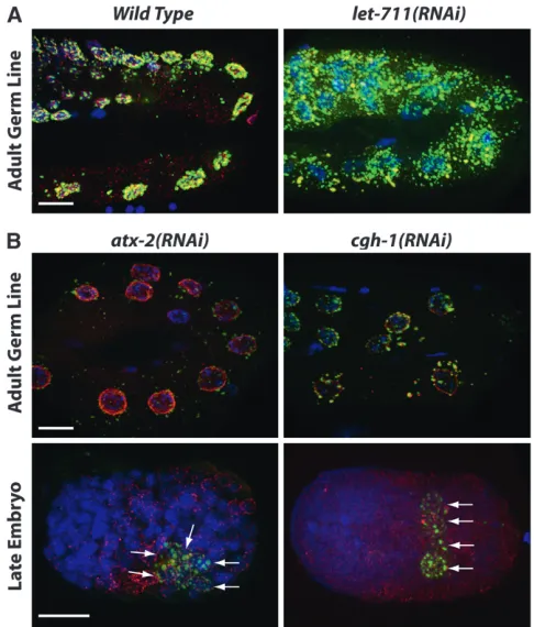
Accumulation of P granules mediated through mRNA homeostasis: In embryos, clusters of poly(A)1
components as being required for regulation ofPGL-1 accumulation. Interestingly, their depletion caused differentPGL-1phenotypes (Figure 6). RNAi depletion of the Not1 componentLET-711resulted in increased PGL-1accumulation in the adult germ line (Figure 6A) and in progeny embryos, while RNAi depletion of the decapping factorsCGH-1andATX-2and the predicted SKI2 exosome homolog F01G4.3 resulted in reduced PGL-1levels (Table 1; Figure 6B). Depletion ofCGH-1 andATX-2 also caused PGL-1 to persist in numerous cells of later embryos (Figure 6B). These findings extend the list of mRNA degradation components that are likely to be shared between P bodies and P granules and support the view that the accumulation of P granules is closely connected to mRNA levels.
CSR-1 downregulates RNA and P-granule accumula-tion through an endo-siRNA pathway in the germ line:
The most strikingPGL-1phenotype observed in the P0 germ line was caused by RNAi ofcsr-1. Depletion ofcsr-1 resulted in very intensePGL-1staining and largePGL-1 aggregates distributed throughout the cytoplasm of the adult germ line and embryos (Figures 1C and 7A). To determine if the largePGL-1aggregates incsr-1(RNAi) germ lines arebona fideP granules, we tested for whether they contain other constitutive components of P gran-ules, such as thePGL-1paralogPGL-3(Kawasakiet al. 2004) and the VASA homolog GLH-1 (Gruidl et al. 1996).PGL-3andGLH-1colocalize with the largePGL-1 aggregates incsr-1(RNAi)adult germ lines (Figure 7C). Quantitative PCR ofpgl-1,pgl-3, andglh-1transcripts in adult hermaphrodites showed a modest increase after csr-1RNAi (1.69-fold forpgl-1,P¼0.0053; 1.12-fold for pgl-3,P¼0.0390; 2.17-fold forglh-1,P¼0.0010). While GFPTPGL-1reporter and antibody-stained endogenous
PGL-1appear dramatically brighter incsr-1(RNAi)worms (Figures 1C and 7A), Western blot analysis revealed a modest 1.4-fold increase inPGL-1protein incsr-1(RNAi) worms compared to wild type (Figure S1), consistent with the 1.7-fold change in pgl-1 transcript noted above. The brighter, larger P granules observed in csr-1(RNAi) embryos may reflect a combination of modestly elevated levels of P-granule proteins and enhanced aggregation of those proteins into granules. We noted an additional phenotype in csr-1(RNAi) embryos. In many RNAi embryos (40%,n¼70),PGL-1 andPGL-3granules were found in somatic blastomeres (Figure 7D). Interestingly,GLH-1did not colocalize with the PGLs in these ectopic granules (Figure 7D). This is reminiscent of the partial P granules (containingPGL-1 and PGL-3; lacking GLH-1) observed in mutant em-bryos defective in autophagy (Zhang et al. 2009), raising the possibility thatCSR-1participates in auto-phagocytic removal of P-granule proteins from somatic cells.
CSR-1is an Argonaute protein with endonucleolytic mRNA cleavage (slicing) activity (Yigitet al.2006; Aoki et al.2007). The slicing activity of Argonautes mediates
have shown that depletion of sperm and arrested ovulation cause RNA to accumulate in the rachis; this phenotype is regulated by the major sperm protein pathway (Schisaet al.2001; Judet al.2008).csr-1(RNAi) animals exhibited no obvious defects in fertilization or ovulation, suggesting that RNA accumulation in csr-1(RNAi)animals is not caused by loss of the major sperm protein pathway. We hypothesize that the accumulation of cytoplasmic RNA and of enlarged P granules
ob-served after depletion ofCSR-1is due to reduced slicing and degradation of mRNAs.
If loss of CSR-1 slicing activity causes enlarged and disorganized P granules, then loss of proteins that function in the CSR-1 pathway should cause similar defects.CSR-1preferentially binds to secondary siRNAs, siRNAs that are produced from primary siRNA-targeted mRNAs by RNA-dependent RNA polymerase (RdRP) (Aoki et al. 2007). We tested whether enlarged and Figure7.—PGL-1 and RNA accumulation in animals depleted of the Argonaute CSR-1. (A) Endogenous PGL-1 staining (green) in wild-type
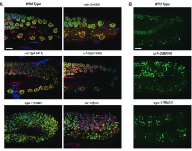
and csr-1(RNAi)germ lines. Depletion of CSR-1
disorganizedPGL-1granules are seen in mutants lack-ing the somatic RdRPRRF-1or either of the two RdRPs that are known to be active in the germ line,EGO-1and RRF-3. We observed an accumulation of large PGL-1 granules in ego-1 mutant germ lines (as observed by Voughtet al.2005) but not inrrf-1orrrf-3mutant germ lines (Figure 8A). ThePGL-1phenotype inego-1andcsr-1 mutants was typically more severe than the phenotype caused by RNAi; in mutant germ lines, the largePGL-1 aggregates were less perinuclear than observed follow-ing RNAi. Mutant germ lines may have less EGO-1 or CSR-1 than can be achieved by RNAi, or alternatively mutant germ lines may have compromised health and show more severe defects. Enlarged PGL-1 granules were not observed in RNAi-defective mutants such as dcr-1 (Voughtet al. 2005) or rde-3(our observations, Figure 8A), suggesting that the phenotype is not due to a general defect in RNAi. Next we examined GFPTPGL-1
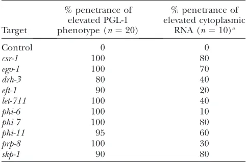
in drh-3(RNAi) germ lines. drh-3 encodes a DExH-box helicase that is required for RdRP activity and shares several phenotypes withego-1andcsr-1(Duchaineet al. 2006; Yigitet al.2006; Aokiet al.2007; Rocheleauet al. 2008; Sheet al.2009). Depletion ofdrh-3phenocopied the PGL-1phenotype observed inego-1andcsr-1RNAi germ lines (Figure 8B), providing further evidence that DRH-3functions in a pathway withEGO-1andCSR-1.
We wanted to see if elevated RNA accumulation correlates with elevated levels ofPGL-1 or if the effect is specific to the depletion ofcsr-1. We found that, like csr-1, depletion of bothego-1anddrh-3also resulted in increased cytoplasmic RNA as detected by SYTO14
staining (Table 3). We also examined other factors whose depletion resulted in elevatedPGL-1in the adult germ line (eft-1,let-711,phi-6,phi-7,phi-11,prp-8, andskp-1). In general, increased PGL-1 levels correlated with increased cytoplasmic RNA. Depletion of csr-1, ego-1, and the splicing factors phi-7, phi-11, and skp-1 had particularly strong effects on RNA patterns (Table 3). At this time we do not know if the accumulation of cytoplasmic RNA results from loss of function of CSR-1,EGO-1,DRH-3, and the handful of RNA-binding proteins whose depletion results in elevatedPGL-1or if the accumulation of RNA results from displaced and elevatedPGL-1itself. A third possibility is that P-granule proteins and cytoplasmic RNAs interact with and stabilize each other.
CSR-1, EGO-1, and DRH-3 are thought to work together in a germ-line-specific endogenous siRNA pathway (Maineet al.2005; Robertet al.2005; Duchaine et al.2006; Yigitet al.2006; Aokiet al.2007; Rocheleau et al.2008; Sheet al.2009). Argonautes, likeCSR-1, have been identified as germ granule components from worms to mammals (Kotaja et al.2006; Batista et al. 2008; Wang and Reinke 2008). In addition, an RGG domain, like that found inPGL-1andPGL-3, in the N terminus ofCSR-1 also suggests that CSR-1is likely to associate with P granules. To test that prediction, we attempted to generate good anti-CSR-1 antibodies and informative GFPTCSR-1 transgenic worms. Although
both antibodies and transgenic worms showed P-granule localization, neither passed our tests for specificity. Nevertheless, on the basis of the specificPGL-1 pheno-Figure 8.—PGL-1 accu-mulates in numerous large granules in the germ line after an endogenous siRNA pathway is compromised. (A) Endogenous PGL-1 staining (green) resembles wild type in the germ lines
ofrde-1,rrf-1, andrrf-3
mu-tants. Large PGL-1 gran-ules accumulate in the germ lines of ego-1 and
csr-1mutants. Most granules
are dissociated from the nuclear periphery. (B) GFPTPGL-1 accumulation
in control(RNAi),
drh-3(RNAi), and ego-1(RNAi)
type of CSR-1 pathway mutants and the predicted P-granule localization of CSR-1, we hypothesize that P granules function as a center for endogenous siRNA silencing in the germ line and that disruption of this endo-siRNA pathway results in not only the accumulation in the germ-line syncytium of RNA transcripts that in wild type are silenced and degraded, but also the accumula-tion of large P granules.
DISCUSSION
Our RNAi screen proved to be an effective method to identify genes whose depletion causes aberrantPGL-1 phenotypes. One advantage of performing an RNAi screen over a forward mutagenesis screen is that mutants are not required to be viable and fertile. This allowed us to identify multiple gene classes, including some essential genes that are required forPGL-1assembly and localiza-tion. One disadvantage of RNAi screens is missing some genes due to ineffective RNAi or incomplete RNAi library coverage. To expedite our screen, RNAi was performed for only 30 hr, and only P0and F1progeny were examined. Because of the maternal contribution of many P-granule proteins, some phenotypes, like that caused byDEPS-1loss, are not observable until the F2 generation after RNAi is started. Our P0/F1screen did not identify any additional genes whose depletion causes thePGL-1phenotypes ofdeps-1andglh-1mutants where PGL-1 is dispersed in all cells of normally developing embryos. We do not know ifDEPS-1andGLH-1are the only proteins whose loss causes that specific phenotype or if an F2generation screen could identify more genes with that phenotype.
Despite the limitations of RNAi screens, this type of genomewide screen serves as an excellent starting point for identification and in-depth analysis of gene families in particular cellular processes. For example, the 5 NPPs identified in our screen drew our attention to nuclear pores as being involved in P-granule localization. Tar-geted retests of the remaining 13 NPP genes represented in the RNAi library identified 4 NPP genes that our screen should have identified, 5 NPP genes that have low-penetrance RNAi phenotypes and therefore require other analysis strategies, and 4 NPP genes that may not participate in regulating P-granule localization. Another example is our identification of one subunit of the SF3b splicing complex. Targeted retests identified two addi-tional subunits. Loss of the remaining three SF3b components, which are not covered or inefficiently covered in the RNAi library, is likely to result inPGL-1 phenotypes as well. Finally, a recent report used a similar RNAi screen and identified a single autophagy compo-nent,lgg-1, which is required for clearing small P granules from somatic cells. Targeted tests of other autophagy components, by injection of double-stranded RNA to achieve stronger RNAi, revealed their involvement in clearing P granules from somatic cells (Zhang et al.
2009). Certainly, the 173 genes identified in our screen are an underestimate. We expect more stringent RNAi procedures, such as injecting or soaking or feeding for longer times, will identify additional genes that affect P granules inC. elegans.
The majority of genes identified in our screen were grouped into large gene classes. While some classes were likely identified because of P-granule segregation or degradation defects (cell cycle/polarity/cell division and proteasome/ubiquitin), most classes associate P granules with post-transcriptional regulation in the germ line. For example, splicing, nuclear pore, and ribosomal classes emphasize the importance of mRNA maturation, nuclear transport, and translational regulation in con-trolling P-granule size and location.
Nascent transcripts are processed in the nucleus by spliceosomes prior to their export as mature mRNAs. Sm proteins are core components of nuclear spliceo-somes and are also concentrated in the cytoplasmic germ granules of multiple species (Moussaet al.1994; Chuma et al. 2003; Barbee et al. 2002; Bilinskiet al. 2004). InC. elegans, Sm proteins are required for proper P-granule localization to the nuclear periphery; it has been proposed that this role is independent of pre-mRNA splicing (Barbee and Evans 2006). We, too, identified the Sm protein,SNR-2, as required for proper PGL-1 granule assembly. We also identified other splicing and mRNA maturation components. These include the cleavage and polyadenylation factor CPF-2 and the putative integrator complex subunitC06A5.1, both of which are involved in maturation of 39ends of mRNA; the putative exon–exon junction factorsMAG-1 and RNP-4; possible snRNP associated factor PRP-8 along with SF3b splicesome components 6, PHI-11, andSAP-49; and other factors whose homologs are required for pre-mRNA splicing, including RSP-7,
TABLE 3
Correlation of elevated PGL-1 and elevated RNA in RNAi germ lines
Target
% penetrance of elevated PGL-1 phenotype (n¼20)
% penetrance of elevated cytoplasmic
RNA (n¼10)a
Control 0 0
csr-1 100 80
ego-1 100 70
drh-3 80 40
eft-1 90 20
let-711 100 40
phi-6 100 10
phi-7 100 80
phi-11 95 60
prp-8 100 30
skp-1 90 80
a
SKP-1, PHI-7, PHI-10, and PHI-12. While the role of Sm proteins in germ granules may still be independent of their role in splicing, our results suggest that proper mRNA maturation in the nucleus affects the assembly and localization of P granules. It will be interesting to determine whether properly assembled P granules require specific features of spliced mRNAs, splicing factors associated with mRNAs, or mRNA export through nuclear pores.
The integrity of nuclear pores appears to be vital for the perinuclear distribution of P granules. At least 14 of 20 nuclear pore components are required forPGL-1’s perinuclear distribution, as are nuclear transport factors such asIMB-1, IMB-5,RAN-1, andRAN-4. Nuclear lamin LMN-1is also required, as is dynein light chain-1,DLC-1. DLC-1’s yeast homolog DYN2 is a nucleoporin (Stelter et al.2007), and interestinglyC. elegansDLC-1was found to interact with PGL-3 in a high-throughput binding assay (Liet al.2004). Therefore, it is possible that some nucleoporins, like the putative nucleoporin DLC-1, provide a physical linkage between P granules and nuclear pores. On that note, we find it interesting that the P-granule componentsGLH-1,GLH-2, andGLH-4 contain multiple Phe–Gly (FG) repeat domains, do-mains that are found in many nucleoporins (Rexach and Blobel 1995). Some FG domains form cohesive hydrophobic interactions with other FG domains in the nuclear pore (Patelet al.2007). The close association between P granules and nuclear pores, as well as similar-ities between GLH proteins and nucleoporins, has led to the prediction that P granules serve as an extension of nuclear pores in the germ line (Kuznicki et al.2000; Pitt et al.2000; Schisa et al. 2001). We are currently testing whether the FG domains ofGLH-1,GLH-2, and GLH-4 interact with each other and with other FG-containing nucleoporins and whether FG proteins in P granules do indeed serve to extend the nuclear pore environment.
As RNAs transcribed in germ nuclei exit through nuclear pores, many transcripts will encounter P gran-ules. Poly(A)1
and developmentally regulated mRNAs have been shown to accumulate in P granules (Seydoux and Fire1994; Schisaet al.2001). P granules may act as a scaffold for multiple RNA processing activities, possi-bly by providing an environment that facilitates germ-line-specific binding interactions and regulates the translation, transport, modification, storage, and/or degradation of RNA. Our screen results support many of these possibilities. For example, we found that multiple ribosomal components are required for proper P-granule accumulation and stability. This find-ing could reflect a need for synthesis of P-granule proteins or a requirement for ongoing translation during the assembly of P granules.
We observed PGL-1 phenotypes when RNA dead-enlyase, decapping, and degradation components were depleted. Depletion ofLET-711, the NOT1 ortholog of
the CCR4/NOT1 mRNA deadenylation complex, had previously been reported to cause PGL-1 localization outside of the embryonic germ line (Galloet al.2008). We identified LET-711 primarily on the basis of the elevated accumulation of transgenic and endogenous PGL-1 in the adult germ line. This phenotype was observed within 30 hr after initiation of RNAi and before germ-line morphology became altered, suggest-ing that thePGL-1phenotype was a direct result of LET-711 depletion. The PGL-1 aggregates inlet-711(RNAi) germ lines are smaller than those observed in csr-1 mutants, but as in the case of csr-1, the enhanced accumulation may be correlated with RNA stabilization. The accumulation of P granules may be sensitive to cytoplasmic RNA levels, requiring RNA destabilizing factors like LET-711 andCSR-1 to control RNA levels through exo- or endonucleolytic activity.
One exciting gene that emerged from our screen was csr-1.CSR-1is an Argonaute that exhibits endonucleo-lytic or slicing activity on RNA targets, showing a preference for using secondary siRNAs generated by RdRP activity rather than primary siRNAs generated by Dicer (Aoki et al.2007). Thus far,C. elegans is unique among animals in its large number of Argonaute-encoding genes: 27 in C. elegans compared to 5 in Drosophila and 8 in humans (Hutvagnerand Simard 2008). The diversity of Argonautes in C. elegans has proven advantageous for dissecting out the multiple roles of these proteins.C. elegansArgonautesALG-1and ALG-2 are essential for miRNA-mediated gene silenc-ing,RDE-1for exogenous RNAi,ERGO-1 for endoge-nous RNAi, andPRG-1andPRG-2for piRNA-mediated gene silencing, and a handful of other Argonautes are thought to bind secondary siRNAs but lack the ability to slice RNA targets (Grishoket al.2001; Yigitet al.2006; Batistaet al.2008; Daset al.2008; Wangand Reinke 2008).csr-1was first obtained in a germ-line cosuppres-sion screen, which suggested that it may be involved in silencing repetitive transgene arrays in the germ line (Robert et al. 2005). csr-1 worms are also partially deficient in RNAi (Yigit et al. 2006). It was recently discovered that csr-1 shares multiple phenotypes with ego-1 and drh-3, which encode an RdRP and an RdRP interacting factor required for small endogenous RNAi in the germ line (Aokiet al.2007; Rocheleauet al.2008; Sheet al.2009).
promote a chromatin state that regulates nuclear pore distribution and that the clumping of nuclear pore material leads to perturbed P-granule assembly (Voughtet al.2005). This model is strengthened by the observation that EGO-1 is found in nuclear fractions and is required for unpaired chromosomal regions to accumulate high levels of histone H3 lysine K9 dime-thylation, a mark associated with heterochromatin assembly and transcriptional silencing (Maine et al. 2005; Sheet al.2009). However, we observed a different PGL-1phenotype when nuclear pore components were depleted (PGL-1granules dissociated from the nuclear periphery in germ-line blastomeres of developing em-bryos) than we did when CSR-1, EGO-1, and DRH-3 were depleted (enhanced accumulation of largePGL-1 granules in the adult germ line). Loss of the nuclear envelope componentLMN-1, which also results in the clustering of nuclear pores (Liuet al.2000), causes a P-granule phenotype that is more similar to that caused by depletion of core nuclear pore proteins than to the large P-granule phenotype caused by depletion of CSR-1, EGO-1, and DRH-3. Another simple model is that genes typically silenced by endo-RNAi in the germ line are desilenced when csr-1 is depleted, allowing these genes’ transcripts to accumulate in the cytoplasm. In normal germ lines, these transcripts could be silenced at the level of chromatin and regulation of transcription or at the level of exiting the nucleus into P granules where they are sliced byCSR-1and subsequently degraded. As all known P-granule components are thought to func-tion in RNA metabolism, the accumulafunc-tion of large P granules incsr-1animals may be due to increased levels of cytoplasmic RNA incsr-1germ lines. Another possi-bility is that transcripts encoding P-granule proteins are normally downregulated by endo-RNAi, and the loss of this downregulation in csr-1 animals leads to the accu-mulation of large P granules. Profiling germ-line tran-scripts incsr-1mutants may reveal which, if any, of these models are correct.
The P-granule components DEPS-1 and PGL-1 are both required for a robust exogenous RNAi response (Robertet al.2005; Spikeet al.2008b). WhilePGL-1has not been implicated in a specific RNAi pathway,deps-1 mutants are thought to resist the effects of RNAi due to a 7- to 10-fold decrease in rde-4 expression (Spike et al. 2008b).RDE-4binds dsRNA and forms a complex with Dicer to produce siRNAs, andrde-4mutants are RNAi defective (Tabara et al. 2002). Another component identified in screens for RNAi-defective mutants, RDE-3, is thought to act downstream of RDE-4and Dicer. Comparison of transcriptional profiles ofrde-3and deps-1mutants showed that nearly 30% of genes upregulated indeps-1mutant germ lines are also upregulated inrde-3 worms, suggesting that RDE-3 and DEPS-1 function together to regulate expression of at least their shared target genes (Spike et al. 2008b). As DEPS-1 is a constitutive P-granule component, it likely participates
in regulation at a post-transcriptional level. Our results withCSR-1,EGO-1, andDRH-3strengthen the emerging view that P granules and RNAi are intimately related: P-granule components participate in regulating RNAi, and RNAi factors influence the structure, localization, and perhaps function of P granules. Better defining the RNAi– P-granule relationship will shed light on mechanisms of post-transcriptional gene regulation used by germ cells.
We thank Colin Thacker for training provided during the screen, use of equipment including the Copas Biosort, and preparation of RNAi colonies from the Ahringer RNAi library; Robb Cundick for the development and use of aC. elegansRNAi database; Susan Mango for the use of lab space and equipment during the first pass of screening; Julie Ahringer for unpublished information on theimb-2,imb-3, and imb-5PGL-1phenotypes; Xingyu She and Eleanor Maine for providing
ego-1strain EL500; Andreas Rechtsteiner for comparison of germ-line
data sets; and the National Bioresource Project at Tokyo Women’s Medical College for providing the FX892 strain. This work was supported by Ruth Kirchstein National Research Service Award postdoctoral fellowship GM084673 (to D.U.) and National Institutes of Health (NIH) grant GM34059 (to S.S.). SomeC. elegansstrains were provided by the Caenorhabditis Genetics Center, which is funded by the NIH National Center for Research Resources.
Note added in proof: Since the acceptance of this article for publication, an additional study was published that shows abnormal P granules incsr-1,drh-3, andego-1mutants ( J. M. Claycomb, P. J.
Batista, K. M. Pang, W. Gu, J. J. Vasaleet al., 2009, The Argonaute CSR-1 and its 22G-RNA cofactors are required for holocentric chromosome segregation. Cell139:123–134). Claycombet al.also
show thatCSR-1,DRH-3, andEGO-1colocalize with P granules.
LITERATURE CITED
Aoki, K., H. Moriguchi, T. Yoshioka, K. Okawaand H. Tabara,
2007 In vitro analyses of the production and activity of second-ary small interfering RNAs in C. elegans. EMBO J.26:5007–5019. Barbee, S. A., and T. C. Evans, 2006 The Sm proteins regulate germ
cell specification during early C. elegans embryogenesis. Dev. Biol.291:132–143.
Barbee, S. A., A. L. Lublinand T. C. Evans, 2002 A novel function
for the Sm proteins in germ granule localization during C. ele-gans embryogenesis. Curr. Biol.12:1502–1506.
Batista, P. J., J. G. Ruby, J. M. Claycomb, R. Chiang, N. Fahlgren et al., 2008 PRG-1 and 21U-RNAs interact to form the piRNA complex required for fertility in C. elegans. Mol. Cell31:67–78. Bilinski, S. M., M. K. Jaglarz, B. Szymanska, L. D. Etkinand M.
Kloc, 2004 Sm proteins, the constituents of the spliceosome,
are components of nuage and mitochondrial cement in Xenopus oocytes. Exp. Cell Res.299:171–178.
Blobel, G., 1985 Gene gating: a hypothesis. Proc. Natl. Acad. Sci.
USA82:8527–8529.
Blow, J. J., and A. Dutta, 2005 Preventing re-replication of
chro-mosomal DNA. Nat. Rev. Mol. Cell. Biol.6:476–486.
Brenner, S., 1974 The genetics ofCaenorhabditis elegans. Genetics
77:71–94.
Cheeks, R. J., J. C. Canman, W. N. Gabriel, N. Meyer, S. Strome et al., 2004 C. elegans PAR proteins function by mobilizing and stabilizing asymmetrically localized protein complexes. Curr. Biol.14:851–862.
Chuma, S., M. Hiyoshi, A. Yamamoto, M. Hosokawa, K. Takamune et al., 2003 Mouse Tudor Repeat-1 (MTR-1) is a novel compo-nent of chromatoid bodies/nuages in male germ cells and forms a complex with snRNPs. Mech. Dev.120:979–990.
Cowan, C. R., and A. A. Hyman, 2007 Acto-myosin reorganization
Curran, S. P., X. Wu, C. G. Riedeland G. Ruvkun, 2009 A
soma-to-germline transformation in long-lived Caenorhabditis elegans mutants. Nature459:1079–1084.
Das, P. P., M. P. Bagijn, L. D. Goldstein, J. R. Woolford, N. J.
Lehrbachet al., 2008 Piwi and piRNAs act upstream of an
en-dogenous siRNA pathway to suppress Tc3 transposon mobility in the Caenorhabditis elegans germline. Mol. Cell31:79–90. DeRenzo, C., K. J. Reeseand G. Seydoux, 2003 Exclusion of germ
plasm proteins from somatic lineages by cullin-dependent degra-dation. Nature424:685–689.
Duchaine, T. F., J. A. Wohlschlegel, S. Kennedy, Y. Bei, D. Conte,
Jr.et al., 2006 Functional proteomics reveals the biochemical
niche of C. elegans DCR-1 in multiple small-RNA-mediated path-ways. Cell124:343–354.
Eddy, E. M., 1975 Germ plasm and the differentiation of the germ
cell line. Int. Rev. Cytol.43:229–280.
Encalada, S. E., P. R. Martin, J. B. Phillips, R. Lyczak, D. R. Hamill et al., 2000 DNA replication defects delay cell division and disrupt cell polarity in early Caenorhabditis elegans embryos. Dev. Biol.
228:225–238.
Ephrussi, A., and R. Lehmann, 1992 Induction of germ cell
forma-tion by oskar. Nature358:387–392.
Gallo, C. M., E. Munro, D. Rasoloson, C. Merrittand G. Seydoux,
2008 Processing bodies and germ granules are distinct RNA granules that interact in C. elegans embryos. Dev. Biol.323:76–87. Geles, K. G., and S. A. Adam, 2001 Germline and developmental
roles of the nuclear transport factor importin alpha3 in C. ele-gans. Development128:1817–1830.
Gonczy, P., and L. S. Rose, 2005 Asymmetric cell division and axis
formation in the embryo. WormBook:1–20. www.wormbase.org Grishok, A., A. E. Pasquinelli, D. Conte, N. Li, S. Parrishet al.,
2001 Genes and mechanisms related to RNA interference reg-ulate expression of the small temporal RNAs that control C. elegans developmental timing. Cell106:23–34.
Gruidl, M. E., P. A. Smith, K. A. Kuznicki, J. S. McCrone, J. Kirchner et al., 1996 Multiple potential germ-line helicases are compo-nents of the germ-line-specific P granules of Caenorhabditis ele-gans. Proc. Natl. Acad. Sci. USA93:13837–13842.
Guedes, S., and J. R. Priess, 1997 The C. elegans MEX-1 protein is
present in germline blastomeres and is a P granule component. Development124:731–739.
Hanazawa, M., I. Kawasaki, H. Kunitomo, K. Gengyo-Ando, K. L.
Bennettet al., 2004 The Caenorhabditis elegans eukaryotic
initiation factor 5A homologue, IFF-1, is required for germ cell proliferation, gametogenesis and localization of the P-granule component PGL-1. Mech. Dev.121:213–224.
Hird, S. N., J. E. Paulsenand S. Strome, 1996 Segregation of germ
granules in living Caenorhabditis elegans embryos: cell-type-spe-cific mechanisms for cytoplasmic localisation. Development122:
1303–1312.
Hutvagner, G., and M. J. Simard, 2008 Argonaute proteins: key
players in RNA silencing. Nat. Rev. Mol. Cell. Biol.9:22–32. Illmensee, K., and A. P. Mahowald, 1974 Transplantation of
pos-terior polar plasm in Drosophila. Induction of germ cells at the anterior pole of the egg. Proc. Natl. Acad. Sci. USA71:1016– 1020.
Jud, M. C., M. J. Czerwinski, M. P. Wood, R. A. Young, C. M. Gallo et al., 2008 Large P body-like RNPs form in C. elegans oocytes in response to arrested ovulation, heat shock, osmotic stress, and anoxia and are regulated by the major sperm protein pathway. Dev. Biol.318:38–51.
Kamath, R. S., M. Martinez-Campos, P. Zipperlen, A. G. Fraserand
J. Ahringer, 2001 Effectiveness of specific RNA-mediated
in-terference through ingested double-stranded RNA in Caeno-rhabditis elegans. Genome Biol.2:RESEARCH0002.
Kamath, R. S., A. G. Fraser, Y. Dong, G. Poulin, R. Durbinet al.,
2003 Systematic functional analysis of the Caenorhabditis ele-gans genome using RNAi. Nature421:231–237.
Kawasaki, I., Y. H. Shim, J. Kirchner, J. Kaminker, W. B. Wood et al., 1998 PGL-1, a predicted RNA-binding component of germ granules, is essential for fertility in C. elegans. Cell94:
635–645.
Kawasaki, I., A. Amiri, Y. Fan, N. Meyer, S. Dunkelbargeret al.,
2004 The PGL family proteins associate with germ granules
and function redundantly inCaenorhabditis elegansgermline de-velopment. Genetics167:645–661.
Kemphues, K. J., J. R. Priess, D. G. Morton and N. S. Cheng,
1988 Identification of genes required for cytoplasmic localiza-tion in early C. elegans embryos. Cell52:311–320.
Kotaja, N., S. N. Bhattacharyya, L. Jaskiewicz, S. Kimmins, M.
Parvinenet al., 2006 The chromatoid body of male germ cells:
similarity with processing bodies and presence of Dicer and mi-croRNA pathway components. Proc. Natl. Acad. Sci. USA103:
2647–2652.
Kuznicki, K. A., P. A. Smith, W. M. Leung-Chiu, A. O. Estevez, H. C.
Scottet al., 2000 Combinatorial RNA interference indicates
GLH-4 can compensate for GLH-1; these two P granule compo-nents are critical for fertility in C. elegans. Development127:
2907–2916.
Lall, S., F. Pianoand R. E. Davis, 2005 Caenorhabditis elegans
de-capping proteins: localization and functional analysis of Dcp1, Dcp2, and DcpS during embryogenesis. Mol. Biol. Cell 16:
5880–5890.
Leacock, S. W., and V. Reinke, 2008 MEG-1 and MEG-2 are
embryo-specific P-granule components required for germline develop-ment inCaenorhabditis elegans. Genetics178:295–306.
Lewis, J. A., and J. T. Fleming, 1995 Basic culture methods.
Meth-ods Cell Biol.48:3–29.
Li, S., C. M. Armstrong, N. Bertin, H. Ge, S. Milstein et al.,
2004 A map of the interactome network of the metazoan C. elegans. Science303:540–543.
Liu, J., T. RolefBen-Shahar, D. Riemer, M. Treinin, P. Spannet al.,
2000 Essential roles for Caenorhabditis elegans lamin gene in nuclear organization, cell cycle progression, and spatial organiza-tion of nuclear pore complexes. Mol. Biol. Cell11:3937–3947. Maine, E. M., J. Hauth, T. Ratliff, V. E. Vought, X. Sheet al.,
2005 EGO-1, a putative RNA-dependent RNA polymerase, is re-quired for heterochromatin assembly on unpaired DNA during C. elegans meiosis. Curr. Biol.15:1972–1978.
Mello, C. C., B. W. Draper, M. Krause, H. Weintrauband J. R. Priess,
1992 The pie-1 and mex-1 genes and maternal control of blasto-mere identity in early C. elegans embryos. Cell70:163–176. Morton, D. G., D. C. Shakes, S. Nugent, D. Dichoso, W. Wanget al.,
2002 The Caenorhabditis elegans par-5 gene encodes a 14-3-3 protein required for cellular asymmetry in the early embryo. Dev. Biol.241:47–58.
Moussa, F., R. Okoand L. Hermo, 1994 The immunolocalization of
small nuclear ribonucleoprotein particles in testicular cells dur-ing the cycle of the seminiferous epithelium of the adult rat. Cell Tissue Res.278:363–378.
Navarro, R. E., E. Y. Shim, Y. Kohara, A. Singsonand T. K. Blackwell,
2001 cgh-1, a conserved predicted RNA helicase required for ga-metogenesis and protection from physiological germline apoptosis in C. elegans. Development128:3221–3232.
Orban, T. I., and E. Izaurralde, 2005 Decay of mRNAs targeted by
RISC requires XRN1, the Ski complex, and the exosome. RNA
11:459–469.
Patel, S. S., B. J. Belmont, J. M. Sante and M. F. Rexach,
2007 Natively unfolded nucleoporins gate protein diffusion across the nuclear pore complex. Cell129:83–96.
Pitt, J. N., J. A. Schisaand J. R. Priess, 2000 P granules in the germ
cells of Caenorhabditis elegans adults are associated with clusters of nuclear pores and contain RNA. Dev. Biol.219:315–333. Reinke, V., I. S. Gil, S. Wardand K. Kazmer, 2004 Genome-wide
germline-enriched and sex-biased expression profiles in Caeno-rhabditis elegans. Development131:311–323.
Rexach, M., and G. Blobel, 1995 Protein import into nuclei:
asso-ciation and dissoasso-ciation reactions involving transport substrate, transport factors, and nucleoporins. Cell83:683–692.
Robert, V. J., T. Sijen, J. vanWolfswinkeland R. H. Plasterk,
2005 Chromatin and RNAi factors protect the C. elegans germ-line against repetitive sequences. Genes Dev.19:782–787. Rocheleau, C. E., K. Cullison, K. Huang, Y. Bernstein, A. C.
Spilker et al., 2008 TheCaenorhabditis elegans ekl (enhancer
of ksr-1 lethality) genes include putative components of a germ-line small RNA pathway. Genetics178:1431–1443.
Rose, L. S., and K. Kemphues, 1998 The let-99 gene is required for