78131
ISOLATION OF BACTERIA IN EFFECTIVE MICROORGANISMS (EM) SUITABLE FOR
DEGRADATION OF THIOSULFATE
ZAINOHA ZAKARIA
MADIHAH MD SALLEH
NUR SUHANAWATI BT ASHAARI
MADZLAN AZIZ
FACULTY OF SCIENCE
UNIVERSITI TEKNOLOGI MALAYSIA
ABSTRACT
A total of 60 potential sulfur-oxidizing bacteria isolated from the Effective
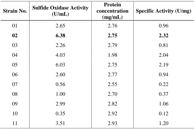
Microorganisms (EM) sample were screened for sulfide oxidase producer. Out of 60
isolates, 11 strains were able to produce sulfide oxidase and the most superior sulfide
oxidase producer was known as strain SO02. Strain SO02 produced 6.83 U/ml sulfide
oxidase with specific activity of 2.32 U/mg. Strain SO02 was later identified as
Serratia
ABSTRAK
Sejumlah 60 bakteria berpotensi mengoksidakan sulfur telah dipencilkan
daripada sampel Mikroorganisma Efektif (EM) dan telah disaring untuk penghasil sulfid
oksida. Daripada 60 isolat, 11 strain menunjukkan keupayaan menghasilkan sulfid
oksida dan penghasil terbaik sulfid oksida dikenali sebagai strain SO02.
TABLE OF CONTENTS
CHAPTER
TITLE
PAGE
1
INTRODUCTION
1.1
Introduction
1
1.2
Objectives of the Study
3
1.3
Scope of the Study
3
2
LITERATURE REVIEW
2.1
Problem Background
5
2.2
Sulfur and the Importance of Sulfur
6
2.3
Sulfur Cycle
8
2.4
Sources of Sulfur Compounds
11
2.5
The Detrimental Effects of Reduced Sulfur
Compounds
14
2.6
Control of Sulfides and Reduced Sulfur
Compounds
15
2.7
Sulfur-Oxidizing Bacteria (SOB)
16
2.8
Sulfide Oxidase
21
2.9
Oxidation of Inorganic Sulfur Compounds
Pathway
22
2.10
Application of Sulfide Oxidase
23
2.11
Concluding Remarks
24
3
GENERAL MATERIALS AND METHODS
3.1
Sulfur-Oxidizer Medium Preparation
25
3.2
Culture Maintenance and Preservation
26
3.3
Experimental Design
26
3.5
Analytical Procedures
28
3.5.1
Determination of Cell Mass Concentration
28
3.5.2
Determination of Specific Growth Rate
and Doubling Time
29
3.5.3
Determination of Kinetic Parameters
30
3.5.4
Sulfate Ion Determination
33
3.5.4.1
Sulfate Calibration Curve
33
3.5.5
Sulfide Oxidase Assay
34
3.5.6
Protein Concentration Determination
35
3.5.7
Thiosulfate Determination
35
4
ISOLATION, SCREENING AND IDENTIFICATION
OF POTENTIAL
SULFUR-OXIDIZER FROM
EFFECTIVE ICROORGANISMS (EM) SAMPLE
4.1
Introduction
36
Experimental Design
37
Materials and Methods
4.3.1
Media Preparation
39
4.3.2
Pre-treatment of Effective Microorganisms (EM)
Sample
39
4.3.3
Isolation of Sulfur-Oxidizing Bacteria
39
4.3.4
Screening of Sulfur-Oxidizing Bacteria
40
4.3.5
Analytical Procedures
40
4.3.6
Morphological Characterization
41
4.3.6.1
Colony Morphology
41
4.3.6.2
Cellular Morphology
41
Gene Analysis
4.3.8.1
DNA Extraction
42
4.3.8.2
Analyzing Genomic DNA
45
4.3.8.3
Polymerase Chain Reaction (PCR)
45
4.3.8.4
Analyzing PCR Products
46
4.3.8.5
Purifying PCR Products
47
4.3.8.6
Sequence Analyses of 16S rRNA
48
4.4
Results and Discussions
48
4.4.1
Isolation and Screening of Sulfur-Oxidizing Bacteria
48
4.4.2
Characterization and Identification of Potential
Sulfur-Oxidizing Bacteria
50
4.4.2.1
Morphological and Cultural Characteristics
50
4.4.2.2
Biochemical Features of strain SO02
52
4.4.2.3
Identification of strain SO02 using 16S
rRNA Gene Analysis
56
4.5
Conclusion
58
5
OPTIMIZATION OF SULFIDE OXIDASE PRODUCTION
BY
Serratia
sp. strain SO02
5.1
Introduction
59
5.2
Experimental Design
60
5.3
Materials and Methods
60
5.3.1
Media Preparation
60
5.3.2
Analytical Procedures
62
5.3.2.1
Tetrathionate, Thiocyanate and Sulfide
Determination
62
5.3.3
Optimization of Sulfide Oxidase Production in
Shake Flask Culture
63
5.3.3.3
Effect of Various Nitrogen Sources
64
5.3.3.4
Effect of Various Peptone Concentrations
65
5.3.3.5
Effect of Various Thiosulfate
Concentrations
65
5.3.3.6
Effect of Various Reduced Inorganic
Sulfur Sources
66
5.4
Results and Discussion
66
5.4.1
Optimization of Sulfide Oxidase Production 66
5.4.1.1
Effect of Initial pH on Sulfide Oxidase
Production
66
5.4.1.2
Effect of Temperature on Sulfide Oxidase
Production
70
5.4.1.3
Effect of Nitrogen Sources on Sulfide
Oxidase Production
73
5.4.1.4
Effect of Peptone Concentration on Sulfide
Oxidase Production
79
5.4.1.5
Effect of Thiosulfate Concentration on
Sulfide Oxidase Production
84
5.4.1.6
Effect of Sulfur Sources on Sulfide
Oxidase Production
89
5.4.1.7
Time Course of Sulfide Oxidase
Production by
Serratia
sp. strain SO02
96
5.4.1.8
Comparative Study of Sulfide Oxidase
Production Before and After the
Optimization Process
99
5.5
Conclusion
102
6
PRODUCTION OF SULFIDE OXIDASE IN BIOREACTOR
BY
Serratia
sp. strain SO02 CULTURE
6.1
Introduction
103
6.2.1
Medium Composition
104
6.2.2
Fermenter Set Up 105
6.2.3
Production of Sulfide Oxidase in Batch Culture
using 2L Fermenter
106
6.2.4
Analytical Procedures 107
6.25
Determination of Oxygen Uptake Rate by Dynamic
Gassing Out Technique
107
6.3
Results and Discussion
108
6.3.1
Sulfide Oxidase Production without Oxygen Control
Strategy
108
6.3.2
Sulfide Oxidase Production with Oxygen Control
Strategy
112
6.3.3
Comparative Performances of
Serratia
sp. strain
SO02 Fermentation between Shake Flask Culture,
without DOT Control and DOT Control Strategy
115
6.4
Conclusion
119
7
CONCLUSION AND FUTURE RESEARCH
7.1
Conclusion
120
7.2
Future Research
121
REFERENCES
123
CHAPTER 1
INTRODUCTION
1.1
Introduction
Sulfur is one of the most important elements for sustaining life on earth. It was
divided into organic and inorganic sulfur compounds (SC). The organic sulfur occurs as
volatile compounds for example dimethyl sulfide (DMS), methanethiol (MT) and
There are many sources of inorganic sulfur compounds be presented on earth.
These include geological, biological and anthropogenic sources of sulfur. Volcanoes,
hot springs and hydrothermal vents are the major natural sources of reduced sulfur
compounds (Madigan
et al
., 2000). Besides that, industrial activity including petroleum
and natural gas extraction and refining, pulp and paper manufacturing, rayon textile
production, chemical manufacturing, agricultural and waste disposal add inorganic
sulfur compounds to the environment (Park
et al
., 2002).
Sulfur undergoes number of biological transformation in nature carried out
exclusively by microorganisms. The transformations of inorganic sulfur compounds in
nature have been formalized in the so-called sulfur cycle. The microorganisms that
participate in sulfur cycle are physiologically diverse and comprise both heterotrophic
and autotrophic organisms (Peck, 1961). Microorganisms that are capable of oxidizing
reduced inorganic sulfur compounds with sulfate as the end product are known as
sulfur-oxidizing bacteria (SOB). Most of the known sulfur-sulfur-oxidizing bacteria belonging to
genera
Thiobacillus, Thiothrix, Beggiatoa, Thiomicrospira
and
Achromatium
(Das
et al
.,
1996). The sulfur-oxidizing bacteria can be isolated from acid, neutral or alkaline
environments, form cold, moderate or hot habitats, as well as from low to highly saline
waters and soils.
In sulfur cycle, nature balances the inorganic sulfur oxidations through the
biological reduction of sulfate to sulfide (Das
et al
., 1996). The process of sulfate
reduction is synonym to hydrogen sulfide (H
2S) production or sulfidogenesis, as a result
Many literatures have been enlightened regarding microbial capacity to
enzymatically oxidize sulfide. Sulfide oxidase is the key enzyme responsible for the
catalysis of sulfide ions oxidation (Mohapatra
et al
., 2006). The aim of this work is to
study the production of sulfide oxidase that was responsible in oxidation of sulfide to
sulfate.
1.2
Objective of the Study
The main objectives of this study are:
To isolate and screen potential sulfur-oxidizing bacteria from Effective
Microorganisms (EM) sample that have a capability to produce sulfide
oxidase.
To identify and characterize the highest sulfide oxidase producer using
biochemical test and 16S rRNA gene analysis.
To optimize sulfide oxidase production using batch culture culture.
1.3
Scope of the Study
Optimization of sulfide oxidase production by SO02 strain was performed in this
study using the shake flask culture system. The parameters involved are:
pH
Temperature
Various nitrogen sources
Various nitrogen source concentrations
Various thiosulfate concentrations
Various reduced inorganic sulfur compounds
CHAPTER 2
LITERATURE REVIEW
2.1
Problem Background
Pulping site, petroleum refinery plant, sewage treatment facility and livestock
rising farm emit very offensive odours and most of them contain sulfur compounds
(Park
et al
., 2002). Among many sulfur-containing compounds, hydrogen sulfide is
notorious compound which is very toxic and corrosive. Hydrogen sulfide production
which gives rotten egg smell can cause community problems and create unpleasant
condition of working area. Besides that, the discharged of untreated industrial waste
containing sulfur compounds into receiving stream will contaminate the habitat and give
rise to toxic impact to the aquatic community. Therefore, removal of sulfur compounds
especially in the waste is necessary.
upon such agents are risky due to potential hazards associated with handling of these
chemicals (Mohapatra
et al
., 2006).
Currently, microbiological bioprocessing is believed to be an efficient and
environmental friendly method for sulfide removal owing to its cost effectiveness, low
energy demand for the process operation and reduced consumptions of hazardous
chemicals (Oprime
et al
., 2001). Microbiological treatment was carried out through the
enzymatic oxidation of sulfide which catalyzes by an enzyme known as sulfide oxidase
(Mohapatra
et al
., 2007).
2.2
Sulfur and the Importance of Sulfur
Sulfur is a major inorganic element that is essential for the entire biological
kingdoms because of its incorporation into amino acids, proteins, enzymes, vitamins,
lipids, carbohydrates and other biomolecules (Madigan
et al
., 2000). In plants, humans
and animals, sulfur occurs in various biological structures. Sulfur is present in body
tissue as part of the amino acids, methionine, cysteine and taurine. Besides that, the
occurrence of sulfur in the living biological organisms can be found as coenzyme A,
glutathione, chondroitin sulfate, heparin, fibrinogen, estrogen, ferredoxin and many
more (Komarnisky
et al
., 2003).
organic compounds in living cells contain sulfur in the lowest oxidation state (-2). This
is the same oxidation state that is found in hydrogen sulfide (H
2S). It is evident that
sulfate or other forms of sulfur in the oxidation state greater than -2 must be reduced to
sulfide before it can be used for the synthesis of amino acids. Unlike plants and certain
microorganism, monogastric animals and human are unable to synthesize
sulfur-containing amino acid such as methionine and cysteine from inorganic sulfur
compounds. Therefore, sulfur has to be supplied through food; and plants are important
sources of sulfur for humans and animals. Sulfur plays many important roles in
industries. Sulfur dioxide and its salts are commonly used as inhibitors of enzymatic
and non-enzymatic browning, broad spectrum antimicrobial agents, dough conditioners,
bleaching agents and antioxidants. Six sulfating agents, approved by the Food and Drug
Administration are used as food preservatives: sodium sulfite, sulfur dioxide, sodium
bisulfite, potassium bisulfite, sodium metabisulfite and potassium metabisulfite. The
antimicrobial properties of sulfur dioxide and sulfite are used to control the growth of
spoilage microorganisms in dried fruits, fruit juices and wine.
Thiosulfate is used in a variety of industrial processes where it serves as a fixing
agent for the complexation of silver in photoprocessing solutions. Thiosulfate also has
been used as a dechlorinating agent for municipal wastewaters, prior to discharge into
receiving stream. In addition, thiosulfate salts are commonly used to control scaling in
scrubbers used for desulfurization units, while in mining operations they enhance the
leaching of precious metals such as gold from their ores (Schreiber and Pavlostathis,
1998).
(S
2O
32-). Production of sulfate from either elemental sulfur or thiosulfate in soils is
believed to be mainly a microbially-mediated process carried out under aerobic
conditions by many different microorganism and fungi (Barbosa-Jefferson
et al
., 1998).
2.3
Sulfur Cycle
Sulfur, in addition to carbon, nitrogen, oxygen, hydrogen and phosphorus is one
of the major metabolic nutrients in the ecosphere. Like other elements, it is cycled and
metabolized through the lithosphere, hydrosphere and atmosphere in the biogeochemical
cycle (Moss, 1978).
The sulfur cycle comprises an interrelated set of oxidation-reduction reactions of
inorganic and organic sulfur compounds with a change in the reduction state of the
sulfur atoms from -2 (sulfide) to +6 (sulfate) through several common intermediates,
such as polysulfide, elemental sulfur, thiosulfate, polythionates and sulfite (Sorokin,
2003). The major available forms of sulfur in nature are sulfate and sulfide in water or
soil, and sulfur dioxide in the atmosphere, whilst thiosulfate, polythionate, sulfoxides
and elemental sulfur play a smaller but significant role. The sulfur chemistry is
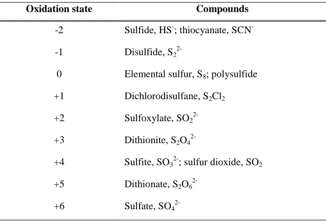
complicated by many oxidation states sulfur can assume. The oxidation states of sulfur
is summarize in Table 2.1.
that involved variety of bacteria found in soil or water and able to oxidize sulfide (IPCS,
1981). However, under aerobic conditions sulfide was oxidized both chemically and
biologically to sulfate. In nature, variety of reduced inorganic sulfur compounds such as
sulfur, thiosulfate and sulfite occur as intermediates for sulfide oxidation to sulfate. As
these compounds are slowly oxidized by a chemical reaction with oxygen, it is obvious
that biological oxidation play an important role in the recycling of reduced inorganic
sulfur compounds under aerobic condition (Kuenen, 1975). Microorganism which are
specialized in the oxidation of sulfur compounds are known as sulfur-oxidizing bacteria
that have the ability to oxidize reduced sulfur compounds and gain energy from these
processes. The end product of oxidative activity is the production of sulfate which is
extremely stable towards further chemical activity in nature (IPCS, 1981).
Table 2.1: Oxidation state of sulfur in common compounds (Komarnisky
et al
., 2003).
Oxidation state Compounds
-2 Sulfide, HS-; thiocyanate, SCN --1 Disulfide, S2
2-0 Elemental sulfur, S8; polysulfide
+1 Dichlorodisulfane, S2Cl2
+2 Sulfoxylate, SO2
2-+3 Dithionite, S2O4
2-+4 Sulfite, SO32-; sulfur dioxide, SO2
+5 Dithionate, S2O6
2.4
Sources of Sulfur Compounds
Release of sulfide to variety of environment media (air, surface water,
groundwater, soil and sediment) occur both naturally and as a result of human activity.
In the natural cycle of sulfur, sulfide and sulfate appears to be the principal sulfur
components in the environment.
Natural sources such as swamps, bogs and volcanoes account for about 90% of
the total amount of H
2S in the atmosphere (ATDSR, 2004). Swamps, bogs and shallow
lakes are the favourable environment for H
2S evolution where there is a highly organic
materials presence, oxygen deficiency and the development of sulfur bacteria (Moss,
1978). Sulfides are also found in petroleum and natural gas as a result of
sulfate-reducing bacteria activity or other sulfidogenic bacteria (Gevertz
et al
., 2000).
A number of bacteria, fungi and actinomycetes (a fungus-like bacteria) are
known to release H
2S during the decomposition of sulfur-containing proteins or by the
direct reduction of sulfate. The heterotroph
Proteus vulgaris
is an example of a common
bacteria that produces H
2S when grown in the presence of protein. The reduction of
sulfate to H
2S can be accomplished by anaerobic sulfate-reducing bacteria such as
Desulfovibrio
and
Desulfotomaculum
(IPCS, 1981). Hence, H
2S production can be
expected in conditions under depletion of oxygen with the availability of organic
material and sulfate.
H
2S was also emitted by some higher plant species as a byproduct of sulfite
metabolism. The H
2S emission was studied in detail in squash and pumpkin plant
(
Cucurbita pepo
L.) by Wilson
et al
. (1978). They found out that the emission was
plant. Rennerberg (1989) has stated that H
2S emission by higher plants seems to be a
significant contribution to the biogeochemical cycle of sulfur.
In addition, H
2S was also produce by living organisms, including human being
through the digestion and metabolism of sulfur-containing materials. Sulfide was
produces as a result of bacterial reduction of inorganic sulfate and sulfite, and also by
fermentation of sulfur-containing amino acids cysteine and methionine.
H
2S was also produced in human large intestine by natural bacteria present in both
intestinal tract and mouth (CICAD, 2003). In addition, sulfide production in human
mouth is a component that causes the bad breath (halitosis) (ATDSR, 2004).
The anthropogenic (man-made) sources of sulfur as a result of human activities
include combustion of fossil fuels, refining petroleum, coking of coal, electric power
generation and other industrial processes (ATDSR, 2004) have a large impact on the
atmospheric sulfur balance. Combustion of sulfur-containing compounds in fossil fuels
emits sulfur oxides, SO
2which produced at the lower atmosphere (Soleimani
et al
.,
2007). Moreover, the SO
2can penetrate soils directly from the atmosphere and becomes
oxidized to sulfate, and soils are available as sources of sulfur compounds. More
commonly, sulfur enters soil system through addition of sulfur-containing fertilizers.
H
2S is also frequently found in industrial settings where it is either used as a
reactant or produces as a by-product of manufacturing or industrial processes. Examples
of these processes are tanneries, wastewater treatment facilities, manure and sewage
facilities, rayon manufacturing plants, Kraft paper mills, drug manufacturing and food
processing plants (IPCS, 1981). H
2S may also enter the environment through accidental
petroleum, it may also be released into the environment during extraction, transport and
refining of these sources.
The anthropogenic emissions of gaseous sulfur compounds are approximately
two or three times as large as those from natural sources (Aneja and Cooper, 1978). The
natural and anthropogenic sources of sulfur are summarized in Table 2.2.
Table 2.2: Main sources of sulfur in environment.
Natural Sources
Anthropogenic Sources
- Environment
Hydrothermal vents
Volcanoes
Petroleum reservoir
Soil
Swamps, bogs
- Bacterial activity
SOB and SRB activity
- Biogenic emissions
From vegetations (higher plants,
wetlands, lands and ocean
- Human metabolism
In large intestine, mouth, muscles
- Sulfur storage
Gypsum, pyrite
Coke oven plants
Coking of coal
Combustion of fossil fuels
Drug manufacturing
Electric power generation
Fertilizers
Food processing
Iron smelters
Kraft paper mills
Livestock raising farm
Rayon manufacturing plants
Refining petroleum,
Tanneries
2.5
The Detrimental Effects of Reduced Sulfur Compounds
Odour from dairy farm, sanitary station and petrochemical plants can
contaminate nearby environments and cause severe complaints. Significant components
present in the odour are sulfur compounds and among these, H
2S is often the most
abundant (Oh
et al
., 1998). H
2S is a colourless and flammable gas with a characteristic
odour of rotten eggs (Wang, 2002). It is produced naturally as well as a result of human
activity. H
2S which is very toxic and corrosive gas are detrimental in the environment
due to many terrible effects towards humans, living organisms, environment and
industrial facilities (Mohapatra
et al
., 2006).
Primary exposure of general population to H
2S is most likely occurs through
inhalation of ambient air, and the gas is rapidly absorbed through the lungs in human
(CICAD, 2003). As H
2S is part of the natural environment, the general population will
have some exposure to H
2S. Populations living in areas of geothermal activity or near
industrial sites may be more likely to be exposed to higher levels of H
2S. Besides that,
workers employed at facilities where H
2S is produce as a byproduct, such as farms with
manure storage pits, petroleum or natural gas drilling operations, landfills and waste
water treatment plants may also be exposed to high level (ATDSR, 2004).
Study by WHO (2000), showed that H
2S poisoning could give rise to respiratory
irritation, pulmonary oedema, and strong simulation in the central nervous system which
leading to convulsions, unconsciousness and death. In fatal human intoxication cases,
brain oedema, degeneration and necrosis of the cerebral cortex and the basal ganglia
have been observed. They also pointed out that workers, who were exposed to H
2S
there is also some evidence that exposure to H
2S may be associated with an increased
rate of spontaneous abortion.
Furthermore, sulfide has hazardous effects on industrial facilities where it brings
damage to plant equipments, concrete and steel structures caused by sulfide-initiated
corrosion. Sulfides are also undesirable during oil production operations because of
their corrosiveness and ability to form insoluble metal sulfides that can lead to a loss of
reservoir permeability (Gevertz
et al
., 2000).
Thiosulfate is likely to become part of the industrial waste-stream and later
discharged into a receiving stream. A number of microorganisms residing in the stream
can use thiosulfate as an energy source, whereby thiosulfate is oxidized and oxygen
being consumed. Therefore, thiosulfate contributes to the biochemical oxygen demand
(BOD) of the stream and potentially impact the community (Schreiber and Pavlostathis,
1998). Thiosulfate is also toxic by causing mortality to the aquatic organisms.
Therefore, treatment of thiosulfate-containing wastewaters is often desirable prior to
final disposal.
2.6
Control of Sulfides and Reduced Sulfur Compound
Study by Oprime
et al
. (2001)shown that, among various reduce inorganic sulfur
compounds exists in environment, H
2S or sulfides are the most common compounds that
associated with human health risk and destructive effects on building structure. The
removal of H
2S from biogas, natural or industrial gases is an important concern of
Conventional physiochemical processes of H
2S removal are mainly based in its
oxidation by air in the presence of several catalysts such as potassium permanganate
(KMnO
4), ferric ion (Fe
3+) and active coal. Besides that, the detrimental activities of
sulfide-producing bacteria can be controlled by the effective use of oxidizing biocides
such as chlorine (Cl
2), hydrogen peroxide (H
2O
2) and
NaClO
(Oprime
et al
., 2001). The
use of these biocides is the most successful method in controlling unwanted activities in
surface facilities. However, processes based upon such agents are expensive due to the
high cost involved in the facility installation, as well as the operational cost due to high
energy demand and toxic chemical usage (Sorokin, 1994).
On the other hand, the biological treatment of sulfides using enzymatic oxidation
of sulfide oxidase has drawn attention since this method is more efficient and
environmental friendly. The bioconversion of sulfide using sulfide oxidase is believed
to be cost effective, higher sulfide removal efficiency, ability to operate at mild
conditions and reduced production of secondary pollution (Mohapatra
et al
., 2007). The
application of sulfide oxidase to remove harmful sulfide may potentially utilize for the
industrial processes and wastewater treatment. Sulfide oxidase has been produce by
number of autotrophic and heterotrophic bacteria, known as sulfur-oxidizing bacteria.
2.7
Sulfur-Oxidizing Bacteria (SOB)
Sulfur-oxidizing bacteria are physiologically diverse and comprise both
autotrophic and heterotrophic microorganisms (Peck, 1961). Sulfur-oxidizing bacteria
are characterized by their ability to catalyze the oxidation of reduced sulfur compounds
(H
2S, thiosulfate, sulfite, polythionate or elemental sulfur) with oxygen as terminal
electron acceptor (Graff and Stubner, 2003). The ability of sulfur-oxidizing bacteria to
oxidize the sulfur compounds under alkaline, neutral and acidic condition has been
reviewed previously (Friedrich
et al
., 2001). The representatives of sulfur-oxidizing
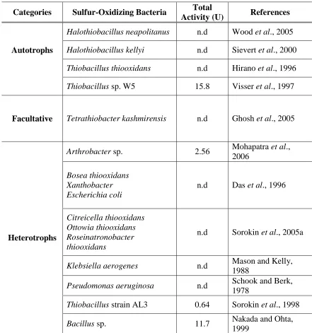
bacteria are displayed in Table 2.3.
The role of autotrophic sulfur bacteria is widely recognized in oxidation of
reduced sulfur compounds in natural and industrial habitats, as well as their physiology,
biochemistry, phylogeny and genetics are well established. Autotrophic sulfur-oxidizing
bacteria are capable of growing chemolithoautotrophically with sulfur compounds as
energy source and carbon dioxide as sole carbon source (Graff and Stubner, 2003). The
autotrophic sulfur-oxidizing bacteria were believed to be able to derive metabolically
useful energy from the oxidation of reduced inorganic sulfur compounds (Kuenen,
1975).
Most of the well known autotrophic sulfur-oxidizing bacteria belongs to the
families of the
Thiobacteriaceae, Beggiatoaceae
and
Achromatiaceae
and commonly
called as colourless sulfur bacteria (Kuenen, 1975). A lot of previous research has
mainly focused on the genus
Thiobacillus
. The thiobacilli are a small group of
Examples of autotrophic sulfur-oxidizing bacteria that has been isolated and
studies are
Halothiobacillus
neapolitanus
(Wood
et al
., 2005),
Halothiobacillus kellyi
(Sievert
et al
., 2000),
Thiobacillus caldus
(Hallberg
et al
., 1996),
Thiobacillus
thioparus
(Cho
et al
., 1991a),
Thiobacillus
thiooxidans
(Hirano
et al
., 1996). Besides that,
numerous studies of sulfur oxidation have been performed on
Thiobacillus denitrificans
and
Thiobacillus ferooxidans
which
utilize nitrate and ferric ions in place of oxygen as
electron acceptor, respectively (Peck, 1961).
However, oxidation of sulfur compounds is not restricted to the true sulfur
bacteria; this process also occurs in heterotrophic bacteria isolated from soil and marine
environment. Most of the heterotrophic bacteria that have such activities belong to
genera
Arthrobacter
(Mohapatra
et al
., 2006)
, Halomonas
(Sorokin, 2003),
Xanthobacter
,
Escherichia coli
(Das
et al
., 1996),
Klebsiella
(Mason and Kelly, 1988)
and
Pseudomonas
(Schook and Berk, 1978).
On the other hand, only few of these heterotrophs have the ability to generate
biologically useful energy from the oxidation of reduced inorganic sulfur compounds.
Few representatives of this type of heterotrophic sulfur-oxidizing bacteria are
Limnobacter thiooxidans
(Spring
et al
., 2001),
Bosea thiooxidans
(Das
et al
., 1996),
Thiobacillus
Q (Gommers and Kuenen, 1988),
Ottowia thiooxidans
and
Roseinatronobacter thiooxidans
. These heterotrophs are increasing their growth yield in
presence of thiosulfate or sulfide, which is indicative of a lithoheterotrophic metabolism
(Sorokin
et al
., 2005a).
(Teske
et al
., 2000). An increase in pH during growth of heterotrophic bacteria during
thiosulfate oxidation was due to tetrathionate production (Sorokin, 2003). Oxidation of
thiosulfate to tetrathionate probably does not supply energy for the growth of cells
capable of catalyzing this oxidation (Peck, 1961).
Ruby
et al
. (1981) have described locally isolated sulfur-oxidizing bacteria from
hydrothermal vents that comprised the heterotrophic base-producing, aerobic or
facultatively anaerobic, rod-shaped bacteria that oxidize thiosulfate to tetrathionate
without measurable enhancement of growth. Amongst the pure cultures of
base-producing sulfur-oxidizing bacteria described in Sorokin’s publication, the organisms
are the members of γ-
Proteobacteria
, which include
Pseudoalteromonas
,
Halomonas
Table 2.3: Sulfur-oxidizing bacteria.
Categories
Sulfur-Oxidizing Bacteria
Total
Activity (U)
References
Autotrophs
Halothiobacillus neapolitanus
n.d
Wood
et al
., 2005
Halothiobacillus kellyi
n.d
Sievert
et al
., 2000
Thiobacillus thiooxidans
n.d
Hirano
et al
., 1996
Thiobacillus
sp. W5
15.8
Visser
et al
., 1997
Facultative
Tetrathiobacter kashmirensis
n.d
Ghosh
et al
., 2005
Heterotrophs
Arthrobacter
sp.
2.56
Mohapatra
et al
.,
2006
Bosea thiooxidans
Xanthobacter
Escherichia coli
n.d
Das
et al
., 1996
Citreicella thiooxidans
Ottowia thiooxidans
Roseinatronobacter
thiooxidans
n.d
Sorokin
et al
., 2005a
Klebsiella aerogenes
n.d
Mason and Kelly,
1988
Pseudomonas aeruginosa
n.d
Schook and Berk,
1978
Thiobacillus
strain AL3
0.64
Sorokin
et al
., 1998
Bacillus
sp.
11.7
Nakada and Ohta,
To our knowledge, the involvement of
Serratia
in the oxidation of sulfur
compounds has never been reported. However, there are reports about
Serratia
regarding sulfate formation from the hydrolysis of organic sulfate by enzymes termed
sulfatase.
Serratia
was reported to synthesize arylsulfatase in the presence of
methionine or taurine as the sulfur source (Murooka
et al
., 1980; Yamada
et al
., 1978).
Arylsulfatase are considered to be important for sulfate ester mineralization in soils that
contain high levels of sulfur in the form of sulfate ester (Murooka
et al
., 1980). This
statement showed the ability of
Serratia
to utilize sulfur compounds in the organic form
and produce sulfate from the process.
The ability of sulfur-oxidizing bacteria to oxidize sulfide and reduced inorganic
sulfur compounds are attributed to an enzyme system present in the cell (Kurosawa
et
al
., 1997), by which sulfide or the reduced inorganic sulfur compounds are biologically
oxidize to sulfate (Das
et al
., 2006). Although in general sulfate is the major oxidation
product of reduced inorganic sulfur compounds oxidation, other end-products or
intermediates may be formed depending on the metabolic pathway used by the particular
organisms (Schreiber and Pavlostathis, 1997). These intermediates compounds include
polythionates, sulfite or elemental sulfur. The mechanism of inorganic sulfur oxidation
appeared to differ radically among different sulfur bacteria (Kelly
et al
., 1997).
2.8
Sulfide Oxidase
There are a few reports on the biological role of the sulfide oxidase in
Arthrobacter
sp. and
Bacillus
sp. BN53-1 and was concentrated in this study (Mohapatra
et al
., 2006; Nakada and Ohta, 1999).
According to the Mohapatra
et al
. (2006) report, the purified sulfide oxidase was
showed to be monomer with a molecular weight of 43 kDa. This molecular weight was
found to be higher compared to the purified enzyme from the
Bacillus
sp. BN53-1 which
is 37 kDa (Nakada and Ohta, 1999). The sulfide oxidase isolated from
Arthrobacter
sp.
was cell-bound and had broad pH activities which are potentially useful in application of
the wastewater treatment process (Mohapatra
et al
., 2006).
2.9
Oxidation of Inorganic Sulfur Compounds Pathway
S-SO
32-(Thiosulfate)
-
O
3S-S-S-SO
3-(Tetrathionate)
5 4
S
2-(Sulfide) ↔ S
o(Sulfur) → SO
32-(Sulfite) → SO
42-(Sulfate)
1
2
3
Figure 2.2: Oxidation of inorganic sulfur compounds pathway. The enzymes
responsible for the reactions were identified as follows: reaction 1- sulfide oxidase;
reaction 2 - sulfur-oxidizing enzyme (sulfur oxygenase); reaction 3 – sulfite oxidase;
reaction 4 – rhodanese (thiosulfate – cleaving enzymes, sulfur transferase); and reaction
5 – thiosulfate-oxidizing enzyme (Suzuki, 1994)
2.10
Application of Sulfide Oxidase
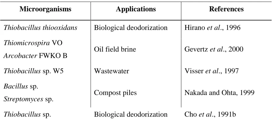
Table 2.4: Application of sulfide oxidase
Microorganisms
Applications
References
Thiobacillus thiooxidans
Biological deodorization
Hirano
et al
., 1996
Thiomicrospira
VO
Arcobacter
FWKO B
Oil field brine
Gevertz
et al
., 2000
Thiobacillus
sp. W5
Wastewater
Visser
et al
., 1997
Bacillus
sp.
Streptomyces
sp.
Compost piles
Nakada and Ohta, 1999
CHAPTER 3
GENERAL MATERIALS AND METHODS
3.1
Sulfur-Oxidizer Medium Preparation
Selective medium containing sodium thiosulfate was used for isolation and
enrichment of microorganism (Asku
et al
., 1991). Thiosulfate was supplemented in this
medium as a sulfur source to the microorganism. It has been used as a preferred sulfur
compound for sulfur-oxidizing bacteria laboratory studies, due to its stability at neutral
pH and metabolized by all sulfur-oxidizing bacteria (Smith and Strohl, 1991). The
medium was prepared by dissolving 10 g Bacto peptone (
Becton-Dickinson
), 1.5 g
di-potassium hydrogen phosphate, 0.75 g ferric ammonium sulfate, 1.0 g sodium
3.2
Culture Maintenance and Preservation
The sulfur-oxidizing bacteria was inoculated into fresh liquid sulfur-oxidizer
medium and grown until the culture reached optical density of 0.5-0.8 at 600 nm. Cells
were then harvested by centrifuging the culture at 4000 rpm for 20 minutes using
refrigerated centrifuge. The cell-free supernatant was discarded and the resulting pellets
were suspended with sulfur-oxidizer medium and mixed homogenously by vortex.
Afterward, the pure culture of sulfur-oxidizing bacteria was transferred into cryovial
containing cryopreservation beads stock and preserved at -20˚C for future experimental
use.
3.3
Experimental Design
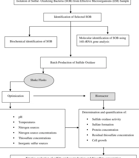
The experimental design of this study was divided into three stages which
include isolation, characterization and identification of selected sulfur-oxidizer,
optimization of sulfide oxidase in shake flasks and lastly the production of sulfide
oxidase in bioreactor (Figure 3.1). This study also assesses the capability of newly
isolated bacteria to break down thiosulfate into sulfate. Besides that, oxygen
Isolation of Sulfur- Oxidizing Bacteria (SOB) from Effective Microorganisms (EM) Sample
Identification of Selected SOB
Biochemical identification of SOB
Batch Production of Sulfide Oxidase
Shake Flasks
Optimization Bioreactor
pH
Temperatures
Nitrogen sources
Nitrogen source concentrations
Thiosulfate concentrations
Inorganic sulfur sources
Determination and quantification of:
Sulfide oxidase activity
Sulfate formation
Protein concentration
Residual thiosulfate concentration
Cell growth
Kinetics evaluation of sulfide oxidase production and thiosulfate consumption
Figure 3.1: Experimental design chart
3.4
Inoculum Preparation
An inoculum was used as a starter culture in all fermentation process carried out
during the optimization studies (section 5.3.3). Cells of sulfur-oxidizing bacteria were
inoculated into 150 mL of sulfur-oxidizer liquid medium in 500 mL flask and incubated
at 30˚C with 200 rpm agitation. The microbial growth was monitored using the turbidity
measurement at 600 nm wavelength with a spectrophotometer. Once the initial OD
reaches 0.5-0.8, the culture was ready to use as inoculum. Inoculation was carried out
by transferring the inoculum into another sulfur-oxidizer broth medium for the
optimization experiments.
3.5
Analytical Procedures
3.5.1
Determination of Cell Mass Concentration
Cell dry weight measurement was used to quantify the cell mass concentration in
samples. About 5 mL aliquots were centrifuged for 20 minutes at 4000 rpm at 4˚C. The
clear supernatant was carefully discarded and the remaining pellet was suspended using
the medium broth. The mixture was mixed using vortex and poured into holding
reservoir fitted on the pre-weighted dried 0.22 µm cellulose acetate filter membrane. The
cell paste was dried in oven until a constant membrane weight was obtained. The
differences in weight between membrane (empty) and membrane with cell paste was
used to determine the dry weight of cell mass as g/L.
L sample of Volume
g membrane of
weight g
paste cell and membrane dried
of Weight g/L
weight dry
3.5.2
Determination of Specific Growth Rate and Doubling Time
In microbiology, the word growth is defined as an increase in the number of
microbial cells in a population, which can also be measured as an increase in microbial
mass (Madigan
et al
., 2000). Growth rate (µ) is the change in cell number or cell mass
per unit time, while doubling time (t
d) is the interval time where the number of cells
doubles during each unit time period.
The specific growth rate was measured from the slope of cell mass curve (dry
weight) by delineating points between the log growth phase, as represents by the
equation below:
t
)
X
ln
X
(ln
μ
t
0…(3.2)
Where;
X
0= cell mass concentration at time 0 (g/L)
X
t= cell mass concentration at time t (g/L)
t = elapsed time between measurement (h)
µ = specific growth rate (h
-1)
The relation between specific growth rate and doubling time of the cell mass is
obtained from equation below:
μ
0.693
μ
2
ln
Where;
µ = specific growth rate (h
-1)
t
d= doubling time (h)
3.5.3
Determination of Kinetic Parameters
Yield coefficient can be used to determine the growth kinetics. A measure of the
overall efficiency of the conversion of substrate to cell mass or specific product is given
by a yield coefficient (Y) which can be stated as Y
x/s, Y
p/sand Y
p/x. The growth and
product yield coefficient, Y
x/sand Y
p/s, are very important parameters since they
represent the efficiency of conversion of the substrate into cell mass or product.
Growth yield coefficient (Y
x/s) can be defined as dry cell weight obtained per
amount of substrate consumed. The growth yield coefficient is an important as a mean
of expressing the quantitative nutrient requirement of microorganism.
ΔS
ΔX
Y
xs
…(3.4)
Where;
Y
x/s= Growth yield coefficient (g dry cell weight /mg substrate consumed)
∆X = Concentration of cell mass (g/L)
Product yield coefficient (Y
p/s) can be defined as:
ΔS
ΔP
Y
sp
…(3.5)
Where;
Y
P/s= Product yield coefficient (g product /mg substrate consumed)
∆P = Concentration of product formed (g/L)
∆S = Concentration of substrate consumed (mg/L)
Product yield coefficient (Y
p/x) that relates product formation with cell mass
production is state as below:
ΔX
ΔP
Y
xp
…(3.6)
Where;
Y
p/x= Product yield coefficient (g product /g cell mass)
∆P = Concentration of product formed (g/L)
∆X = Concentration of cell mass (g/L)
The productivity of a culture system maybe described as the output of cell mass
per unit time of the fermentation. Thus, the productivity of a batch culture maybe
represented as:
initial final
0 max batch
t
t
X
X
R
Where;
R
batch= cell productivity (g/L/h)
X
0= initial cell concentration at inoculation (g/L)
X
max= maximum cell concentration (g/L)
t
initial= initial time (h)
t
final= time at maximum cell concentration (h)
Sulfate productivity, R
sulfatecan be defined as:
initial final
0 max sulfate
t
t
P
P
R
…(3.8)
Where;
R
sulfate= sulfate productivity (g/L/h)
P
0= initial sulfate production (g/L)
P
max= maximum sulfate production (g/L)
t
initial= initial time (h)
t
final= time at maximum cell concentration (h)
The rate of growth associated products formation in a fermentation process is
directly related to cells growth. The patterns of cells growth and product accumulation
can be described by measuring the specific product formation rate (q
p). It shows that
when product formation is growth associated, the specific rate of product formation (q
p)
increase with specific growth rate.
μ
x
Y
q
x p
Where;
q
sulfate=
specific rate of sulfate formation (g/g/h)
Y
p/x= product yield coefficient base on cell concentration (g product /g cell mass)
µ = specific growth rate (h
-1)
3.5.4
Sulfate Ion Determination
The amount of sulfate ion (SO
42-) produced during growth of sulfur-oxidizing
bacteria on thiosulfate was determined spectrophotometrically. Sulfate was measured by
adding 1:1 barium chloride solution (10% w/v) with supernatant followed by mixing the
suspensions vigorously (Cha
et al
., 1999). A resulting white turbidity due to barium
sulfate formation was measured at 450 nm with a Jenway 6300 spectrophotometer, U.K
.
3.5.4.1
Sulfate Calibration Curve
Potassium sulfate (K
2SO
4) was used as standard to construct a sulfate calibration
curve according to Kolmert
et al
. (2000). Standard sulfate solutions were made by
dissolving K
2SO
4in deionized water to known concentrations in the range 0 to 3 mM.
3.5.5
Sulfide Oxidase Assay
The sulfide oxidase activity was determined by measuring the product of
enzymatic reaction, sulfate (SO
42-) in the reaction mixture. The enzyme assay protocol
was originated from Hirano
et al
. (1996) method and modified in this study to fit with
the sulfate measurement.
The reaction was initiated by addition of 0.5 mL of sodium sulfide (Na
2S)
solution into the reaction mixture that contain 4.5 mL of 0.1 M sodium acetate buffer
(pH 5.6) and 1 mL supernatant. The Na
2S solution was prepared by dissolving 0.06 g
Na
2S in an alkaline solution consisting of 0.16 g NaOH, 0.02 g EDTA Na
2.2H
2O
(sodium ethylenediaminetetraacetic acid), 2 mL glycerol and 40 mL distilled water. The
Na
2S solution was freshly prepared prior to use. The mixture was incubated for 30
minutes at 30˚C and the reaction was subsequently terminated by the addition of 1.5 mL
NaOH (1.0 M) followed by thorough mixing.
3.5.6
Protein Concentration Determination
Protein concentration in the supernatant sample was colorimetrically determined
according to Lowry method (Lowry, 1951). In this assay, when protein is placed in an
alkaline system containing copper ion (Cu
2+), a colored complex forms between the
peptide bonds of the protein and the copper ions. The Lowry protein assay applied the
Folin-Ciocalteau phenol reagent to enhance the color response resulting a blue color
complex. The absorbance of the complex was measured spectrophotometrically at
wavelength of 750 nm. The concentration of protein was calculated from a standard
curve generated using the Bovine Serum Albumin (BSA) as standard. Reagent
preparation and the standard curve (0-1 mg/mL) for protein quantification is presented in
Appendix A2.
3.5.7
Thiosulfate Determination
CHAPTER 4
ISOLATION, SCREENING AND IDENTIFICATION OF POTENTIAL
SULFUR-OXIDIZER FROM EFFECTIVE MICROORGANISMS (EM) SAMPLE
4.1 Introduction
Sulfide and thiosulfate are the most abundant reduced inorganic sulfur species in
the environment and converted to sulfate primarily by bacterial action in the sulfur
cycle. Sulfides are detrimental in the environment because of the toxicity to human
health and corrosive effect on concrete and metallic infrastructure, besides it strong
unpleasant smell (Oprime et al., 2001). While thiosulfate is one of the intermediate
product of sulfide oxidation process in sulfur cycle and considered as important in
nature.
The use of microorganisms able to remove reduced inorganic sulfur compounds
in nature has been considered as a potential alternative. Oxidation of reduced inorganic
sulfur compounds especially sulfide and thiosulfate was exclusively carried out by
contributed to an enzyme system present in the cell known as sulfide oxidase and sulfate
is the expected end product of reduced inorganic sulfur compounds oxidation by the
sulfur-oxidizing bacteria (Visser et al., 1997).
The potential for oxidation of inorganic sulfur compounds, such as sulfide and
thiosulfate is widely spread among the bacteria. Most of the known sulfur-oxidizing
bacteria are members of the so-called ‘colourless sulfur bacteria’ belong to genera
Thiobacillus, Thiothrix, Thiomicrospira, Beggiatoa and Achromatium (Gommers and
Kuenen, 1988). However, there are many heterotrophic bacteria especially those belonging to the γ-subdivision of the Proteobacteria that are capable of oxidizing
thiosulfate and sulfide (Sorokin et al., 2005b). Most of the example of heterotrophic
bacteria belongs to the genera Pseudomonas, Xanthomonas, Halomonas, Escherichia
coli and Klebsiella (Gommers and Kuenen, 1988).
This study describes the isolation work of potential sulfur-oxidizing bacteria
from Effective Microorganisms (EM) sample. The isolates were then screened for
sulfide oxidase production. The superior sulfur-oxidizing bacteria was identified using
biochemical tests and 16S rRNA gene analysis.
4.2 Experimental Design
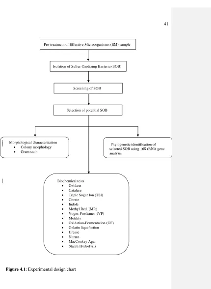
The flowchart of the experimental design carried out in this study is summarized
Pre-treatment of Effective Microorganisms (EM) sample
Isolation of Sulfur Oxidizing Bacteria (SOB)
Screening of SOB
Selection of potential SOB
Figure 4.1: Experimental design chart
Morphological characterization
Colony morphology
Gram stain
Biochemical tests
Oxidase
Catalase
Triple Sugar Ion (TSI)
Citrate
Indole
Methyl Red (MR)
Voges-Proskauer (VP)
Motility
Oxidation-Fermentation (OF)
Gelatin liquefaction
Urease
Nitrate
MacConkey Agar
Starch Hydrolysis
4.3 Materials and Methods
4.3.1 Media Preparation
The composition and preparation of sulfur-oxidizer medium (SOM) used
throughout this study was listed in section 3.1.
4.3.2 Pre-treatment of Effective Microorganisms (EM) Sample
The EM sample that has been used as a source for isolation of potential
sulfur-oxidizing bacteria was kindly donated from Associate Professor Dr. Zainoha Zakaria
from Chemistry Department, UTM. In order to ascertain the recovery of viable
microorganisms in the sample, the EM was pre-treated prior to cultivation.
Pre-treatment was done by mixing 1 mL of EM with 1 mL molasses and 18 mL of sterile
distilled water (method from previous work). The treated sample was then incubated at
30˚C overnight to enhance the population of microorganisms in the EM sample.
4.3.3 Isolation of Sulfur-Oxidizing Bacteria
Isolation of sulfur-oxidizing bacteria was performed by using direct plating
agar. The sample was spread evenly on the sulfur-oxidizer medium plate using the
sterile glass spreader. The plates were incubated at 30˚C for 24 h. The well defined
isolated colonies appeared on the plate were picked up by wire loop and restreak on the
other sulfur-oxidizer medium agar plate for purity conformation.
4.3.4 Screening of Sulfur-Oxidizing Bacteria
Screening of potential sulfur-oxidizing bacteria was performed by inoculating 10
% (v/v) inoculum into sulfur-oxidizer medium broth formulated by Asku et al. (1991).
The cultures were then incubated at 30˚C and agitated at 200 rpm until the optical
density of the cultures reached 0.6 – 0.8. The cultures were centrifuged at 4000 rpm, 4˚C for 30 minutes. The supernatant was analyzed for sulfide oxidase activity and
protein concentration (section 3.5.4 - 3.5.6). Culture that gives the highest sulfide
oxidase activity was selected and used throughout the studies.
4.3.5 Analytical Procedures
Quantification of sulfide oxidase activity and protein concentration that was
4.3.6 Morphological Characterization
4.3.6.1 Colony Morphology
Morphological characteristics of strain SO02 that include configuration, margin,
elevation and colour of the bacteria were examined using 24 h culture that was
previously cultivated on sulfur-oxidizer medium agar. The morphology of the culture
colony was observed using a stereomicroscope.
4.3.6.2 Cellular Morphology
The overnight-grown culture of strain SO02 was used for the observation of
gram stained under 1000x immersion oil objective of bright-field microscope (Olympus
C-35 AD-4). The procedures for gram staining can be reviewed in Appendix A7.
4.3.7 Biochemical Tests
Biochemical characterizations test of strain SO02 were performed using 24 h
culture. The tests were include the carbohydrate metabolism test (starch hydrolysis,
Methyl Red, Vogues Proskauer, Simmons citrate and triple sugar iron), nitrogen
(catalase and oxidase) and growth on differential medium (MacConkey agar). The
biochemical tests were performed as described by MacFaddin (1980). The procedure of
biochemical tests is presented Appendix A7. The biochemical test results along with
morphological and staining results are the key for the bacterial identification process. Bergey’s Manual of Determinative Bacteriology has been used as reference for
classification and identification of the SO02 bacteria (Holt et al., 1994).
4.3.8 Identification of Potential Microbe using 16S rRNA Gene Analysis
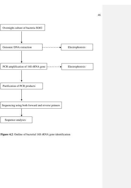
Strain SO02 showing the superior sulfide oxidase activity was identified at 16S
rRNA gene level according to the steps shown in Figure 4.2.
4.3.8.1 DNA Extraction
The potential sulfur-oxidizing bacteria which known as SO02 strain in this work
was grow on sulfur-oxidizer medium agar. The cell suspension for DNA extraction was
prepared from 24 h culture incubated at 30˚C with agitation of 200 rpm. The 16S rRNA
gene analysis began with DNA extraction of SO02 bacteria. The solutions needed in
16S rRNA gene analysis were provided in the Wizard® Genomic DNA Purification Kit
Overnight culture of bacteria SO02
Genomic DNA extraction Electrophoresis
PCR amplification of 16S rRNA gene Electrophoresis
Purification of PCR products
Sequencing using both forward and reverse primers
Sequence analyses
The overnight-grown culture with optical density, OD600nm of 0.5 - 0.8 was
transferred into microcentrifuge tube and centrifuged at 14,000 rpm for 5 min.
Approximately 600 µl Nuclei Lysis Solution was added to the pellet and resuspended
gently. The mixture was incubated at 80˚C for 5 min, and allowed to cool at room
temperature. Afterward, 3 µL RNase solution was added to the cell lysate, mixed by
inverted the tube 2-5 times and incubated at 37˚C for 30 min. After the sample cool at
room temperature, 200 µL of Protein Precipitation Solution was added to the
RNase-treated cell lysate. The sample was vortex vigorously at high speed for 20 seconds to
mix the Protein Precipitation Solution with the cell lysate and incubated on ice for 5
min. After that, the sample was centrifuged at 14 000 rpm for 5 min.
The next step on DNA extraction was known as DNA precipitation and
rehydration. The supernatant containing DNA obtained from the previous spin was
transferred into a new clean microcentrifuge tube containing 600 µL of room
temperature isopropanol. The sample was gently mix by inversion until the thread-like
strands of DNA form a visible mass and centrifuged at 14,000 rpm for 5 min. The
supernatant was carefully poured off and the tube was drain on a clean absorbent paper.
The pellet was added with 600 µL of room temperature 70% (v/v) ethanol and gently
inverts several times to wash the DNA pellet. The sample was centrifuged for 5 min and
supernatant containing ethanol was discarded from the tube. The tube was drain on
absorbent paper and the pellet was allowed to air-dry for 15 min. Approximately 100
µL Rehydration Solution was added and pellet containing DNA was rehydrate by
incubating at 65˚C for 1 h. The isolated genomic DNA was stores at -20˚C until further
4.3.8.2 Analyzing Genomic DNA
The extracted genomic DNA was analyzed via agarose gel electrophoresis to
prove the presence of DNA after the extraction procedures. The agarose gel was
prepared by dissolving 1.0 % (w/v) agarose in 1X Tris-Acetate-EDTA (TAE) buffer by
heating the agarose solution in a microwave oven. TAE buffer was used because it
provides faster electrophoretic migration of linear DNA and better resolution of
supercoiled DNA. Ethidium bromide (10 mg/mL) was added to the molten agarose and
poured slowly into a casting tray that was previously inserted with comb to form wells.
The gel was left to cool and solidify for 20-40 min before removing the comb from the
solidified gel. The tray was then placed onto the electrophoresis tank with sample wells
are near to the cathode (black). DNA sample will migrate toward the anode (red) during
the electrophoresis process. The electrophoresis tank was filled up with 1X TAE buffer
until the gel was submerged under the buffer. Sample of genomic DNA (5 µL) was
prepared by mixing with 2 µL loading dye before loading the mixture into respective
wells on the agarose gel. The agarose gel electrophoresis was performed at 70 volts for
1 h or until the blue dye has migrated about two-third the length of the gel. The gel was
then removed from the tray and place on the Gene-Flash to visualize and photographed
the observed DNA band.
4.3.8.3 Polymerase Chain Reaction (PCR)
The PCR amplification for the nearly full-length 16S rRNA gene of SO02 bacteria was conducted using the universal primers, pA and pH’ (Edwards et al., 1989).
Table 4.1: Sequences of universal primers (pA and pH’) (Edwards et al., 1989)
Primers Sequences
pA (Forward) 5’-AGA GTT TGA TCC TGG CTC AG-3’
pH’ (Reverse) 5’-AAG GAG GTG ATC CAG CC-3’
The PCR reaction mixture was prepared by adding 10 µL of template DNA into
tube containing 2.0 µL of each forward and reverse primer (10 pmol of each primer), 25
µL of 2X PCR master mix and topped up with 11 µL distilled water. Amplification was
performed in a GeneAmp PCR system 9700 thermal cycler (Perkin-Elmer Corp.,
Emeryville, Calif.), under the following parameters: an initial denaturation at 94˚C for 3
min; 25 cycles, denaturation at 94˚C for 1 min, annealing at 50˚C for 1 min, and extension at 72˚C for 2 min; with 8 min final extension at 72˚C. The expected PCR
products (1.5 kb) were cooled at 4˚C until removed and can be stored at -20˚C until
further uses. The amplified 16S rRNA gene was now continued with purification
procedures before sequencing can be done.
4.3.8.4 Analyzing PCR Products
The presence of PCR products can be distinguished using agarose gel
electrophoresis (section 4.3.8.2). The PCR products were electrophoresed through 1.0
% (w/v) agarose gels containing ethidium bromide in 1X TAE buffer as the running
buffer. A GeneRuler™ 1kb DNA Ladder Plus (Fermentas) was used as marker for
sizing and approximate quantification of PCR products. Five microlitres of PCR