1
Nanoparticles for Bioapplications: Study of the Cytotoxicity of Water
Dispersible Quantum Dots
Fatemeh Mirnajafizadeh**1, Deborah Ramsey**1, Shelli McAlpine1, Fan Wang2,3, Peter Reece2 and John Arron Stride*1
1. School of Chemistry, University of New South Wales, Sydney, NSW 2052, Australia 2. School of Physics, University of New South Wales, Sydney, NSW 2052, Australia
3. School of Mathematical and Physical Sciences, University of Technology Sydney, Ultimo, Sydney, NSW 2007, Australia
Email: j.stride@unsw.edu.au
* Corresponding Author: A/Prof. John Arron Stride
** These authors contributed equally in this work.
Abstract: Semiconductor nanocrystals or quantum dots (QDs), have unique optical and
physical properties that make them potential imaging tools in biological and medical
applications. However, concerns such as the aqueous dispersivity, toxicity to cells and
stability in biological environments may limit the use of QDs in bioapplications. Here, we
report an investigation into the cytotoxicity of aqueously dispersed CdSe(S) and
CdSe(S)/ZnO core/shell QDs in the presence of human colorectal carcinoma cells (HCT-116)
and a human skin fibroblast cell line (WS-1). The cytotoxicity of the precursor solutions used
in the synthesis of the CdSe(S) QDs was also determined in the presence of HCT-116 cells
and compared to that of the heat-shock protein (Hsp90) inhibitor, 17-AAG. CdSe(S) QDs
were found to have a low toxicity at concentrations up to 100 µg/ml, with a decreased cell
viability at higher concentrations, indicating a highly dose-dependent response. Meanwhile,
CdSe(S)/ZnO core/shell QDs exhibited lower toxicity than uncoated QDs at higher
concentrations. Confocal microscopy images of HCT-116 cells after incubation with CdSe(S)
and CdSe(S)/ZnO QDs showed that the cells were stable in aqueous concentrations of 100 µg
of QDs per ml, with no sign of cell necrosis, confirming the cytotoxicity data.
Key words: HCT-116, WS1,water dispersive QDs, aqueous synthesis, cytotoxicity of QDs.
2 1. Introduction
Semiconductor nanocrystals or quantum dots (QDs) have received a great deal of attention
over the last decade due to their unique optical and physical properties. This has led to them
being classified as a powerful new class of bio-imaging tools [1-8]. Despite the desirability of
using QDs as intense fluorescent nanoscale markers in biological applications, cytotoxicity is
a serious constraint that limits potential uses [9-11]. QDs can be considered as being
potentially toxic due to both their nanoscale size and the presence of heavy metals [10, 11].
Nanoscale particles can have a greater toxicity than bulk counterparts, with the small size
enabling greater penetration into cells than corresponding bulk chemical materials [12]. In
addition, the generation of free radicals such as reactive oxygenated species (ROS) upon cell
exposure to nanomaterials, is a significant cause of nanoparticle cytotoxicity [12-15].
Interactions of QDs with cell mitochondria and nuclei can result in the disruption of cell
function, inhibition of cell proliferation and decreased cell viability due to ROS production,
ultimately resulting in cell death, mutation, or induced immunotoxicity [13, 16-18]. The
long-term toxicity of QDs has also been attributed to bioaccumulation in organs, leading to organ
damage and chronic illnesses [19]. The high ratio of reactive surface area relative to the bulk
generally increases the chemical activity of nanoparticles, which can result in an
accumulation of nanoparticles in tissues and associated organ toxicity [20], particularly those
having high blood flow such as spleen, kidneys, liver and lungs, along with the blood
circulation system itself [21]. QDs have therefore largely been classified as poisonous to both
human and animal cells and as such, cytotoxicity assays of QDs are an essential requirement
before applying QDs to cellular environments.
There are various methods to determine the toxicity of QDs in both in-vivo and in-vitro
studies. In-vivo studies may involve introducing QDs to microorganisms [22] or the use of
3
with QDs in order to investigate the cytotoxicity of nanocrystals in mammalian cell lines
[28-37]. A common feature in all reports of cadmium-based QDs, including CdTe, CdSe and
CdS, is that Cd is a primary source of toxicity. Accordingly, core/shell QDs such as
CdTe/CdS, CdTe/CdS/ZnS and CdSe/ZnS have been shown to exhibit less toxicity than core
QDs alone [11, 31]. However, the cytotoxicity of QDs depends on a number of parameters
including the surface modifications of the QDs, cell type, cellular morphology, cell growth
and the interaction of QDs with cell membranes [36, 30].
There are extensive reports detailing cytotoxicity assays of QDs synthesized in organic media
[25, 29-39], but few papers have reported in detail the cytotoxicity of QDs directly obtained
from aqueous solution [11, 40-43]. Bhatia and co-workers investigated the in-vitro
cytotoxicity of organically synthesized CdSe QDs on liver cells and showed that the release
of free cadmium ions from the QDs resulted in cell death [30]. Meanwhile, Zhu et al. studied
both the in-vivo and in-vitro cytotoxicity of aqueous CdSe and CdSe/CdS QDs and reported
that the toxicity of QDs depends on both target cells and physicochemical properties of the
QDs [42]. Plank et al. investigated the cytotoxicity of CdSe and CdSe/ZnS QDs obtained in
organic solvents, demonstrating that QD cytotoxicity is related to both particle size and the
surface covering of functional groups such as amines and carboxylic acids used to disperse
them in water [32]. Fan et al. investigated the cytotoxicity of a wide range of QDs (CdTe,
CdTe/CdS and CdTe/CdS/ZnS) synthesized in aqueous reactions, concluding that free
cadmium ions are a major source of toxicity in CdTe-based QDs [11], also reporting that
CdTe/CdS/ZnS QDs were slightly less toxic than CdTe in their experiments [11]. The present
work reports on the cytotoxicity of CdSe(S) and CdSe(S)/ZnO QDs, synthesized in wholly
aqueous reactions, to human colorectal carcinoma cells (HCT-116) and human skin fibroblast
cells (WS1). The water dispersible QDs were synthesized in a modified literature method
4
incubation of HCT-116 cells with the QDs over a wide range of concentrations (25 to 500
µg/ml). Cytotoxicity of CdSe(S) QDs was benchmarked against the heat-shock protein
(Hsp90) inhibitor, 17-AAG, and confocal images of HCT-116 cells after treatment with QDs
were recorded. Cytotoxicity of QDs to WS1 cells was also studied to explore the action of
QDs toward normal, resilient cell types.
2. Experimental methods
2.1 Materials
3-mercaptopropionic acid (MPA) was obtained from Fluka. CdCl2.2.5H2O,
Zn(CH3COO)2.2H2O, NaBH4, Se (powder), NaOH, bisbenzimide (Hoechst 33342),
4′,6-diamidino-2-phenylindole (DAPI) and 17-allyamino-17-demethoxy-geldanamycine (Hsp90
inhibitor 17-AAG) were all obtained from Sigma-Aldrich. Human colorectal carcinoma cells
(HCT-116) and human skin fibroblast cells (WS1) were obtained from ATCC (Manassas,
Virginia, USA). Dulbecco’s modified eagle medium (DMEM), fetal bovine serum (FBS),
penicillin, streptomycin, L-glutamine and nonessential amino acids were received from
Invitrogen. All reagents were used as supplied, without additional purification. Ultra-pure
water was used in all syntheses.
2.2.1 Synthesis of QDs: Water dispersible CdSe(S) and CdSe(S)/ZnO QDs were synthesized
and characterized using optimized experimental parameters in a modified literature method
[44], as detailed in Supplementary Data S1.
2.2.2. Characterization of QDs: UV-vis absorption spectra were measured with a Varian
Cary UV spectrometer. Photoluminescence spectra were measured on a Carry Eclipse
fluorometer using an excitation wavelength of 350 nm. X-ray powder diffraction patterns
(PXRD) were recorded on an X'pert PRO Multi-purpose X-ray diffraction system (MPD
5
was performed using an Escalab 250Xi spectrometer and a monochromated Al Kα X-ray
source (hν = 1486.6 eV) operated at 10 kV and 10 mA. High resolution transmission electron
micrographs (HRTEM) were obtained using a Philips CM200 instrument.
2.2.3 Preparation of aqueous solutions of QDs: A total of twenty different solutions of
CdSe(S) and CdSe(S)/ZnO QDs, Cd-MPA (Sample 1, Supplementary Data S1) and
Cd-Se-MPA (Sample 2, Supplementary Data S1) were prepared by diluting the aqueous solutions
with ultra-pure water to achieve concentrations of 25, 50, 100, 250, 500 (µg/ml). Each sample
was used in cytotoxicity assays without further purification. In order to remove excess
cadmium ions and capping agent from the aqueous solution, a Slide-A-Lyzer dialysis cassette
was used in the QD stock solution and the cytotoxicity of dialyzed CdSe(S) QDs in the
presence of HCT-116 cell line was investigated in five samples at concentrations of 25, 50,
100, 250, 500 µg/ml.
2.2.4 Cytotoxicity assays: Cell cultures of human HCT-116 and WS-1 cell lines were
obtained according to the standard protocol [45], as detailed in Supplementary Data S2. The
cells were then separately seeded in two 96-well plates (3000 cells/well) and allowed to
adhere to the dish for 24 hours in a humidified incubator. As a control, both the cell media
alone (3000 cell/well) and cell media in the presence of Hsp90 inhibitor 17-AAG (100 nM =
0.06 µg/ml) were also seeded. Finally, 10 µl of each aqueous solution was added to the
plates. The plates were incubated in the presence of the QDs for 72 hours at 37ºC with 5%
CO2, after which the samples were analysed to determine the cell proliferation using a Cell
Counting Kit-8 assay.
2.2.5 Confocal microscopy studies: first two samples of either fixed or live HCT-116 cells-
QDs were prepared (Supplementary Data S3). Images of the cells in the presence of QDs
were recorded under Leica TCS SP5 CW STED and Zias LSM 780 confocal microscopes,
6
before recording images, whilst the fixed cells were stained using DAPI. In addition, the
emission profiles of QDs in both cell media and aqueous solution were recorded using a Zias
LSM 780 confocal microscope.
3. Results and discussion
3.1 Synthesis of QDs: the method of synthesising QDs can play an important role in the
overall toxicity, with literature reports indicating that QDs synthesized in organic solvents
tend to be more toxic than those synthesized by aqueous pathways [46-48]. However, the
toxicity of Cd-based QDs has largely been attributed to the free Cd ions existing in
equilibrium with the QDs in solution [11, 30, 49]. Coating the QD cores with appropriate
shell materials may prevent the core from oxidation, thereby reducing the number of free Cd
ions released [42, 50-54]. In this work, an aqueous hydrothermal method reported by Aldeek
and co-workers [44] was used to synthesise CdSe(S) and CdSe(S)/ZnO QDs using optimized
experimental parameters, leading to the formation of highly crystalline QDs.
PXRD patterns of the synthesized QDs showed that both CdSe(S) and CdSe(S)/ZnO QD
cores had the cubic zinc blende structure (Figure 1), according to the standard CdSe cubic
pattern [55]. The diffraction peaks of the QDs were found to be consistent with standard
patterns of the cubic phases of CdSe [55] and CdS [56] and are characteristic of alloyed
CdSe-CdS, Table S1, Supplementary data. As ZnO is amorphous, there was no peak
observed in PXRD that can be attributed to ZnO. Meanwhile, the cubic structure of CdSe(S)
QDs remained intact after coating with an amorphous ZnO shell, in accord with a previous
report [44]. The size of the core was estimated using the Scherrer equation as 3.6±0.1 and
3.0±0.1 nm in CdSe(S) QDs and CdSe(S)/ZnO QDs, respectively. This indicates a potential
decrease in the size of the QD core during the heating process in the formation of the zinc
7
in accord with our previous studies on CdSe nanoparticles [57]. However, the total size of
CdSe(S)/ZnO QDs cannot be estimated by using the Scherrer equation because the ZnO shell
is amorphous.
Figure 1. PXRD of as- synthesized QDs: (a) CdSe QDs & (b) CdSe(S)/ZnO QDs.
HRTEM images of the crystalline NPs showed evidence of atomic planes with a d-spacing
equal to 3.51±0.01 Å in CdSe(S) QDs and 3.5±0.01 Å in CdSe(S)/ZnO QDs. These relate to
the (111) planes in cubic CdSe, in accord with the PXRD data (Figure 2), which relate to the
(111) planes in cubic CdSe, in accord with the PXRD data (Figure 2). Similar to previous
reports [44, 54], HRTEM did not show existence of the shell due to the small size of
8
Figure2: HRTEM images of QDs: (a), (b) CdSe QDs and (c) CdSe(S)/ZnO QDs.
The obtained QDs were found to be highly luminescent, with narrow emission peaks at 560
and 550 nm for CdSe(S) and CdSe(S)/ZnO QDs, along with broad excitation bands starting at
530 nm for CdSe(S) and 520 nm for CdSe(S)/ZnO QDs, respectively (Figure 3). The optical
properties of the QDs are related wholly to the CdSe(S) QD cores, with the ZnO present as an
9
Figure 3. Optical spectra of CdSe(S) and CdSe(S)/ZnO QDs.
The emission wavelength of the CdSe(S) QDs (λ = 560 nm) (particle size = 3.6±0.1 nm) was
found to be slightly different to that of CdSe QDs (λ =515 nm) (particle size = 2.5±0.5 nm)
reported previously [44], consistent with both the larger particle size and modified Cd:MPA
molar ratio used. Moreover, the emission wavelengths of CdSe(S)/ZnO QDs (λ = 550 nm)
were found to shift to lower wavelengths relative to the cores CdSe(S) QDs (λ = 560 nm), in
agreement with the estimated particle size (3.0±0.1 nm) but in contrast to a previous report
[44]. This is primarily due to the sensitivity of QDs on changes in environmental or
experimental parameters. For example, Aldeek and co-workers [44] reported that the
emission wavelengths of CdSe(S)/ZnO QDs shifted to lower wavelengths compared to
uncoated CdSe(S) QDs after 4 minutes of UV exposure [44], indicating that even small
changes in temperature or illumination can lead to shifts in emission and excitation
wavelengths of the QDs.
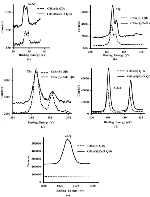
The XPS spectra of both CdSe(S) and CdSe(S)/ZnO QDs (Figure 4), confirmed the existence
of the peaks assigned to selenium, cadmium, sulfur and carbon, indicating that the shell
growth does not influence the structure of CdSe(S) QD cores. The presence of sulfur was
10
also in agreement with previous reports in the literature where MPA releases sulfur at high
temperature [58, 59], resulting in sulfur from the 3-mercaptopropionic acid (MPA)
participating the growth of CdSe(S) particles. Indeed, XPS data of CdSe(S)/ ZnO QDs
confirmed the existence of both Zn and O in the core/shell CdSe(S)/ZnO QDs, with the
observation of a new peak related to Zn2p at 1022 eV (Figure 4e), indicating the formation of
a ZnO shell around the CdSe(S) cores; this is consistent with the standard X-ray photo
electron spectrum of zinc oxide [60] and in accord with previous work [44].
XPS analysis was also used as quantitative method to determine elemental composition. The
atomic percentages of as-prepared QDs, have been summarized in Table 1. This confirmed
that CdSe(S) QDs were coated with a ZnO shell. The CdSe(S) QDs contained cadmium
(13.7%), selenium (0.7%), sulfur (13.1%), oxygen (29.9%) and carbon (42.5%), with the
ratio of atomic percentages C:O:S = 3.2:2.3:1, fully consistent with the molecular
composition of the capping agent MPA, C3O2SH6. The atomic percentage of Cd was found to
be approximately equal to the sum of the atomic percentages of the Se and S contributions,
indicating that at the surface of the CdSe QDs, the MPA coordinates by substitution of S at
the Se sites. The low intensity of the Se peak suggests a low atomic percentage of Se relative
to that of S (1:18.2) and is due to the dominance of surface atoms in the XPS data, essentially
MPA and the outer surface of the CdSe(S) QDs. Besides, according to quantitative analysis
data (Table 1), the most abundant element in the CdSe(S)/ZnO QD spectrum was O (37.7
atomic %). Each MPA molecule accounts for two O and one S atom; the atomic % of S was
found to be 12.3% and so MPA accounts for 24.6% of the O contribution, leaving 13.1%,
which closely matches the 12.0% contribution of Zn. The Cd:(Se+S) ratio of 1:0.8 and Cd:Zn
of 1:0.7 are fully consistent with a ZnO shell around the CdSe(S) core. In addition, the peaks
of Cd and Se shift with the incorporation of ZnO, highlighting the modified bonding
11
Figure 4. XPS spectra of CdSe(S) and CdSe(S)/ZnO QDs. Binding energy of (a) Se3d,
12
Table 1. Elemental analysis of QDs.
3.2 Cytotoxicity assays: The response of HCT-116 and WS1 cell lines upon exposure to the
QDs was studied in order to investigate toxicity. Cancer cells such as HCT-116 cells have an
irregular DNA pattern, making them more sensitive than healthy cells to free heavy metals,
including cadmium. In contrast, normal skin cells (WS1) are known to be some of the most
resistant cell types to free metal ions. In addition, 50% of cancer cells, including HCT-116,
depend upon a heat-shock protein (such as Hsp90) to survive [61]. The viability of these
cancer cells decreases in the presence of Hsp90 inhibitor 17-AAG, which is a common
anti-tumour compound [62, 63]. The viability of HCT-116 cells was therefore investigated both in
the presence of 17-AAG and after treatment of the cells with QDs.
3.2.1 Cytotoxicity of CdSe(S) QDs toward HCT-116 cells: The lethal concentration
corresponding to the death of 50% of cells (LC50) was determined as 105 µg/ml for the
HCT-116 cell line upon exposure to CdSe(S) QDs. CdSe(S) QDs were found to have a low toxicity
at the highest dilutions studied (25, and 50 µg/ml), with 100.0±0.2%, 91.5±2.6% cell viability
respectively, but have a cell mortality rate of ≥50% at concentrations of ~100 µg/ml and
13
Figure 5. The results of cytotoxicity assays of CdSe(S) QDs, Cd-MPA and Cd-Se-MPA precursors toward HCT-116 cell line. Error bars indicate standard error of the mean
and the concentration of 17-AAG was 0.06 µg/ml
3.2.2 Relative toxicity of CdSe(S) QDs in comparison to 17-AAG: Hsp90 inhibitor
17-AAG is an anti-tumour drug that inhibits the activity of Hsp90, a protein necessary for the
growth of the cells [62, 63]. The cytotoxicity results showed that CdSe(S) QDs inhibit the
cell viability by 64.4±0.5% at a concentration of 500 µg/ml; a similar value to that of
17-AAG (66.4±2.7%) at a concentration of only 0.06 µg/ml (Figure 5). Hence, CdSe(S) QDs are
much less cytotoxic than Hsp90 inhibitor 17-AAG, presumably as a result of non-specific cell
activity.
3.2.3 Cytotoxicity of precursor solutions: The CdSe(S) QDs were synthesised by
hydrothermal reactions of two precursor solutions containing Cd-MPA and Cd-Se-MPA. The
viability of HCT-116 cells was measured to both of the precursor solutions (Figure 5), which
were found to have no significant toxicity at concentrations of 25 and 50 µg/ml, but to be
cytotoxic at a concentration 500 µg/ml, similar to that of CdSe(S) QDs. The Cd-MPA
solution was found to be more toxic than the Cd-MPA-Se precursor solution. In addition,
14
µg/ml than the MPA precursor (97.0±8.3%), indicating that formation of the
Cd-Se-MPA complex decreases the toxicity, presumably due to a decreased number of free
cadmium ions.
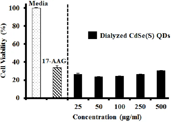
3.2.4 Cytotoxicity of dialyzed CdSe(S) QDs: A dialysis cassette was used to remove excess
capping agent and cadmium ions from solution, however the results showed that the CdSe(S)
QD solutions were in fact more toxic after dialysis than before (Figure 6). This is consistent
with a report of CdTe QDs with thiol capping agents, in which a surface cadmium-thiol shell
protected the QDs against oxidation, resulting in greater stability of the QDs [64]. In aqueous
solution, QDs are in an active equilibrium with excess cations and thiol in solution. During
dialysis, excess thiol and cadmium ions are removed from solution, resulting in the
destabilisation of the QD surface, thereby leaching more free cadmium ions into solution and
so increasing toxicity.
Figure 6. The results of cytotoxicity assays of dialyzed CdSe(S) QDs. Error bars indicate standard error of the mean and the concentration of 17-AAG was 0.06 µg/ml.
3.2.5 Cytotoxicity of CdSe(S)/ZnO core/shell QDs: Core/shell QD structures, in which a
relatively inert shell encapsulates the core, are widely considered to be an efficient method of
15
43, 50, 52, 53, 64]. The toxicity of core/shell CdSe(S)/ZnO QDs towards HCT-116 cancer
cells indicated that the core/shell QDs exhibited low toxicity at all concentrations studied. As
shown in Figure 7, the viability of the cells was determined to lie between 72.5±1.0% and
56.9±1.0% across the concentration range of 25 to 500 µg/ml, indicating that the LC50 of the
cells is not reached even at a concentration of 500 µg/ml, the highest concentration used in
this series of experiments. Clearly the shell inhibits release of free cadmium ions, limiting the
cell death.
Figure 7. The results of cytotoxicity assays of CdSe(S)/ZnO core/shell QDs toward HCT-116 cell line. Error bars indicate standard error of the mean and the
concentration of 17-AAG was 0.06 µg/ml
3.2.6 Cytotoxicity assays toward WS1 cells: Human skin fibroblast (WS1) cells were used
to determine the cytotoxicity of the as-synthesized QDs toward normal, resilient cells. The
results showed that both CdSe(S) and CdSe(S)/ZnO QDs exhibited low toxicity at all of the
concentrations studied (Figure 8), whilst the viability of HCT-116 cells was significantly
decreased after incubation with both CdSe(S) and CdSe(S)/ZnO QDs at all concentrations
(Figures 6 & 7). This indicates that cell resistance is another important factor in cytotoxicity
16
Figure 8. The results of cytotoxicity assays of CdSe(S) and CdSe(S)/ZnO QDs toward WS1 cell line. Error bars indicate standard error of the mean and the concentration of
17-AAG was 0.06 µg/ml
3.3 Confocal microscopy studies: Images of HCT-116 cells after treatment with QDs
confirmed the cytotoxicity data. As can be seen in Figures 9a & b, where only the cell nuclei
are visible by Hoechst or DAPI staining, many cells remained stable after treatment with
CdSe(S) QDs at 100 µg/ml. The images show highly luminescent CdSe(S) QDs present in
both the fixed and live cells, with no determinable loss of brightness. Live HCT-116 cells
were also incubated with CdSe(S)/ZnO QDs, confocal images of which showed that whilst
CdSe(S)/ZnO QDs aggregated in the biological media, many cells remained stable in
17
Figure 9. Confocal image of HCT-116 cells) -QDs: (a) fixed cells: cells (red) and CdSe(S) QDs (green), (b) live cells: cells (blue) and CdSe(S) QDs (green), (c) live cells (blue) and
CdSe(S)/ZnO QDs (green).
The emission profiles of QDs in both the cell media and water were recorded using a
confocal microscope in order to determine any changes in photoluminescence spectra of QDs
in cell media (Figure 10a). The photoluminescence emission of CdSe(S) QDs were found
with no change in cell media, in contrast to literature reports which have indicated that water
soluble MPA-capped CdTe QDs had an altered emission profile in cell growth media [65].
Emission spectra indicated that CdSe(S) QDs have a maximum emission at 548 nm in the cell
media, with no significant change or shift in photoluminescence relative to that in water. The
emission of CdSe(S)/ZnO QDs was found to have a lower intensity and shift to 546 nm in
18
Figure 10. Photoluminescence spectra of CdSe(S) QDs (A) and CdSe(S)/ZnO QDs (B): (a) in water and (b) in cell media. The excitation wavelength of the instrument was
adjusted at 405 nm in time of measuring PL.
4. Conclusions
We have shown that the cytotoxicity of QDscan be controlled with the aqueous synthesis of
stable QDs. It was determined that coating of QDs with a ZnO shell protects the core against
oxidation and the production of toxic free radicals, resulting in decreased cytotoxicity of
QDs, even at high concentrations. Images of cells after incubation with QDs at a
concentration of 100 µg/ml indicated that the cells remained viable, confirming the
cytotoxicity data. The stability of the QD cores in the cell media was found to be related to
the overall toxicity of QDs, which is largely governed by the free Cd-ion concentration.
Acknowledgments
We would like to thank School of Chemistry and the Mark Wainwright Analytical Centre of
University of New South Wales for facilities and Ms. Katerina Bendova and Dr. Renee Wan
19 References
[1]. B.A. Kairdolf, A.M. Smith, T.H. Stokes, M.D. Wang, A.N. Young, S. Nie,
Semiconductor quantum dots for bioimaging and biodiagnostic applications, Annu. Rev.
Anal. Chem., 6 (2013) 143-162.
[2]. A.M. Smith, G. Ruan, M.N. Rhyner, S. Nie, Engineering luminescent quantum dots for
in vivo molecular and cellular imaging, Ann Biomed Eng., 34 (2006) 3-14.
[3]. I.L. Medintz, H.T. Uyeda, E.R. Goldman, H. Mattoussi, Quantum dot bioconjugates for
imaging, labelling and sensing, Nat. Mater., 4 (2005) 435-446.
[4]. X. Yu, L. Chen, K. Li, Y. Li, S. Xiao, X. Luo, J. Liu, L. Zhou, Y. Deng, D. Pang, Q.
Wang, Immunofluorescence detection with quantum dot bioconjugates for hepatoma in
vivo, J. Biomed. Opt., 12 (2007) 014008/014001-014008/014005.
[5]. I.L. Medintz, A.R. Clapp, H. Mattoussi, E.R. Goldman, B. Fisher, J.M. Mauro,
Self-assembled nanoscale biosensors based on quantum dot FRET donors, Nat. Mater., 2
(2003) 630-638.
[6]. X. Michalet, F.F. Pinaud, L.A. Bentolila, J.M. Tsay, S. Doose, J.J. Li, G. Sundaresan,
A.M. Wu, S.S. Gambhir, S. Weiss, Quantum Dots for Live Cells, in Vivo Imaging, and
Diagnostics, Science, 307 (2005) 538-544.
[7]. E. Petryayeva, W.R. Algar, I.L. Medintz, Quantum dots in bioanalysis: a review of
applications across various platforms for fluorescence spectroscopy and imaging, Appl.
Spectrosc., 67 (2013) 215-252.
[8]. L. Qi, X. Gao, Emerging application of quantum dots for drug delivery and therapy,
Expert Opin. Drug Delivery, 5 (2008) 263-267.
[9]. A. Sadaf, B. Zeshan, Z. Wang, R. Zhang, S. Xu, C. Wang, Y. Cui, Toxicity evaluation of
hydrophilic CdTe quantum dots and CdTe@SiO2 nanoparticles in mice, J. Nanosci.
Nanotechnol., 12 (2012) 8287-8292.
[10]. A. Nel, T. Xia, L. Maedler, N. Li, Toxic Potential of Materials at the Nanolevel,
Science, 311 (2006) 622-627.
[11]. N. Chen, Y. He, Y. Su, X. Li, Q. Huang, H. Wang, X. Zhang, R. Tai, C. Fan, The
cytotoxicity of cadmium-based quantum dots, Biomaterials, 33 (2012) 1238-1244.
[12]. V. Labhasetwar, D. Leslie-Plecky, Biomedical Applications of Nanotechnology, Wiley
20
[13]. K. Unfried, C. Albrecht, L.-O. Klotz, A. Von Mikecz, S. Grether-Beck, R.P.F. Schins,
Cellular responses to nanoparticles: Target structures and mechanisms, Nanotoxicology,
1 (2007) 52-71.
[14]. H.C. Fischer, W.C.W. Chan, Nanotoxicity: the growing need for in vivo study, Curr.
Opin. Biotechnol., 18 (2007) 565-571.
[15]. K.W. Powers, S.C. Brown, V.B. Krishna, S.C. Wasdo, B.M. Moudgil, S.M. Roberts,
Research strategies for safety evaluation of nanomaterials. Part VI. Characterization of
nanoscale particles for toxicological evaluation, Toxicol. Sci., 90 (2006) 296-303.
[16]. J. Lovric, S.J. Cho, F.M. Winnik, D. Maysinger, Unmodified Cadmium Telluride
Quantum Dots Induce Reactive Oxygen Species Formation Leading to Multiple
Organelle Damage and Cell Death, Chem. Biol, 12 (2005) 1227-1234.
[17]. N. Singh, B. Manshian, G.J.S. Jenkins, S.M. Griffiths, P.M. Williams, T.G.G. Maffeis,
C.J. Wright, S.H. Doak, NanoGenotoxicology: The DNA damaging potential of
engineered nanomaterials, Biomaterials, 30 (2009) 3891-3914.
[18]. N. Li, C. Sioutas, A. Cho, D. Schmitz, C. Misra, J. Sempf, M. Wang, T. Oberley, J.
Froines, A. Nel, Ultrafine particulate pollutants induce oxidative stress and
mitochondrial damage, Environ. Health Perspect., 111 (2003) 455-460.
[19]. R. Mejias, L. Gutierrez, G. Salas, S. Perez-Yague, T.M. Zotes, F.J. Lazaro, M.P.
Morales, D.F. Barber, Long term biotransformation and toxicity of dimercaptosuccinic
acid-coated magnetic nanoparticles support their use in biomedical applications, J.
Controlled Release, 171 (2013) 225-233.
[20]. W.G. Kreyling, M. Semmler-Behnke, W. Moeller, Health implications of nanoparticles,
J. Nanopart. Res., 8 (2006) 543-562.
[21]. K.L. Aillon, Y. Xie, N. El-Gendy, C.J. Berkland, M.L. Forrest, Effects of nanomaterial
physicochemical properties on in vivo toxicity, Adv. Drug Delivery Rev., 61 (2009)
457-466.
[22]. L. Wang, H. Zheng, Y. Long, M. Gao, J. Hao, J. Du, X. Mao, D. Zhou, Rapid
determination of the toxicity of quantum dots with luminous bacteria, J. Hazard. Mater.,
177 (2010) 1134-1137.
[23]. T.S. Hauck, R.E. Anderson, H.C. Fischer, S. Newbigging, W.C.W. Chan, In vivo
Quantum-Dot Toxicity Assessment, Small, 6 (2010) 138-144.
[24]. B. Ballou, B.C. Lagerholm, L.A. Ernst, M.P. Bruchez, A.S. Waggoner, Noninvasive
21
[25]. W.-h. Chan, N.-h. Shiao, Cytotoxic effect of CdSe quantum dots on mouse embryonic
development, Acta Pharmacol Sin, 29 (2008) 259-266.
[26]. A. Valipoor, G. Amiri, K. Parivar, M. Modaresi, J. Taheri, A. Kazemi, M. Abasi, A.
Mirzakhani, A comparative study about toxicity of CdSe quantum dots on reproductive
system development of mice and controlling this toxicity by ZnS coverage,
Nanomedicine Journal, 2 (2015) 261-268.
[27]. R. Li, F. Jiang, Q. Xiao, J. Li, X. Liu, Q. Yu, Y. Liu, C. Zeng, Microcalorimetric,
spectroscopic and microscopic investigation on the toxic effects of CdTe quantum dots
on Halobacterium halobium R1, Nanotechnology, 21 (2010)
475102/475101-475102/475110.
[28]. M. Yan, Y. Zhang, K. Xu, T. Fu, H. Qin, X. Zheng, An in vitro study of vascular
endothelial toxicity of CdTe quantum dots, Toxicology, 282 (2011) 94-103.
[29]. V. Brunetti, H. Chibli, R. Fiammengo, A. Galeone, M.A. Malvindi, G. Vecchio, R.
Cingolani, J.L. Nadeau, P.P. Pompa, InP/ZnS as a safer alternative to CdSe/ZnS
core/shell quantum dots: in vitro and in vivo toxicity assessment, Nanoscale, 5 (2013)
307-317
[30]. A.M. Derfus, W.C.W. Chan, S.N. Bhatia, Probing the Cytotoxicity of Semiconductor
Quantum Dots, Nano Lett., 4 (2004) 11-18.
[31]. S. Chang, D. Chen, B. Kang, Y. Dai, UV-enhanced cytotoxicity of CdTe quantum dots
in PANC-1 cells depend on their size distribution and surface modification, J. Nanosci.
Nanotechnol., 13 (2013) 751-754.
[32]. W.J. Parak, T. Pellegrino, C. Plank, Labelling of cells with quantum dots,
Nanotechnology, 16 (2005) R9-R25.
[33]. Q. Wang, T. Fang, P. Liu, X. Min, X. Li, Study of the bioeffects of CdTe quantum dots
on Escherichia coli cells, J. Colloid Interface Sci., 363 (2011) 476-480.
[34]. M. Ando, M. Horie, Y. Akazawa-Ogawa, Y. Hagihara, N. Murase, Y. Shigeri,
Cytotoxicity of CdSe-based quantum dots incorporated in glass nanoparticles evaluated
using human keratinocyte HaCaT cells, Biosci., Biotechnol., Biochem., 80 (2016)
210-213.
[35]. L. Lai, J.-C. Jin, Z.-Q. Xu, P. Mei, F.-L. Jiang, Y. Liu, Necrotic cell death induced by
the protein-mediated intercellular uptake of CdTe quantum dots, Chemosphere, 135
22
[36]. R. Schneider, C. Wolpert, H. Guilloteau, L. Balan, J. Lambert, C. Merlin, The exposure
of bacteria to CdTe-core quantum dots: the importance of surface chemistry on
cytotoxicity, Nanotechnology, 20 (2009) 225101/225101-225101/225110.
[37]. X. Li, Z. Yan, J. Xiao, G. Liu, Y. Li, Y. Xiu, Cytotoxicity of CdSe quantum dots and
corresponding comparison with FITC in cell imaging efficiency, Int J Clin Exp Med, 10
(2017) 753-759.
[38]. G. Guo, W. Liu, J. Liang, Z. He, H. Xu, X. Yang, Probing the cytotoxicity of CdSe
quantum dots with surface modification, Materials Letters, 61 (2007) 1641-1644.
[39]. E. Oh, R. Liu, A. Nel, K.B. Gemill, M. Bilal, Y. Cohen, I.L. Medintz, Meta-analysis of
cellular toxicity for cadmium-containing quantum dots, Nat Nano, 11 (2016) 479-486.
[40]. Y. Su, M. Hu, C. Fan, Y. He, Q. Li, W. Li, L.-h. Wang, P. Shen, Q. Huang, The
cytotoxicity of CdTe quantum dots and the relative contributions from released cadmium
ions and nanoparticle properties, Biomaterials, 31 (2010) 4829-4834.
[41]. X. Li, N. Chen, Y. Su, Y. He, M. Yin, M. Wei, L. Wang, W. Huang, C. Fan, Q. Huang,
Autophagy-Sensitized Cytotoxicity of Quantum Dots in PC12 Cells, Adv. Healthcare
Mater., 3 (2014) 354-359.
[42]. Y.J. Bao, J.J. Li, Y.T. Wang, L. Yu, L. Lou, W.J. Du, Z.Q. Zhu, H. Peng, J.Z. Zhu,
Probing cytotoxicity of CdSe and CdSe/CdS quantum dots, Chin. Chem. Lett., 22 (2011)
843-846.
[43]. Y. Su, Y. He, H. Lu, L. Sai, Q. Li, W. Li, L. Wang, P. Shen, Q. Huang, C. Fan, The
cytotoxicity of cadmium based, aqueous phase - Synthesized, quantum dots and its
modulation by surface coating, Biomaterials, 30 (2008) 19-25.
[44]. F. Aldeek, C. Mustin, L. Balan, G. Medjahdi, T. Roques-Carmes, P. Arnoux, R.
Schneider, Enhanced Photostability from CdSe(S)/ZnO Core/Shell Quantum Dots and
their use in Biolabeling, Eur. J. Inorg. Chem., (2011) 794-801.
[45]. M. C. Phelan, In Current Protocols in Cell Biology, John Wiley & Sons, Inc., 2007, pp.
1.1.1-1.1.18.
[46]. Q. Wang, X. Zhou, T. Fang, P. Liu, X. Li, X. Min, One-step growth of high-quality
CdTe quantum dots via hydrothermal method and cytotoxicity evaluation, Powder
Technol., 247 (2013) 81-86.
[47]. Y. Li, L. Jing, R. Qiao, M. Gao, Aqueous synthesis of CdTe nanocrystals: progresses
23
[48]. F. Mirnajafizadeh, D. Ramsey, S. McAlpine, F. Wang, P. Reece, J.A. Stride,
Hydrothermal synthesis of highly luminescent blue-emitting ZnSe(S) quantum dots
exhibiting low toxicity, Mater. Sci. Eng., C, 64 (2016) 167-172.
[49]. M. Cirillo, T. Aubert, R. Gomes, R. Van Deun, P. Emplit, A. Biermann, H. Lange, C.
Thomsen, E. Brainis, Z. Hens, “Flash” Synthesis of CdSe/CdS Core–Shell Quantum
Dots, Chemistry of Materials, 26 (2014) 1154-1160.
[50]. C. Kirchner, T. Liedl, S. Kudera, T. Pellegrino, A.M. Javier, H.E. Gaub, S. Stoelzle, N.
Fertig, W.J. Parak, Cytotoxicity of Colloidal CdSe and CdSe/ZnS Nanoparticles, Nano
Lett., 5 (2005) 331-338.
[51]. F. Yang, P. Yang, Effect of CdS Interlayer on Properties of CdTe Based Quantum Dots,
J. Cluster Sci., 24 (2013) 643-656.
[52]. Y. Liu, P. Wang, Y. Wang, Z. Zhu, F. Lao, X. Liu, W. Cong, C. Chen, Y. Gao, Y. Liu,
The Influence on Cell Cycle and Cell Division by Various Cadmium-Containing
Quantum Dots, Small, 9 (2013) 2440-2451.
[53]. B.A. Rzigalinski, J.S. Strobl, Cadmium-containing nanoparticles: Perspectives on
pharmacology and toxicology of quantum dots, Toxicology and Applied Pharmacology,
238 (2009) 280-288.
[54]. F. Mirnajafizadeh, F. Wang, P. Reece, J.A. Stride, Synthesis of type-II CdSe(S)/Fe2O3
core/shell quantum dots: the effect of shell on the properties of core/shell quantum dots,
J. Mater. Sci., 51 (2016) 5252-5258.
[55]. The International Centre for Diffraction Data (ICDD), 1978, JCPDs files, No.
00.19-0191.
[56]. The International Centre for Diffraction Data (ICDD), 1978, JCPDs files, No.
00-010-0454.
[57]. F. Mirnajafizadeh, Synthesis and investigation of the properties of water soluble
quantum dots for bioapplications., PhD Thesis, University of New South Wales, 2015.
[58]. H. Qian, X. Qiu, L. Li, J. Ren, Microwave-assisted aqueous synthesis: a rapid approach
to prepare highly luminescent ZnSe(S) alloyed quantum dots, J Phys Chem B, 110
(2006) 9034-9040.
[59]. A.L. Rogach, T. Franzl, T.A. Klar, J. Feldmann, N. Gaponik, V. Lesnyak, A. Shavel, A.
Eychmueller, Y.P. Rakovich, J.F. Donegan, Aqueous Synthesis of Thiol-Capped CdTe
Nanocrystals: State-of-the-Art, J. Phys. Chem. C, 111 (2007) 14628-14637.
[60]. F. J. Moulder, F. W. Stickle, E. P. Sobol, D. K. Bomben, Hand book of X-ray
24
[61]. Z. Solarova, J. Mojzis, P. Solar, Hsp90 inhibitor as a sensitizer of cancer cells to
different therapies, Int. J. Oncol., 46 (2015) 907-926.
[62]. L.H. Pearl, C. Prodromou, Structure and in vivo function of Hsp90, Curr. Opin. Struct.
Biol., 10 (2000) 46-51.
[63]. T. Horibe, A. Torisawa, M. Kohno, K. Kawakami, Molecular mechanism of
cytotoxicity induced by Hsp90-targeted Antp-TPR hybrid peptide in glioblastoma cells,
Mol. Cancer, 11 (2012) 59.
[64]. M. Gao, S. Kirstein, H. Möhwald, A.L. Rogach, A. Kornowski, A. Eychmüller, H.
Weller, Strongly Photoluminescent CdTe Nanocrystals by Proper Surface Modification,
The Journal of Physical Chemistry B, 102 (1998) 8360-8363.
25
Supplementary Data
Nanoparticles for Bioapplications: Study of the Cytotoxicity of Water
Dispersible Quantum Dots
Fatemeh Mirnajafizadeh**1, Deborah Ramsey**1, Shelli McAlpine1, Fan Wang2, Peter Reece2 and John Arron Stride1*
1. School of Chemistry, University of New South Wales, Sydney, NSW 2052, Australia 2. School of Physics, University of New South Wales, Sydney, NSW 2052, Australia
3. School of Mathematical and Physical Sciences, University of Technology Sydney, Ultimo, Sydney, NSW 2007, Australia
* Corresponding Author: A/Prof. John Arron Stride ** These authors contributed equally in this work.
S1. Synthesis of Water Dispersible QDs
S.1.1. Synthesis of CdSe(S) QDs: CdSe(S) QDs were prepared according to a modified
literature method [1]. Briefly, a Cd-MPA precursor solution was prepared by dissolving 1.48
g (6.5 mmol) CdCl2 in 100 ml ultrapure water and adding MPA (0.7 ml) to the solution
making the molar ratio of Cd:MPA 6.5:8; the pH was adjusted to 12.5 using 1M NaOH, this
was labelled Sample 1. Sample 1 was then placed under nitrogen for 10 minutes and NaHSe
(1 mmol, 20 ml), was injected to the solution to form a Cd-Se-MPA complex as a clear
yellow liquid; this was labelled as Sample 2. Sample 2 was transferred to a Teflon-lined
autoclave and placed in a conventional oven for 1 hour, under hydrothermal treatment at T =
150ºC. Finally, CdSe-MPA capped QDs were formed in an aqueous orange solution and
labelled as Sample 3. The as-prepared QDs were orange in colour in contrast to that
previously reported [1], with a yellow emission colour under UV light.
S.1.2. Synthesis of CdSe(S)/ZnO QDs: CdSe(S) QDs were prepared, as described in section
S.1.1 and coated with ZnO according to previous literature report [1], based upon the
controlled hydrolysis of zinc salts at basic pH. First, an aqueous suspension of as-prepared
26
concentration of CdSe(S) QDs was 1.7 mg in 5 ml and the pH was adjusted to 13 using 1M
NaOH. This solution was refluxed for 2 hours before injecting an aqueous solution of
Zn(OAC)2.2 H2O (18 ml, 0.01M) at T = 100ºC. Reflux was continued for another hour and
CdSe(S)/ZnO core/shell QDs were obtained as orange nanoparticles.
S2. Cell Culture of HCT-116 and WS1 Cells
Cell cultures of human colorectal carcinoma cells (HCT-116) and human skin fibroblast cell
line (WS-1) were obtained according to the standard protocol [2]. As cells are usually
received frozen in the culture medium with 5-10% dimethyl sulfoxide (DMSO) in the vapour
of liquid nitrogen at 77 K, they were first defrosted to 37ºC and the culture medium discarded
by using a centrifuge at 1500 rpm for 5 minutes. Then, the HCT-116 and WS-1 cells were
separately resuspended in Dulbecco’s Modified Eagle Medium (DMEM) supplemented with
10% fetal bovine serum (FBS), 1% penicillin/streptomycin, (1%) nonessential amino acid
and L-glutamine (2 mM) as growth medium and transferred in two 75 cm2 flasks. Finally,
the cells were grown in a humidified incubator at 37ºC with 5% CO2 and as cells reached
confluence; they were immediately used for cytotoxicity tests in separate experiments.
S3. Preparation of HCT-116 cells-CdSe(S) QDs Samples
S.3.1. Fixed HCT 116 cells-CdSe(S) QDs sample: cells were cultured (as described in S2)
and were seeded in 35 mm fluorodish cell culture dishes at a density of 20000 cell/dish for 24
hours. The cells were then treated with an aqueous solution of CdSe(S) QDs (250 µg/ml) and
incubated at 37ºC with 5% CO2 for 24 hours. After incubation, the cells were fixed with 4%
paraformaldehyde in PBS for 1 hour, before finally being washed with PBS and stained with
5 µg/ml bisbenzimide (Hoechst 33342) for 20 minutes in room temperature.
S.3.2. Live HCT 116 cells-QDs samples: cells were cultured according to the standard
protocol [2] similar to the fixed cells and were seeded in a 24-well glass bottom plate at a
27
CdSe(S) QDs (100 µg/ml) and incubated at 37ºC with 5% CO2 for 24 hours. As-prepared
samples were used for confocal microscopy studies.
S4. The comparison of PXRD of CdSe(S) QDs with cubic CdSe and CdS standard peaks
The diffraction peaks of CdSe(S) QDs were observed between standard cubic CdSe [3] and
CdS [4], as shown in Table S1.
Table S1. The comparison of diffraction peaks of as-synthesized CdSe(S) QDs with
cubic CdSe and CdS standard peaks
h k l CdSe cubic
(Standard pattern)
As –synthesized
CdSe(S) QDs
CdS cubic
(Standard pattern)
1 1 1 25.35 26.29 26.50
2 2 0 42.00 43.66 43.96
3 1 1 49.69 51.67 52.13
References
[1]. F. Aldeek, C. Mustin, L. Balan, G. Medjahdi, T. Roques-Carmes, P. Arnoux, R.
Schneider, Enhanced Photostability from CdSe(S)/ZnO Core/Shell Quantum Dots and
their use in Biolabeling, Eur. J. Inorg. Chem., (2011) 794-801.
[2]. M. C. Phelan, In Current Protocols in Cell Biology, John Wiley & Sons, Inc., 2007, pp.
1.1.1-1.1.18.
[3]. The International Centre for Diffraction Data (ICDD), 1978, JCPDs files, No.
00.19-0191.
[4]. The International Centre for Diffraction Data (ICDD), 1978, JCPDs files, No.