letters
to
the editor
Pharmacology of cranial dystonia
To the Editor: We read with interest the articles by Tolosa and h i 1 and Casey,2 who suggested that Meige’s disease is due to “striatal dopaminergic preponderance,” and the more recent papers by Micheli et aP and Tanner et a14 on the pharmacology of this condition. The major diffi- culty with all these otherwise excellent and interesting reports is that they seek to establish a consistent phar- macology to this disorder on relatively small numbers of patients.
Tolosa and Lai reported inconsistent results in 5 patients, Casey described 2 patients, Micheli et a1 treated 2 individuals with the disease (and reported 2 others in an addendum), and Tanner et a1 studied 13 patients. Our own pharmacologic observations of 56 patients with cranial dystonia do not suggest that there is any uniform phar- macologic profile for this curious illness. All of the patients were treated with maximum tolerated doses of the drugs indicated in the table for periods of weeks to months. Slow titration of each drug also allowed evaluation of patient response to low doses, particularly in the case of lisuride, where a dosage of 1.2 to 3.0 mg was given for extended periods. Improvement refers to a degree of benefit suffi- cient to persuade the patient and the physician that it was worthwhile continuing the medication. It is also worth noting that, in our own double-blind studies of drug therapy for this condition, we have found that placebo treatment may cause an appreciable improvement of up to 20% in disability scores.
Tolosa and Lai observed a decrease in the intensity of cranial dystonia after apomorphine. They interpret this as indicating “striatal dopamine preponderance,’’ because apomorphine, in the doses given, may reduce the chorea of Huntington’s disease and the orofacial movements of tar- dive dyskinesia, perhaps by actions on presynaptic dopamine receptors. Micheli et aP confirmed the effect of apomorphine in their two cases. However, other dopamine agonists, in general, have little or no effect on cranial dystonia. Tolosa and Lai were unable to demonstrate any
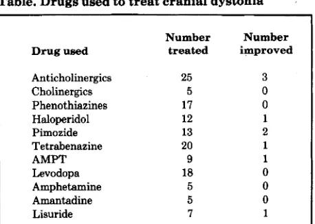
Table. D r u g s used to treat cranial dystonia
Number Number
Drug used treated improved
Anticholinergics Cholinergics Phenothiazines Haloperidol Pimozide Tetrabenazine
AMP”
Levodopa Amphetamine Amantadine Lisuride
25
5 17 12 13 20 9 18 5 5
7
3 0 0 1
2
1
1
0 0 0
1
Based on the experience of a personal series of 56 patients with blepharospasm and/or ommandibular dystonia. All drugs were used
up to maximum tolerated dosage.
effect of levodopa in their small series ofpatients, and none of our 18 patients so treated improved; indeed, 2 got slightly worse. The other dopamine agonists shown in the table had no effect whatsoever, except lisuride, which improved one of our patients. Micheli et aP reported improvement in four patients treated with lisuride (two of whom were men- tioned in an addendum). This inconsistent response to lisuride may reflect variations in its absorption when given orally. However, even if lisuride does sometimes provide benefit, pharmacologic conclusions are difficult because it is a powerful serotonin agonist as well as a dopamine ago- nist. Furthermore, the effects of apomorphine may reflect its sedative properties rather than any selective presynap- tic dopamine action.
Tblosa and Lai, and Casey, based their pharmacologic arguments on the effects of dopamine antagonists for haloperidol’ and perphenazine2 improved cranial dystonia in their patients. However, we have treated many patients with such drugs and with presynaptically acting dopamine antagonists such as tetrabenazine and alpha-methyl-para- tyrosine (AMPT)? but with no consistent effects (table). Indeed, we have seen the involuntary movements of vari- ous facial areas respond in opposing directions, as in one patient treated with AMPT in whom the jaw and tongue movements improved but blepharospasm increased.5 Less than 10% of patients treated with phenothiazines, haloperidol, pimozide, tetrabenazine, or AMPT gained useful benefit. Unwanted side effects of these drugs were common and disabling, particularly pseudoparkinsonism, akathisia, and depression.
Both Tolosa and Lai, and Casey, also used observations on the effects of anticholinergic and cholinergic drugs as subsidiary evidence to support their arguments for “striatal dopamine preponderance” in cranial dystonia. In fact, Tolosa and Lai found that physostigmine aggravated the condition in their patients, which is the reverse of what might be expected. They avoided this problem, however, by pointing out similar inconsistent responses to physostig- mine i n Huntington’s disease. Casey described the opposite effect of deanol, a supposed cholinergic agent of dubious reputation which, in two of his patients, appar- ently improved matters; however, the anticholinergic drug benztropine had little effect. Tanner et a1,4 in acute studies, found that intramuscular (IM) scopolomine improved their patients, an action that was reversed by subsequent IM physostigmine. In a subsequent chronic oral study using benztropine or trihexyphenidyl for some weeks or months, Tanner et a1 found that 12 of 13 patients improved. They conclude that “acetylcholine plays a role in the pathophysiology of Meige syndrome.”
Our
own experience with cholinergics (limited to cho- line chloride) and anticholinergic drugs has produced no consistent results (table). In a further study: we were unable to show any beneficial effect of intravenous atro- pine, benztropine, or chlorpheniramine when compared with placebo in six patients with cranial dystonia.In summary, our own large experience of the treatment of cranial dystonia leads us to conclude that there is no consistent pharmacologic response to drugs manipulating either the dopamine or acetylcholine systems of the brain. This seems to be the case in all forms of torsion dystonia, including torticollis and writer’s cramp. Unfortunately, the treatment of these distressing and disabling illnesses with
drugs remains unsatisfactory and empirical.
C.D. Marsden, M D A.E. Lang, M D M.P. Sheehy, M D
Denmark Hill, London
References
1. Tolosa ES, Lai C. Meige disease: striatal dopaminergic pre- ponderance. Neurology (NY) 1979;29:1126-30.
2. Casey DE. Pharmacology of blepharospasm-oromandibular
dystonia syndrome. Neurology (NY) 1980;30:690-5. 3. Micheli F, Fernhdez Pardel MM, Leiguarda RC. Beneficial
effects of lisuride in Meige disease. Neurology (NY)
4. Tanner CM, Glantz RH, Klawans HL. Meige disease: acute a n d chronic cholinergic effects. Neurology ( N Y ) 1982;32:783-5.
5 . Lang AE, Marsden CD. Alphamethylparatyrosine and tetrabenazine in movement disorders. Clin Neuropharmacol (in press).
6. Lang AE, Sheehy MP, Marsden CD. Anticholinergics in adult-onset focal dystonia. Can J Neurol Sci 1982;9:313-9. 1982;32:432-4.
To the Editor: The report of Tanner e t all on Meige’s disease did not mention the drug dosage used in their successfully treated patients. This information would be helpful. The following case illustrates the long-term daily use of 10 mg benztropine, a higher than usual dose. Starting at age 31, this woman successively noted
a 4-inch amplitude flapping tremor of the right hand, tautness of shoulder muscles, flapping tremor of the left hand, blepharospasm, grimacing, facial spasms, tightness of throat and neck muscles, flexor spasms of the neck, and dysphonia. Benztropine was prescribed in small doses but the patient, on her own initiative, increased the dose to 10 mg daily with “magical” 95% relief. She began to live normally, stopped visiting physicians, and did not report the beneficial effect. After 14 years, at age 51, she discontinued the benztropine because of pupillary dilatation in her cataractous left eye.
Within 5 weeks all of the old complaints returned, but disappeared when medication was resumed. I first saw her at this time (19761, and the neurologic exam- ination was normal. At her request, another trial without medication was undertaken, and in 5 days the eyes were again continuously closed and tremor reappeared. Re- sumption of benztropine therapy brought relief, which has continued for another 6 years, some 20 years in all.
Because of this experience, we used benztropine, 8 mg daily, in another woman, aged 75, who had been functionally blind and helpless because of blepharospasm for 4 years. Her eyes remained open, and she resumed walking outdoors. Patients with less severe, but dis- abling, blepharospasm have been relieved by 4 to 6 mg daily.
C.M. Fisher, MD
Boston, MA
Reference
1. Tanner CM, Glantz RH, and Klawans HL. Meige disease: acute and chronic cholinergic effects. Neurology ( N Y ) 1982;32:783-5.
Reply from the Authors: “Improvement” as defined by Lang and Marsden fails to separate three distinct phar- macologic properties: effective dose, toxic dose, and therapeutic index. The failure of an agent with known effects on a specific neurotransmitter system to provide a “degree of benefit sufficient to persuade the patient and the physician that it was worthwhile continuing with the med- icat‘ion” does not negate the importance of the neu- rotransmitter to the pathophysiology of the disorder being treated. A drug may affect the appropriate neurotransmit- ter system, but its usefulness may be limited by its low therapeutic index.
Dose-limiting side effects are common when anti- cholinergic drugs are administered, especially in the older age group a t risk for blepharospasm/oromandibular dys- tonia. In our chronically treated patients, side effects prompted dosage reduction in 50% of those whose move- ment disorder had improved with anticholinergic therapy. Despite the therapeutic difficulties posed by frequent side effects of anticholinergic therapy, the pharmacologic effect of this class of dystonic movements was beneficial.
Clinical studies aimed at defining the pharmacology ofa disorder are frequently limited by unwanted side effects. Nevertheless, a distinct pharmacologic effect, if specially sought, may often be appreciated. We maintain from our data t h a t the cholinergic system plays a role in the pathophysiology of blepharospasm/oromandibular dys- tonia.
We appreciate Dr. Fisher’s addition of cases of bleph- arospasm responding to high dose benztropine treatment. Of our nine patients with Meige’s disease who sustained a clinical response to anticholinergics, five were taking benztropine in daily doses of 2 mg to 8 mg (mean dose, 4.5 mg per day), and four were taking tri- hexyphenidyl in daily doses of 4 to 8 mg (mean dose, 6.5 mg) at 12 months follow-up. The one patient who responded to 8 mg per day benztropine was similar to Dr. Fisher’s patients. Further increase in anticholinergic dosage was limited by side effects (particularly, disabling memory loss and confusion) in these older patients. We have observed several young adults w i t h partial dys-
tonias who responded to benztropine 10 mg per day to 16 mg per day. Marsden and Fahnl have reported dystonic children who responded well to 80 mg per day with few side effects.
Caroline M . Tanner, MD Russell H . Glantz, MD Harold L. Kalwans, MD
Chicago, I L
Reference
In: Marsden CD, Fahn S, eds. Movement Disorders. London:
Butterworth Scientific, 1982;191-5.
Neurologists are like cops
. . .
To
the Editor: A physician friend once told me, on the day I agreed to see his ailing mother, “You neurologists are like cops. .
.
you’re all over the place until you need one.”Much has been written recently about the optimal num- ber of neurologists, based on demographic trends and pro- jected health care needs for the The most recent survey by Dr. Menken projects a lugubrious future for our specialty and its related training program^.^.^ Specifically, he calls for closing neurologic residencies as a means of controlling numbers of neurologists, since oversupply leads to competition with primary care physicians, to the over- use of tests, and dilutes the impact and effectiveness of neurologic programs at academic centers.
His overview of the supply-side economics of neurology in the 80s is hardly a forecast-rather, a statement of the status quo in many localities. Neither does it go far enough in its implications for both neurologists and nonneurolo- gists, because it examines the symptoms rather than the underlying disease.
But who needs neurologists, anyway? How do we account for this paradoxical scarcity of individuals in the midst of collective plenty?
Dr. Menken touches on the truth in his commentary. Clearly, the job description of the neurologist has changed and continues to evolve.
Marketing, in our present health care system, is based on the notion of “technofix.” Health problems, like all problems, are a matter of technical troubleshooting until the source of the disease, pain, or discomfiture is found, application of appropriate treatment is made and prompt subsequent relief of symptoms, reversal of disease, and quiescence of discomfiture is obtained. While the paradigm apparently works for the worried well and minimally ill, neurologic diseases and symptoms rarely fit that model. Nonetheless, the public seems to have the “need to know” not only the source of every symptom, but the need to know today, what will become obvious tomorrow or the next day. Much of modern marketing of health care services is based on that need to know. As a consequence, the time-honored tradition of classical neurology- “Let the pathology mature; the diagnosis will declare itself”-is no longer permitted to us, at least not in America. The more time the physician has to make a diagnosis, the fewer the tests needed. On the other hand, the price of finding out the potential seriousness of any symptom or imtation on the day it occurs is astronomical indeed, and requires the use of every bit of technology available to the physician. Neurolo- gists in many communities function precisely in the latter mode, using sophisticated technologic approaches to meet the demand for quick, accurate service by sophisticated consumers with high expectations. In the process, eclectic neurology has given way to electrical empiricism.
Of course, the consumers have plenty of help in the quest for medical technofix. They have emergency room physicians telling them they need to see a neurologist as
soon as possible for symptoms and complaints of brief duration. They have primary care doctors telling them that the routine tests done to evaluate their symptoms have failed to disclose their nature and now they must see a specialist. And there are those who have seen a series of specialists-otolaryngologists, cardiologists, a n d ophthalmologists-who have done numerous sensitive and expensive tests that establish the cause of symptoms to be definitely outside their field of expertise. To some, hysteria has been contagious; they caught it in their doctor’s office. Still others have even seen one or more neurologists, have had all the neurotechnology, and have either a diagnosis or no diagnosis. Either way, they are bewildered and unhappy, because there is no quick fix; now they just need someone to talk to, to answer a few questions.
. .
about their future.Academic neurologists are in a particularly uncomforta- ble crossfire. They are perceived by referring physicians, including other neurologists; as having plenty of time to engage patients, to discuss and analyze difficult diagnostic and therapeutic problems. Yet they are seen by their administrators as competitors of their own progeny in the private sector, who need to see patients in high volume to compensate for their poor income-generating capacity. Space-occupying lesions treated by neurosurgeons gener- ate high professional fees and use multiple reimbursable hospital resources as well; in the neurology clinic, however, time-occupying lesions can be seen only in limited num- bers, and often without third-party coverage.
One-on-one professional patient care is the most efficacious thing that doctors do. Unfortunately, in today’s marketplace, it is least valuable as compared with the standard set by high-priced technology. Neurologic patients presenting to tertiary care centers have had all the technology done, all the routine therapies tried. When the more elaborate, expensive, and financially rewarding tech- nologies are repeated, the economic benefits accrue to departments other than neurology. Tertiary care neurolo- gists are asked to see ever-increasing numbers of patients, whether those patients have MS, intractable migraines, “low-pressure hydrocephalus,” or proctalgia fugax. They are expected to do this at low cost and high volume, at overhead rates identical to those of other physicians, whose practices require high-cost milieus like radiology suites and operating rooms. Then, they are asked to either justify the continued existence of their cost-inefficient spe- cialty or acquire some forms of technology to generate fees disproportionate to the time required to perform them.
Sooner or later, someone must play the traditional doc- tor’s role without pretentions and merely minister to the sick. Technology was transferred to the point of ubiquity during the 70s; neurologists are among those to whom responsibility for technologies’ failures will be transferred in the 80s.
Surely, there will be no dearth of referrals for neurolo- gists’ services. Despite packaging modern medicine as “health care” delivery, we in neurology know that even- tually preventive medicine is bound to disappoint even its most staunch true believers. Even if all effective medica- tions and surgeries do accomplish their goals as advertised, and prevent death from heart disease and even cancer, they are also deferring an increased load to the neurologist of the future, who must eventually deal with disease, complica-
tion, or side effects in the organ system that does not repair itself, is irreplaceable, and is not transplantable. Further, every therapeutic novelty (like hyperbaric oxygenation) and innovation (like chronic renal dialysis) seems to create a series of new neurologic syndromes or diseases.
Therefore, in contrast to Dr. Menken’s hypothesis that there are too many, we will probably need more and more neurologists-or a t least classical physicians who can func- tion in a power failure, who can take a history and apply deductive and dialectical reasoning with the wisdom of talmudic scholars, yet who will serve as apologists, public relations men, even cheerleaders for the institutions they represent and for the physicians who refer patients to them. Someone has to maintain the cost efficiency of the more lucrative high-volume machines and services.
The problem is not one of need, the problem is one of value systems and how these neurologists will make a liv- ing! Their colleagues, their administrators, their patients, and “health” insurance carriers seem to have trouble estab- lishing what they’re worth.
Some aspects of medicine are simply and intrinsically not cost effective by modern standards. Traditional or classical neurology is one of them. The most cost-efficient method of dealing with the chronically ill and the symp- tomatically disabled is euthanasia. Since that is unlikely to become a socially or morally acceptable alternative for conscious patients, the future need for neurologists or their substitutes is guaranteed. What their services are worth, and to whom, has to be rethought!
Meanwhile, neurologists must respond to the demands of anxious patients and their equally anxious doctors. After all the tests, whatever the results, somebody has to sit down and engage the patient, to find out what’s wrong with him, and, like a detective, to distinguish the natural history of the disease from its unnatural modifications by previous professionals.
.
.
and to answer questions. That takes time. Where are all the neurologists when you need one? They’re doing “emergency” EMGs, trying to distinguish acute paresthesias due to hyperventilation from incipient Guillain-Barr6 syndrome-or they’re seeing the doctor’s mother.Gaetano F. Molinari, M D
Washington, DC
References
1. Yahr M. Summary report of the Joint Commission on Neu- rology. Neurology (Minneap) 1975;25:497-501.
2. Goldstein M. The neurologists as a health care resource: facts, estimates, and aspirations for the supply of neurologists. Guest editorial. Neurology (Minneap) 1977;27:901-4. 3. Menken M. The coming oversupply of neurologists in the
1980s: implications for neurology and primary care. JAMA
4. Menken M. The coming oversupply of neurologists in the
1980s: implications for neurology training programs. Neurol- ogy (NY) 1982;32:510-2.
5. O’Doherty DS. “An ounce of prevention
. . . .”
Neurology (NY) 1982;32:513.6. Dyken ML. The continuing undersupply of neurologists in
198 1;2452401-3.
the 1980s: impressions based on data from three studies. Neurology (NY) 1982;32:651-6.
Reply from an Author: Professor Molinari’s thoughtful and provocative letter addresses a keystone issue in man- power research namely, the need to identify the role of each group of physicians in the provision of health care services. He asks: What is a neurologist?
I would suggest that, in our society, a neurologist is a medical specialist who has learned to approach disorders of the nervous system with the skills and attitudes of a clinical neuroscientist. He or she has acquired this approach in a neurology training program that teaches exclusively the biomedical science of neurologic care, and measures the quality of that care by the completeness of “work-ups.” Accordingly, when practicing neurologists develop patient care protocols that make technology the master of medi- cine, and not its servant, it seems to me they are doing exactly what they have been trained to do.
Along with a n overuse of technology, Professor Molinari has identified a serious shortfall of essential pri- mary care services-such as counseling and the com- prehensiveness and coordination of care. Could this shortage have emerged because the matrix and organiza- tion of care must now accommodate large numbers of neu- rologists to serve as principal care providers for large numbers of patients?
I question the general assumption among neurologists that those most knowledgeable about an organ system are, inter alia, most highly qualified to provide care for virtually all patients whose symptoms fall within their area of scien- tific expertise. We seem to have forgotten that most of the care required most of the time by the majority of patients with neurologic symptoms, signs, and diagnoses is primary care. (The same thing may be said of all organ systems and all medical specialties.) I would therefore suggest that many patients in need of neurologic care are best off when that care is provided by well-trained primary care physi- cians, who use the services of neurologists as needed to improve the quality, effectiveness, and economy of care.
Matthew Menken, M D
New Brunswick, NJ
Cerebral embolism and anticoagulation
To the Editor: We read with interest the papers by Furlan et all and Koller2 dealing with cerebral embolism and anticoagulant therapy. They differ about the prog- nosis for recurrent emboli: fairly good in the Furlan et a1 report but a high mortality rate in Koller’s report. Furlan e t all found only one case of hemorrhagic in- farction in their CT series of 54 cases, but they did not specify whether the recurrent embolic events were stud- ied by CT or not (maybe some of these cases could have been hemorrhagic infarctions). The low frequency of hemorrhagic infarction is not in accordance with the higher incidence of hemorrhagic infarction in other series of cerebral embolism3-j or our own experience.
ease (without endocarditis). All of them were studied within 24 hours of the stroke, and at least one CT was performed in each case. Only the first week’s outcome was noted. Seventeen patients were treated with IV heparin within 24 hours of admission. Fifteen patients did not receive anticoagulant therapy during the first week. There were no recurrent embolic events in any patient. CT showed hemorrhagic infarction in five pa- tients. Four of these five patients followed a similar clinical and CT pattern: admission CT was normal or showed ischemic infarction (low-density lesion); and within 48 hours of the first stroke, a new stroke (or sudden worsening of the previous neurologic status) occurred. A new CT at this time showed a large hem- orrhage on the same side as the first stroke. The first stroke had provoked a severe neurologic disability in two patients, moderate disability in one patient, and minimal disability in one patient. Two of the four patients who were on anticoagulant therapy died in the first days. The other two patients were left with severe and mild disability. In another patient, CT showed a hem- orrhagic infarct with no clinical variation. Thus, a new stroke after a cerebral embolus can be due not only to
a recurrent emboli, but also to a hemorrhage into the infarct. The global outcome of the 32 patients at the end of the first week was as follows: 4 were dead, 14 showed severe disability, 7 moderate disability, and 8 mild or no disability. We did not intend to compare the outcome of patients treated and those not treated with anticoagulants. The main conclusion from our series is that hemorrhagic infarction is not rare after a n embolic stroke; it usually happens within the first 48 hours, and it carries a poor prognosis (probably worsened by an- ticoagulant therapy, a s reported in experimental stud- ies% In deciding about anticoagulant therapy in embolic stroke, the danger of early hemorrhagic infarction should be weighted against the risk of recurrent embolus.
L. Calandre, MD J . Fernandez Ortega, MD F. Bermejo, MD A . Portera, MD Madrid, Spain
References
1. Furlan AJ, Cavalier SJ, Hobbs RE, Weinstein MA, Modic MT. Hemorrhage and anticoagulation after nonseptic embolic brain infarction. Neurology (NY) 1982;32:280-2.
2. Koller RL. Recurrent embolic cerebral infarction and an- ticoagulation. Neurology (NY) 1982;32:283-5.
3. Fisher CM, Adams RD. Observations on brain embolism with special reference to the mechanism of hemorrhagic infarction. J Neuropathol Exp Neurol 1950;10:92-3.
4. Jorgensen L, Torvik A. Ischemic cerebrovascular diseases in an autopsy series. Part 2. Prevalence, location, pathogenesis and clinical course of cerebral infarcts. J Neurol Sci
5 . Hayman LA, Evans RA, Bastion FO, Hinck VC. Delayed high dose contrast CT identifying patients a t risk of massive hemorrhagic infarction. Am J Roent 1981;136:1151-9.
6. Wood MW, Wakim KG, Sayre GP, Millikan CH, Whisnant
JP. Relationship between anticoagulants and hemorrhagic 1969;9:258-320.
cerebral infarction in experimental animals. Arch Neurol 1958;79:390-6.
To the Editor: The papers by Furlan et all and Kollerz on the risk of re-embolization after cerebral embolism of cardiac origin and the possible benefit of immediate anticoagulation are of great interest to us. Recently one of us (J.S.) reviewed the clinical records of 126 patients with embolic brain infarction admitted between January 1976 and July 1980.5 All patients had a definite cardiac source of emboli. Fifty-five patients (44%) had rheumatic heart disease (RHD) (Mitral53, Aortic 2), 54 (43%) had nonrheumatic atrial fibrillation (NRAF) (chronic 50, paroxysmal 41, 11 (9%) had a prosthetic valve, and 6 (5%) a recent myocardial infarction (MI). Cerebral hem- orrhage was ruled out by CT in 40 patients and an- giography in 26. Lumbar puncture was not done rou- tinely. At one time, our policy was to start anticoagulation 1 to 3 weeks after the cerebral embolism.
In patients with RHD, re-embolization in the first week occurred in six (11%) with nine events (six sys- temic). In patients with NRAF, re-embolization occurred in four (7%), all but one systemic. On the other hand, only one of the patients with a prosthetic valve and none with MI suffered early recurrence.
Re-embolization, especially in the first week, has hardly ever been considered in the literature; there is controversy about its real incidence and when it appears. Whisnant4 suggested t h a t it was not necessary to start anticoagulation immediately to prevent re-embolization in the first week. However, the high incidence found in our study and those of Furlan e t all and Koller* suggests that immediate anticoagulation would be more beneficial than dangerous.
Early re-embolization was lower in patients with NRAF, but 11% of them had suffered a previous embolism in the year before admission. These results, although not described by Furlan et all and Koller,2 agree with Bharucha e t al,5 who found recurrence of cerebral em- bolism in 10% of cases within 1 year. Later embolization in patients with NRAF adds more difficulties when one considers anticoagulant therapy. First, the incidence of hemorrhagic complications increases with time. Second, the possibility of re-embolization does not disappear completely; 20% of the patients of Furlan e t all had the first cerebral embolism while they were receiving ad- equate anticoagulation. Although immediate anticoag- ulation seems to prevent early re-embolization, further studies are needed to assess whether long-term anti- coagulation can prevent late recurrence and if benefits outweigh the risks of hemorrhagic complications.
Joan Santamaria, MD Francesc Gram, MD Jaume Peres, MD
Barcelona, Spain.
References
1. Furlan AJ, Cavalier SJ, Hobbs RE, Weinstein MA, Modic MT. Hemorrhage and anticoagulation after nonseptic embolic brain infarction. Neurology (NY) 1982;32:280-2.
2. Koller RL. Recurrent embolic cerebral infarction and an- ticoagulation. Neurology (NY) 1982;32:283-5.
3. Santamaria J. Embolismo cerebral de origen cardiac0 (chap 11). In: Peres J, ed. Enfermedades vaseulares del cerebro. Barcelona: Sandoz S.A.E., 1981:127-32.
4. Whisnant JP. Indications for medical and surgical therapy for ischemic stroke. In: Thompson RA, Green JR, eds. Stroke, advances in neurology, vol 16. New York: Raven Press,
1977:133-44.
5. Bharucha NE, Wolf PA, Kannel WB, McNamara PM. Ep- idemiological study of cerebral embolism: the Framingham study. Ann Neurol 1981;10:105.
Reply from the Authors: The data provided by Dr. Santamaria and her colleagues from Barcelona are most interesting and add to the mounting evidence that early recurrent embolization after nonseptic embolic brain infarction is not rare. We suspect t h a t the relative risk of early recurrence parallels the relative risk of initial embolization for each cardiac condition. Although such a trend was not evident in our data, the small number ofpatients within each cardiac subgroup makes it difficult to draw firm conclusions. For example, there was only one early recurrence among the 14 patients with rheu- matic valve disease, whereas there were 3 recurrences in the group with cardiomyopathy. However, many of the patieqts with rheumatic valve disease received im- mediate anticoagulation therapy. The only common risk factor appeared to be atrial fibrillation. Of the seven patients with early recurrence, six had atrial fibrillation, and in two the only cardiac problem was chronic atrial fibrillation. Examination of larger numbers of patients within each cardiac group will be necessary t o clarify the relative risk of early recurrence.
As Doctor Santamaria points out, long-term antico- agulant therapy lowers, but does not eliminate, the risk of subsequent embolization. Such therapy carries definite risks of hemorrhage that are related to the duration of therapy, the patient’s age, and several other variables. The efficacy of long-term anticoagulation is accepted for a number of cardiac conditions (mitral stenosis with atrial fibrillation), but is less clear for conditions such as chronic atrial fibrillation. Further studies are nec- essary to determine the long-term benefits and risks of anticoagulation therapy for patients with each of these different cardiac conditions. Nonetheless, once a patient with a persistent cardiac source of emboli is judged a candidate for long-term anticoagulant therapy, we rec- ommend that it be instituted immediately after nonseptic embolic brain infarction if there is no evidence for brain hemorrhage.
Anthony J . Furlan, M D Steven J . Cavalier, M D Robert E . Hobbs, M D
Cleveland, OH
The letter from Dr. Santamaria and colleagues addresses a crucial issue in the long-term treatment of patients with a predisposition for cardiac embolization.
Easton and Sherman] reviewed the literature to ad- dress this issue. They stated that “in rheumatic heart disease the natural recurrence rate of approximately
50% is reduced to from 5 to 25%.” Likewise, in patients with myocardial infarction, “the incidence of clinical cerebral embolism is diminished t o approximately one- quarter of the natural incidence.” While they were unable to cite specific figures concerning nonrheumatic atrial fibrillation, they suggest that here, too, recurrence rates are lowered by anticoagulation.
In regard to prosthetic valves, both Bonchek e t a12 and Klostel.3 believe that the combination of cloth-covered valves and chronic anticoagulation has significantly re- duced the risk of embolic events.
Wright et a12 in a randomized prospective study, found t h a t hemorrhagic complications occurred in approxi- mately 6% of patients randomized to the dicumarol- treated group. Eaton e t a1 cite complication rates of up to 20%, most of which were minor or transient.
In properly selected patients, it seems that long-term anticoagulation does reduce embolic events, and this benefit outweighs the risk of hemorrhagic complication.
Richard L. Koller, M D
Minneapolis, M N
References
1. Easton JD, Sherman DG. Management ofcerebral embolism of cardiac origin. Stroke 1980;11:433-42.
2. Bonchek LI, Starr A. Ball valve prosthesis: current appraisal of late results. Am J Cardiol 1975;35:843-54.
3. Kloster FE. Diagnosis and management of complications of prosthetic heart valves. Am J Cardiol 1975;35:872-85. 4. Wright IS, Marple CD, Fahs Beck D. Report of the Committee
for the Evaluation of Anticoagulants in the Treatment of Coronary Thrombosis with Myocardial Infarction. Am Heart J 1948;38:801-15.
Since the publication of our report, we have been informed about patients who were taking heparin and suffered hemorrhage into a n embolic infarct. We never implied t h a t immediate anticoagulant therapy is without risk, but rather t h a t the risk (ie, early recurrent emboli) of delaying therapy in carefully selected patients is far greater. Our subsequent experience and the data provided by Calandre e t a1 do not change that opinion.
Pathologic evidence of microscopic hemorrhagic in- farction should not be confused with macroscopic hem- orrhagic infarction that is visible on CT. Furthermore, macroscopic hemorrhagic infarction without mass effect must be distinguished from frank intracerebral hem- orrhage. CT evidence of hemorrhagic infarction following cerebral embolization is uncommon, occurring in about 2% of cases. In our extensive CT files we found that hemorrhagic infarction resulting from any cause was rather infrequent.
to selection factors can be expected to result in a hem- orrhagic catastrophe. However, in patients with non- hemorrhagic infarction who are candidates for long- term anticoagulant therapy, there seems to be little rationale for delaying treatment for a n arbitrary number of days. There is considerable evidence t h a t the risk of early recurrent embolization is between 10 and 15% in these patients. Careful monitoring of heparin dose, ac- tivated partial thromboplastin time, and blood pressure will minimize the risk of immediate heparin therapy.
All of the patients in our series with a recurrent event had repeat CT, and none of these episodes was hem- orrhagic in nature.
More information is needed about the relative risks of hemorrhage and recurrent embolization for patient subgroups, and treatment must always be individualized. Until data are forthcoming, we intend to continue im- mediate anticoagulant therapy according to our stated guidelines.
Anthony
J.
Furlan, MD StevenJ.
Cavalier, MD Robert E . Hobbs, MDMeredith Weinstein, MD
Cleveland, OH
Global aphasia without hemiparesis
To the Editor: Although global aphasia without hemi- paresis can be produced by two separate infarcts of Broca’s and Wernicke’s areas a s reported by Van Horn and Hawes,’ there are other anatomic possibilities.
Case reports. Patient 1. A 58-year-old, right-handed hypertensive man sustained a left middle cerebral artery (MCA) occlusion in January 1979 (figure). Right hemi- paresis cleared quickly and he regained full use of his right hand. However, at 3 years after stroke, he remained a global aphasic. When tested with a standardized aphasia battery2.3 speech was nonfluent with occasional single word production, aural comprehension was dis- turbed (5 of 8 commands), as were visual naming (1 of
Figure. CT o f patient 1 (left) and 2 (right).
1106 NEUROLOGY 33 August 1983
16) and word repetition (10/30). His Token Test (TT) performance was very poor (1122).
Patient 2. A 61-year-old, right-handed man with atrial fibrillation presented in January 1980 with large left MCA and small right temporal infarcts (figure). He had a transient right hemiparesis but now he uses his pre- ferred hand in everyday life. Despite good motor recovery his aphasia picture remained stable; 1 year after stroke he still presented a global aphasia. Speech was nonfluent with some single word production, naming and repetition were impossible, and comprehension disturbed (418).
Performance on the TT was poor (5/22).
I n both patients a single large infarct of both Broca’s and Wernicke’s area, partly sparing the posterior limb of the internal capsule and the corona radiata, caused by MCA occlusion without involvement of the lentic- ulostriate or the anterior choroidal arteries produced a stable global aphasia but only transient motor signs.
Jose
M.
Ferro,M1)
Lisbon, Portugal
References
1. Van Horn G, Hawes A. Global aphasia without hemiparesis: a sign of embolic encephalopathy. Neurology (NY)
2. Castro Caldas A. Diagn6stico e E v o l u ~ I o das Afasias de Causa Vascular. Doctoral Thesis. Faculdade de Medicina de Lisboa. 1979.
3. Ferro JM, Santos ME, Castro Caldas A, Mariano MG. Gesture recognition in aphasia. J Clin Neuropsychol 1980;2:277-92. 1982;32:403-6.
Reply from the Authors: We appreciate Dr. Ferro’s thoughtful letter and would like to hear from other physicians who have seen global aphasia without hemi- paresis.
The original purpose in reporting our three patients was to point out a possible early sign of embolic cerebral infarcts. Our concluding statement could have read: It
is suggested t h a t global aphasia without hemiparesis, appearing acutely, is caused by two discrete lesions in the dominant hemisphere and is a sign of embolic en- cephalopathy. (Italics denote added phrase.)
Both of Dr. Ferro’s patients had some degree of hemi- paresis at the onset of their illnesses. Therefore, his patients were not strictly comparable to our three, each of whom had no hemiparesis a t the onset. Neither the severity nor the duration of the associated hemiparesis was mentioned, but Dr. Ferro’s experience appears to support our original conclusion.
Gage Van Horn, MD Anne C . Hawes, MD
Houston, TX
Transient global amnesia and migraine
sociated with migraine. A syndrome similar to or iden- tical with transient global amnesia in children with migraine has been called “acute confusional migraine”’ or transient global amnesia in childhood.2 Episodes are frequently but not always precipitated by trivial head injury, as in the following case.
Case report. A 14-year-old boy was playing football and sustained a mild blow to the head. He thought he was struck by the knee of an opponent but he was not unconscious. A few minutes later he had bifrontal pounding headache t h a t was accompanied by 5 minutes of tingling in the right face, arm, and leg. He walked home, where his parents found that he was confused. He was disoriented for time and gave incorrect responses to questions; for example, “When do you get out of school?”-‘When I am tired.” Changing out of his football clothes, he tried to put a shirt on his legs. (For the past 4 years he had had pounding headaches about once a month, unilateral or bilateral, lasting up to several hours. Several times, there had been accompanying numbness on the right side. A maternal a u n t had migraine; the mother and a brother had osteogenesis imperfecta.)
Because of persistent disorientation, confusion, and memory defect he was admitted to the hospital. Neu- rologic examination was otherwise normal and the headache was subsiding. By the next morning, when I saw him, his mental state was normal although he was partially amnesic for events of the previous evening.
Croft et a13 described a patient who from age 14, had amnesic episodes and migraine that were precipitated by playing football, “and especially after heading the ball”; episodes recurred for several years.
It is tempting to invoke benign vascular spasm as the explanation of the isolated transient global amnesia in older adults. However, Mazzucchi e t a14 found deficits in verbal longterm memory and verbal I& in 16 patients, who had had one episode of transient global amnesia; this suggests a less benign pathogenesis.
Andrew L . Hahn, MD
New Brunswick, NJ
References
1. Ehyai A, Fenichel GM. The natural history of acute con- fusional migraine. Arch Neurol 1978;35:368-9.
2. Jensen TS. Transient global amnesia in childhood. Develop Med Child Neurol 1980;22:654-67.
3. Croft PB, Heathfield KWG, Swash M. Differential diagnosis of transient amnesia. Br Med J 1973;4:593-6.
4. Mazzuchi A, Moretti G, Caffarra P, Parma M. Neuropsy- chological functions in the follow-up of transient global am- nesia. Brain 1980:103:161-78.
Reply from the Author: Close interaction between adult and child neurologists has undoubtedly proven mutually helpful. An example is the recognition of neu- robehavioral syndromes such as transient global amnesia (TGA) in children. Once a disorder of higher cortical
function is well characterized in adults, the distinctive features can be later dissected out in children in whom cognitive testing is more difficult. Gascon and Barlow’ first called the attention of the pediatric community to acute confusional migraine in children, some of whom had definite amnesia. As Hahn indicates, the phenom- enon is probably more common in childhood than is currently recognized.
TGA remains a disorder of uncertain cause. I would classify our brief article on this subject as hypothetical speculation. Until the cause is clarified, I would argue for two general precautions.
(1) Phenomenologic; let’s reserve the term TGA for pure amnestic attacks of a n acute nature uncontaminated by other neurologic symptoms or signs and without fixed deficits. Hahn’s case had associated tingling and Mathews and Meyer’s2 cases had multiple associated symptoms and some patients had fixed deficits, that is, amnestic strokes.
(2) Etiologic; trauma needs to be considered separately. It has long been recognized t h a t temporary amnesia frequently results from head injury and in fact temporary retrograde or anterograde amnesia is one of the criteria for the diagnosis of concussion.
Louis R. Caplan, MD Chicago, ZL
References
1. Gascon G, Barlow C. Juvenile migraine presenting as an
2. Mathews N, Meyer J. Pathogenesis and natural history of acute confusional state. Pediatrics 1970;45:628-35.
transient global amnesia. Stroke 1974;5:303-11.
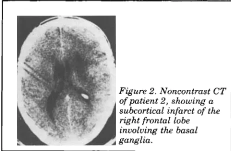
Correction
In “Classical ‘parietal’ neglect syndrome after subcortical right frontal lobe infarction” by Stein and Volpe, June 1983, page 798, figure 2 has been placed upside down. The correct position is shown here.
I Figure 2. Noncontrast CT
DOI 10.1212/WNL.33.8.1107-a
1983;33;1107-1107-a
Neurology
infarction
Classical 'parietal' neglect syndrome after subcortical right frontal lobe
This information is current as of August 1, 1983
Services
Updated Information &
http://n.neurology.org/content/33/8/1107.2.citation.full including high resolution figures, can be found at:
Permissions & Licensing
ssions
http://www.neurology.org/about/about_the_journal#permi (figures,tables) or in its entirety can be found online at: Information about reproducing this article in parts
Reprints
http://n.neurology.org/subscribers/advertise
Information about ordering reprints can be found online:
1526-632X.
American Academy of Neurology. All rights reserved. Print ISSN: 0028-3878. Online ISSN: continuously since 1951, it is now a weekly with 48 issues per year. Copyright © 1983 by the