Open Access
Review
Barrett's oesophagus and adenocarcinoma
Christine PJ Caygill*
1, Anthony Watson
2, Pierre Lao-Sirieix
3and
Rebecca C Fitzgerald
4Address: 1Registrar UK National Barrett's Oesophagus Registry (UKBOR), and Honorary Senior Lecturer, University Department of Surgery, Royal Free Hospital, Rowland Hill Street, London NW3 2PF, UK, 2Joint director UK National Barrett's Oesophagus Registry (UKBOR), and visiting Professor, University Department of Surgery, Royal Free Hospital, Rowland Hill Street, London NW3 2PF, UK, 3MRC Cancer Cell Unit, Hutchison Research Centre, Cambridge, CB2 2XZ, UK and 4Joint director UK National Barrett's Oesophagus Registry (UKBOR) and Group Leader MRC Cancer cell Unit, Hutchison Research Centre, Cambridge, CB2 2XZ, UK
Email: Christine PJ Caygill* - c.caygill@rfc.ucl.ac.uk; Anthony Watson - profwatson@tinyworld.co.uk; Pierre Lao-Sirieix - pss@hutchison-mrc.cam.ac.uk; Rebecca C Fitzgerald - rcf@hutchison-mrc.cam.ac.uk
* Corresponding author
Norman Barrett, a surgeon from St. Thomas' Hospital, first described Barrett's oesophagus in 1950 [1]. He described two variants of columnar-lined (Barrett's) oesophagus (CLO): a congenitally short oesophagus with intra-thoracic gastric epithelium and congenital gastric heterotopia in the oesophagus, with ulceration. Three years later, Allison, a surgeon from Oxford, provided sound anatomical reasons why a columnar lining could occur in the oesophagus as an acquired condition that appeared to be prevalent in patients with gastro-oesopha-geal reflux [2]. Subsequently, several authors confirmed the association of CLO with clinical gastro-oesophageal reflux [3,4] and subsequent studies confirmed the devel-opment of CLO following induction of gastro-oesopha-geal reflux in an animal model [5].
It became apparent from the histological standpoint that the columnar lined oesophagus embraced a spectrum of different cellular types, principally comprising a gastric fundic type epithelium, a junctional type epithelium, which had similarities to gastric mucosa but did not secrete digestive juices, although possessing the ability to withstand acid-peptic digestion, and a distinctive type of intestinal metaplasia, characterised by the presence of goblet cells [6]. The malignant potential of the columnar lined oesophagus was subsequently described [7,8], which conferred great importance on the condition and consequently on its accurate diagnosis. For this reason, and in order to eliminate any confusion between CLO and the normal junctional columnar epithelium, as well as
difficulty in identifying the precise oesophago-gastric junction in cases of hiatal hernia, an arbitrary minimal length of 3 cm of CLO from the oesophago-gastric junc-tion was recommended before the diagnosis of CLO should be made [9]. Until the last few years, Barrett's oesophagus was defined as any histological type of columnar epithelium with a minimum length of 3 cm above the oesophago-gastric junction.
If viewed from the standpoint of the risk of developing adenocarcinoma, it became apparent that this applied only to CLO with intestinal metaplasia (IM) and that CLO with fundic epithelium had no malignant potential [10,11]. However, endoscopic appearances did not distin-guish between the various histological types and all com-prised "Barrett's oesophagus" and were all included in the initial surveillance programmes, which resulted in a much lower incidence of adenocarcinoma than more recent series which have documented the risk in patients with intestinal metaplasia. The problem of definition has become more clouded with the realisation that short seg-ments of columnar lined oesophagus with intestinal metaplasia, less than 3 cm in length, can be associated with the development of adenocarcinoma and even in short, non-circumferential tongues of columnarisation [12]. These two entities have each been referred to as "short segment Barrett's" since the length of these seg-ments, which have malignant potential, fall short of the 3 cm required to fulfil the traditional definition. Subse-quent studies have shown that such short and usually
Published: 07 May 2004
World Journal of Surgical Oncology 2004, 2:12
Received: 03 March 2004 Accepted: 07 May 2004
This article is available from: http://www.wjso.com/content/2/1/12
circumferential segments of columnar lined oesophagus with intestinal metaplasia are visible in 42% of adenocar-cinoma of the cardia when detailed pathological examina-tion is undertaken [13,14]. Furthermore, pathophysiological studies have shown that patients with these short segments of columnarisation have gastro-oesophageal reflux disease, the pathophysiological sever-ity of which is intermediate between that in patients with erosive oesophagitis and those with "traditional Barrett's CLO" [15].
The problem of definition has been further compounded by numerous reports of microscopic intestinal metaplasia around the oesophago-gastric junction, present in up to 36% of patients undergoing endoscopy for a variety of gastro-intestinal symptoms, and some have referred to this phenomenon also as "short-segment Barrett's or "ultra-short segment Barrett's" [16-18]. In Spechler's series [16], only patients with "traditional Barrett's oesophagus" and those with microscopic intestinal metaplasia at the cardia were studied, those patients with confluent or cir-cumferential columnarisation seen endoscopically were excluded from the study. The bulk of evidence suggests that microscopic intestinal metaplasia at the cardia is not associated with gastro-oesophageal reflux disease, but associated principally with increasing age and Helico-bacter infection. It is believed to have a different histogen-esis from intestinal metaplasia in confluent and circumferential areas of columnarisation in the oesopha-gus, and its risk of malignant change appears to be extremely low [19]. In these circumstances, there is confu-sion in using the term "short segment Barrett's" inter-changeably between endoscopically visible confluent or circumferential columnarisation with intestinal metapla-sia and microscopic intestinal metaplametapla-sia around the car-dia, and furthermore it would appear entirely inappropriate to apply the term "Barrett's oesophagus" at all to the latter group, in the absence of endoscopically visible columnarisation, gastro-oesophageal reflux dis-ease and a significant malignant risk.
Pathophysiology
It is now well established that CLO is a complication of severe and long-standing gastro-oesophageal reflux and is found in 10–16% of such patients at endoscopy [20]. Pathophysiological studies have shown that patients with Barrett's CLO show a higher proportion of lower oesopha-geal sphincter failure, and peristaltic dysfunction than patients with erosive oesophagitis and over 90% have an associated hiatal hernia [21]. CLO is also associated with higher levels of acid exposure than erosive oesophagitis and duodeno-gastro-oesophageal exposure as measured by Bilitec monitoring, particularly in the presence of com-plications [22,23]. Therefore, patients with CLO are at the extreme end of the pathophysiological spectrum of
gastro-oesophageal reflux disease. This is compounded by the fact that symptoms may be minimal or absent due to impaired sensitivity of the columnar lining to acid per-fusion [24]. As a consequence of this, many cases of CLO remain undiagnosed. In a clinical and autopsy study per-formed in the USA, the incidence at endoscopy was 18 per 100,000 population, whereas at autopsy the correspond-ing figure was 376 per 100,000, with only 5% becomcorrespond-ing clinically apparent [25]. However, this figure was subse-quently revised to 20% with increasing use of endoscopy [26].
Epidemiology
Barrett's (columnar-lined) oesophagus (CLO) is an important condition because, together with gastroesopha-geal reflux disease (GORD), it is the only known precursor of oesophageal adenocarcinoma (AC) [27,28]. Like AC, the prevalence of CLO has also been rising in Europe and North America, whereas in the USA the increase in CLO parallels the increase in the number of upper gastrointes-tinal endoscopies [29], in the UK there has been a real increase in the numbers diagnosed which exceeds the increased performance of upper gastrointestinal endos-copy [30-32]. Although the majority of CLO will not progress to malignancy it is important to identify relevant risk factors associated with such progression.
In an analysis of 5317 CLO cases in the UK it was found that fewer than 5% developed AC [33]. Most of these (approximately 80%) were prevalent cancers, i.e. cancer arising within one year of CLO diagnosis and about 20% were incident cancers, i.e. those arising more than one year after CLO diagnosis. It is not known whether AC can develop without passing through the CLO stage. A recent study [28] has shown only a modest increase in the oesophageal cancer risk in GORD patients having no record of CLO. The rate at which CLO progresses through increasingly severe dysplasia to AC is between 1 in 44 and 1 in 441 patient years [34,27]. This is 30 – 125 times the rate of AC development in the general population [35].
H-pylori infection
are discussed in reviews by Sharma (2001) [37] and Koop (2002) [38].
Characteristics of Barrett's oesophagus patients
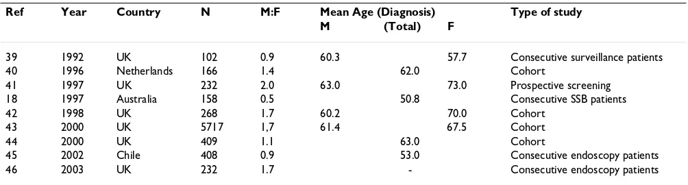
There are a number of studies showing the basic character-istics of CLO patients and some of these are summarised in Table 1.
Most European studies, from the UK and The Nether-lands, show a male predominance of CLO, whereas the studies from Australia and Chile do not, and all but three of these studies have fewer than 300 CLO patients. The mean age at diagnosis is over 60 years in Europe, but 50.8 years and 53.0 years respectively in Australia and Chile. It is not possible at this stage to speculate as to the reason for this. In all but one study the mean age at diagnosis was greater for females than males by almost a decade. In an analysis of CLO from a single UK centre over 15 years it was found that prevalence rose incrementally from age 20–29 years, from 0.16% in males and 0% in females, to a maximum at age 70–79 years, of 4.89% in males and 3.75% in females. Although there was a steep rise in prev-alence with age in both sexes, it was slower in females between the ages of 20–59 than in males, and this was reflected in a 10-year delay in the onset of CLO in females. One could speculate that premenopausal females are pro-tected to some degree against the development of CLO by their hormones [47].
Life style factors affecting CLO development
There are very few studies on lifestyle factors and CLO thus making it impossible to say anything concrete at this stage. The available evidence suggests that neither alcohol consumption nor tobacco use have an effect (Table 2). One study [50] found the past smoking to be moderately connected with CLO development, possibly as a result of the effect of smoking on promoting gastroesophageal reflux. Another study [48] suggested a role for obesity in young CLO patients. In this context it is of interest that
CLO occurs as a complication of long standing GORD [20] which, itself, is a complication of obesity.
The UK National Barrett's Oesophagus Registry
Because of growing concern about the rise in the inci-dence of both AC and CLO The UK National Barrett's Oesophagus Registry (UKBOR) was established in June 1996. The aims of the Registry were to establish a national database of all cases of CLO in the UK in order to learn more about the aetiology, epidemiology and natural his-tory of CLO and to provide a co-ordinating infrastructure for prospective studies. The primary aim being the identi-fication of those sub-groups of CLO most at risk of devel-oping AC so that targeted surveillance strategies can be implemented. This is the world's first such registry, and was set up as a joint initiative of the Oesophageal Section of the British Society of Gastroenterology and the Euro-pean Cancer Prevention Organization (ECP) [51]. Since then almost 9500 CLO patients have been registered from 42 hospitals nationwide. In the following section we give an overview of the data, for the UK, from studying UKBOR patients.
Results of studies from UKBOR
The results of studies using the expanding UKBOR data-base are summarised in table 3.
A single centre's 20 years' experience of columnar-lined (Barrett's) oesophagus diagnosis
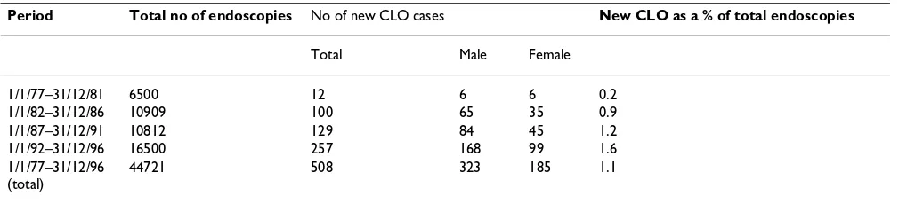
All upper GI endoscopy and histology reports from Wex-ham Park Hospital, Slough, Southern England between Jan 1977 and Dec 1996 were reviewed and data from patients with histologically proven CLO analysed in 5-year bands [30]. The results are summarised in Table 3. It is to be noted that there was an increasing number of endoscopies and CLO diagnoses over time, CLO being diagnosed more frequently in the last quinquenium com-pared with the first.
Table 1: Characteristics of CLO patients
Ref Year Country N M:F Mean Age (Diagnosis) Type of study
M (Total) F
39 1992 UK 102 0.9 60.3 57.7 Consecutive surveillance patients
40 1996 Netherlands 166 1.4 62.0 Cohort
41 1997 UK 232 2.0 63.0 73.0 Prospective screening
18 1997 Australia 158 0.5 50.8 Consecutive SSB patients
42 1998 UK 268 1.7 60.2 70.0 Cohort
43 2000 UK 5717 1,7 61.4 67.5 Cohort
44 2000 UK 409 1.1 63.0 Cohort
45 2002 Chile 408 0.9 53.0 Consecutive endoscopy patients
Characteristics of CLO patients in the UK
Demographic data of 5,717 CLO patients from 27 UK centres (each registering >50 patients with UKBOR) were analysed [43]. All 27 hospitals provided data on sex and date of birth; of these 23 also supplied date of diagnosis of CLO and therefore, age at diagnosis could be calcu-lated. Only 13 of the 27 hospitals were able to supply cur-rent data on numbers of AC. The 27 centres were spread geographically throughout the UK with 3 in Scotland, 3 in Wales, 6 in Northern England, 4 in the Midlands and 11 in Southern England.
Table 4 shows the characteristics of CLO patients by geo-graphical area in UK. There was little variation in M: F ratio (1.3 – 1.7) and in mean age at diagnosis between the centres, except for in males in Scotland where there was a trend towards a lower age at diagnosis. Peak age at diag-nosis of CLO in the 23 centres for males varied from 40– 49 to 70–79 years and in females varied from 60–69 to 70–79 years.
It is also worthy of note that these basic characteristics changed little whether the analysis was done using 9 cen-tres (2130 CLO patients) [52], 20 cencen-tres (4261 CLO patients) [53] or 27 centres (5717 CLO Patients) [43], in spite of the greatly increased numbers and greater geo-graphical coverage.
Adenocarcinoma in CLO
Data for AC were available in 3880 (67.9%) CLO patients from 13 centres. AC was confirmed in 136 (3.5%) (102
males and 34 females). The patient characteristics are shown in Table 5. The M: F ratio of those with AC was 3.0, almost twice that of CLO (1.7), suggesting that differences in the rate of progression from CLO to AC are different in the two sexes. Alternatively as males develop CLO at a younger age their risk of progression to AC may be greater as they have a longer time for the carcinogenic changes to occur. We hope that studies currently in progress at UKBOR will help to clarify this point.
Lifestyle Factors and CLO
This analysis was based at two centres in the UK and on two separate studies. The reason for this was that at one centre (Dundee, Scotland), both heights and weights were available for nearly all patients enabling us to calculate the BMI and thus it was decided to study lifestyle factors with an observational study [48]. At the other centre (Slough, southern England) height was almost never recorded so it was decided that a case-control study was the most appropriate study to give us information on life-style factors.
Observational study – Dundee (Scotland)
The medical records of 136 CLO patients diagnosed between March 1985 and October 1998 were examined. Data recorded included height, weight, tobacco consump-tion and alcohol intake. Body Mass Index (BMI) was cal-culated (kg/m2) using the body weight nearest to and
preceding CLO diagnosis (Table 6). For analysis, tobacco consumption and alcohol intake were graded to give a score using the scoring system in the paper by Caygill et al Table 2: Lifestyle risk factors for CLO
Ref Year Country Tobacco Alcohol Obesity
[48] 2002 UK - - +
[49] 1993 UK - - n/a
[50] 1990 UK + - n/a
Table 3: Detection rate of Barrett's oesophagus over a 20-year period at a single UK hospital.
Period Total no of endoscopies No of new CLO cases New CLO as a % of total endoscopies
Total Male Female
1/1/77–31/12/81 6500 12 6 6 0.2
1/1/82–31/12/86 10909 100 65 35 0.9
1/1/87–31/12/91 10812 129 84 45 1.2
1/1/92–31/12/96 16500 257 168 99 1.6
1/1/77–31/12/96 (total)
(2002) [48]. A previous study of nine UK centres [52] had shown that in Dundee there was a higher proportion (43%) of young (<50 years) male CLO patients compared to the other eight centres. The reasons for this are unclear and remain to be established. Therefore in the above study lifestyle factors were compared between those below and above 50 years of age. The percentages of men and women with either a tobacco score of 3 or more (i.e. smokers, either current or within the last 10 years) or an alcohol score of 3 or more (i.e. those drinking more than that recommended by the government guidelines of 21 units for men and 14 units for women) were calculated and subdivided into the two age groups. The results showed that there did not appear to be a difference in smoking or drinking habits between the older and younger age groups, and these aspects of lifestyle do not
appear to be the cause of the high proportion of young male CLO patients in Dundee.
It is generally accepted that individuals with a BMI of 30 or more are considered to be obese. In the general UK population 11% of men and 13 % of women fulfil this cri-terion [54] (The health of the nation: One year on). We, therefore, calculated the BMI in the Dundee CLO patients and divided them into the two age groups as for tobacco and alcohol consumption. In this cohort of CLO patients 31% of men and 71% of women aged less than 50 years were obese compared with 11% and 13% respectively in the general population. In contrast, those aged more than 50 years had BMI, which were very similar to the general population.
Table 4: CLO patient characteristics by geographical area in UK.
Geographical Area Total Males Females M:F
n mean age (yrs) mean age (yrs)
Scotland 563 57.4 65.3 1.4
Wales 388 61.4 66.4 1.9
England
North 1157 61.6 67.6 1.6
Midlands 1269 63.8 68.1 1.3
South 2340 61.6 67.7 1.7
Total 5717 62.0 67.5 1.7
Table 5: Adenocarcinoma in CLO
Total Males Females
No of AC 136 102 34
No of CLO 3880 2530 1350
Prevalence of AC in CLO (%) 3.5% 4.0% 2.5%
Mean age at diagnosis of AC (years) 67.0 64.7 74.0
Mean age at diagnosis of CLO (years) 63.5 61.4 67.5
Table 6: BMI for Barrett's oesophagus patients in dundee
Age
BMI > 30 (%) 0 – 49 50+ All
M 31 14 20
F 71 19 30
Case – control study, Slough
Data on weight, alcohol and tobacco use were recorded in case notes from this centre but not height, thus making it impossible to calculate BMI. Accordingly a case-control study was set up to study the influence of these lifestyle factors on the development of CLO. Cases were CLO patients and controls were reflux oesophagitis (RO) patients (principally Savary – Miller grade I). In each group there were 50 males and 48 females, and the two groups were matched for gender, age (±3 yr) and year of diagnosis (±3 yr). The data recorded were weight, alcohol intake and tobacco consumption. Alcohol and tobacco were scored as before. There were no significant differ-ences in any of the lifestyle factors studied between CLO and RO patients.
Length of Barrett's oesophagus segment: demographic associations and cancer risk
Some reports have suggested a higher incidence of AC in longer CLO segments, yet AC has also been described in short CLO segments (≤3 cm) [55]. We therefore reviewed 1000 medical records of CLO patients on our database. Data on age, gender, BMI, tobacco and alcohol use, seg-ment length at CLO diagnosis and presence of AC were extracted. Histology and segment length were available from 625 records. The distribution of AC according to seg-ment length is shown in Table 7. The risk of overall or incident cancers was greater for short segment CLO (≤3 cm) than for longer segment CLO (3 – 6 cm) but the great-est risk is for segments >6 cms (Pearson A2 p = 0.04). There
was a small non-significant increase in CLO length with age but no correlation between gender, BMI, or tobacco and alcohol consumption and segment length.
Conclusions from UKBOR studies
There are a number of conclusions that can be drawn from the analyses of the UKCLOR database, namely:
-1. That the Male to female ratio of CLO patients (1.7) is approximately double that of AC patients (3.0)
2. Mean age at diagnosis in male CLO patients is lower (62.0 years) than in female CLO patients (67.5 years). The
same applies to AC, mean ages at diagnosis being 64.7 years in males and 74.0 years in females.
3. Peak age at diagnosis of CLO is 60–69 years in males and 70–79 years in females.
4. Overall prevalence of AC in CLO is 3.5%, being 4.0% in males and 2.5% in females.
5. There appears to be very little geographical variation in CLO patient characteristics throughout the UK. The excep-tion is Scotland, where both the mean and the peak age at diagnosis are lower.
6. Previous alcohol and tobacco use do not appear to affect the risk of developing CLO; obesity may be a risk factor in younger CLO patients.
7. Risk of AC is greatest for >6 cm segments of CLO but is greater for short (≤3 cm) segment Barrett's than for seg-ments >3 ≤ 6 cm.
The malignant risk
The overriding importance of Barrett's CLO is the risk of development of oesophageal adenocarcinoma (AC). Between 5–10% of patients with CLO will develop aden-ocarcinoma, the annual risk in surveillance programmes being 0.5–1%, which is 30–125 times that of the general population [56]. Of particular concern is the escalating incidence of AC, which has increased eightfold in Western Europe in the last three decade, a rate of increase greater than that of any solid tumour. Three decades ago AC com-prised less than 10% of oesophageal tumours, a dramatic increase in this proportion first described in the 1980's, has occurred such that AC now represents over 50% of oesophageal tumours in most UK units [57]. Not every-one with CLO has a similar risk of developing AC and much work has been done to identify risk factors that increase the risk. The risk factors which have been identi-fied are broadly divisible into demographic, pathophysio-logical, environmental, histopathological and molecular genetic.
Table 7: Distribution of AC according to CLO segment length
CLO segment length Overall ≤3 cm >3 ≤ 6 cm >6 cm
No of CLO 625 170 253 202
No of AC 28 10 4 14
All AC (% CLO) 4.5 5.8 1.6 7.1
Demographic
AC occurs almost exclusively in white males. In the USA, AC occurs in 2.5 per 100,000 males and 0.3 per 100,000 females, giving a M: F ratio of 8:1 [56]. In the UK, where there are several epidemiological differences from the USA, the M: F ratio is 3–4:1 [33]. Obesity is a risk factor in several series, the risk being proportional to the degree of obesity, with an odds ratio (OR) of 3.0 in the fourth quar-tile of the BMI range [58,59]. There is an increasing inci-dence of genetic factors in the genesis of both CLO and AC, familial cases of both having been documented, and reflux is more prevalent among siblings than spouse con-trols for both CLO and AC [60].
Pathophysiological
Whilst is it accepted that gastro-oesophageal reflux disease (GORD) is a precursor lesion of CLO and CLO is a pre-malignant lesion. A Swedish population-based study showed chronic, long-standing GORD to be a risk factor for AC independent of CLO, with an overall OR of 7.7 for chronic GORD and 43.5 for severe GORD of more than 20 years duration [61]. Despite the fact that over 90% of CLO patients have a hiatal hernia (HH), the presence of an HH was found to be a risk factor for AC in two case-controlled studies with multi-variate analysis, the risk being proportional to the HH length [62,63]. In terms of reflux parameters, those with AC had higher levels of acid exposure (mean % TT pH < 4, 20 v 16) and lower resting lower oesophageal sphincter pressure (LOSP) (mean LOSP 6 v 10 mmHg) than those with uncomplicated CLO [63]. The combination of duodeno-gastric reflux (DGR) and GORD appears to be a strong risk factor, culminating in oesophageal exposure to alkaline duodenal contents. An increased risk of AC following partial gastrectomy has been documented for some time [39] and measurement of duodenal content reflux by Bilitec monitoring has shown a progressive increase in those refluxers developing CLO, and those CLO patients developing AC [64].
Environmental
Dietary factors have been studied extensively, with con-flicting evidence of low fruit intake as a risk factor [58] although one study has demonstrated increased AC risk in females with dietary deficiencies of vitamins A, C, E and folate [65]. More recent studies have postulated that die-tary nitrate present in fertilisers may be a risk factor [66].
Smoking and alcohol ingestion have been studied exten-sively as putative risk factors with conflicting results. Few series have reported smoking as a risk factor with an OR of around 2.5, but proportional to the extent and duration of exposure [65,67], although some failed to find increased risk [32,35]. In most series alcohol was not a risk factor [61,63,67] whilst in one series which studies wine consumption, it had a protective effect [68].
Drugs, which relax the LOS, such as nitro-glycerine, ami-nophylline, anticholinergics and calcium channel block-ers, have been found to increase risk with an OR of 3.8 in several studies [59,69]. The effect of acid-suppressing drugs is controversial, with an OR of 3–4 in two series [61,70], but two series showing no increased risk [69,71].
Several series have reported infection with H. pylori as a negative risk factor, particularly the CagA strain [70,72].
Histopathological
Many series document increasing risk of AC with increas-ing length of Barrett's segment, particularly so when the segment length exceeds 6 cm [62,63,67]. As a conse-quence, it is generally assumed that the AC risk in short segment CLO (SSBO) is relatively low. However, in a study conducted by the UKBOR which looked at AC risk in 625 patients, 27% of whom had SSBO, the greatest risk in segments >6 cm was confirmed, but, somewhat surpris-ingly, the risk in SSBO was three times higher than that in CLO segments between 3–6 cm in length [55].
The most important risk factor of all in CLO is the pres-ence of dysplasia, and particularly high-grade dysplasia (HGD) in the presence of which between 16–59% develop AC [73,74]. Within HGD, the presence of a raised lesion, ulceration and multifocality of HGD all increase the risk of AC. Multifocality of HGD is associated with an OR of 5.4 of AC, the corresponding figure for a raised lesion being 3.8. If HGD is complicated by ulceration, 80% develop AC [72-76]. In of low-grade dysplasia (LGD) overall risk is less than that with HGD at 5–28% [72,73]. However, the diagnosis of LGD is very subjective with sig-nificant inter-observer variation, but it has been shown that where two pathologists agree a diagnosis of LGD, the incidence of AC is 41% and when three agree, its 80% [75].
The challenge for the future is to develop identifiers of high-risk comparable to dysplasia and its complications, but at a much earlier stage in the process of genomic insta-bility and the principal hope lies in the identification of appropriate molecular markers.
majority of CLO patients who will not develop cancer. It still remains unclear whether it is predominantly environ-mental or genetic factors that determine the progression to adenocarcinoma.
Development of Barrett's oesophagus
There is plenty of patient data to suggest that gastro-oesophageal reflux has an important role to play in the development of metaplasia in the distal oesophagus [80,20]. Three theories have emerged: (1) transdifferenti-ation of stem cells from the basal squamous cells, migra-tion of (2) submucosal gland cells or (3) gastric cells to colonise the damaged squamous mucosa. Nevertheless, very little evidence is available to test these hypotheses or to determine the signalling pathways implicated in the metaplastic process. The lack of reliable animal or in vitro
models does not facilitate the research.
The morphological characterisation of CLO when com-pared to other glandular epithelia such as gastric and duo-denal mucosa is complex. Barrett's oesophagus is a mosaic of three main types of columnar epithelium: a junctional zone of gastric cardia type epithelium, a gastric fundic type and a columnar epithelium with intestinal features characterised by goblet cells (sometimes termed special-ised intestinal metaplasia) [6]. In 1996 Spechler suggested that there should be an alternative classification for the metaplasia depending on the presence or absence of spe-cialised intestinal metaplasia since it is the intestinal metaplasia type which confers the highest risk for malig-nancy [81,11]. Intestinal metaplasia is classified in several ways depending on how strongly it resembles the small intestinal epithelium [82,83]. Metaplasia which is very similar to the small intestine is referred to as complete, whereas that which differs from it is termed incomplete. The complete type (or type 1) should contain absorptive cells that do not secrete mucus, have a well-defined brush border containing enzymes such as disaccharides. There may also be occasional paneth cells. In contrast, incom-plete intestinal metaplasia (types 11 and 111/11B, which is more common) is composed mainly of 'intermediate' columnar cells that secrete mucus. It also contains goblet cells that secrete sulphomucins, sialomucins or both. The clinical relevance of these histopathological subtypes in terms of predicting cancer risk is not clear and does not currently contribute to the diagnosis or patient manage-ment. CLO is also regarded to be a hyperproliferative epi-thelium, although a review of the literature is somewhat conflicting [84-88]. The proliferative status may be impor-tant in determining the likelihood of cancer progression.
Molecular factors implicated in dysplasia development (Table 8)
Growth factors
Epidermal growth factor (EGF), its receptor (EGFR) and transforming growth factor alpha (TGFα) have mitogenic activities and have been widely implicated in cancer development. Increased expression of EGFR and TGFα has been demonstrated as CLO progresses to AC [89-91], and patients over expressing TGFα and EGFR [92] have been shown to have lymph nodes metastasis and a poor prognosis [93]. EGF was found to be over expressed in intestinal metaplasia compared with cardiac, fundic meta-plasia and normal gastric mucosa [94,95] with maximal expression in oesophageal AC [94-96]. c-erb-B2 (Her-2/ Neu), a truncated version of EGFR, involved in cell prolif-eration and differentiation [97] is expressed in late stages of carcinogenesis [98-102] in a subset of the cancer patients [103] and is associated with a poor prognosis [98]. Transforming growth factor beta (TGFβ) is a potent anti-proliferative agent acting through two receptors (TGFβRI and TGFβRII) and its signalling molecules called Smad 2, 3 and 4. The loss of function of TGFβ signalling in Barrett's associated adenocarcinoma could be related to Smad 4 mutations [104,105] or to the loss of mRNA expression of TGFβRII [106,107].
The increase of growth factors and their associated recep-tors suggests the possibility of an autocrine stimulation of growth, unimpaired by negative regulators.
Oncogenes
The products of the ras family (H, K and N) of oncogenes are believed to modulate cell growth by abrogation of cell growth requirements [108]. Investigators were unable to identify c-ras mutations in dysplastic and non-dysplastic CLO mucosal biopsy or in cancer samples [95,109]. Muta-tions of codon 12 of the k-ras gene were found to be rare in CLO but increased in frequency along the progression to AC [110,111]. Amplification of k- and h-ras were only seen in established AC, suggesting that this is a late event in carcinogenesis [112].
The diverse abnormalities in oncogenes will result in abnormal epithelial cell proliferation independent of the usual growth requirements.
Cyclo-oxygenase-2 and inducible nitric oxide
Expression of Cyclooxygenase-2 (cox-2) and inducible nitric oxide (iNOS) and mediators of were shown to be high in CLO and low grade dysplasia when compared to normal squamous oesophagus, gastric mucosa and aden-ocarcinoma [117-121]. Cox-2 expression was shown to be
higher in the distal end of the oesophagus, where AC tends to occur, when compared to the proximal end [122]. Furthermore, Cox-2 stimulation was shown to induce proliferation in an in vivo system [124]. This sug-gests an early involvement in the carcinogenesis process by stimulation of mucosal proliferation.
Luminal factors
Gastro-oesophageal refluxate, the major factor associated with the development of CLO contains bile salts and Table 8: Summary of the molecular alterations studied in the progression of Barrett's associated carcinogenesis (list none exhaustive).
Increased proliferation
Growth factors Receptors/effectors
TGFα
EGF EGFR
C-erb-B2 (Her-2/Neu)
TGFβ TGFβR 2
Smad 4 p27 Mediators of inflammation
Cox-2 PKC
INOS
TNFα β catenin, c-myc
Luminal components
Gastrin CCK2
Bile salts Cox-2
Acid Cox-2
PKC p38MAPK JUNK p44ERK Oncogenes
Ras Fos Jun c-myc
Cell cycle proteins Cyclin D1, E, B1 Rb
p16
Avoidance of apoptosis
p53 p21(cip1/waf1)
Bcl-2 Bax
Increased invasive potential
Angiogenesis FGF 1&2 VEGF
hydrochloric acid. In vitro studies in AC adenocarcinoma cell lines, demonstrated that proliferation could be induced through involvement of the sodium-hydrogen exchanger (Na+-H+), a major acid-extruder in CLO cells, as
well as via activation of the p38MAPK, p44ERK1 and
Jun-N-terminal kinase (JNK) enzymes [124,125]. Pulsatile acid exposure of cell lines was shown to suppress apoptosis via the p53 pathway and to stimulate proliferation possibly through the MAP kinase pathway [126]. Cox-2 expression was also induced by acid and bile salts in ex vivo experi-ments [127,128] in a protein kinase C (PKC) dependant manner. As well as this in vitro work, ex vivo studies have also suggested a hyerproliferative effect of acid and bile exposure on the Barrett's epithelium [129,130]. This has implications for the use of reflux-suppressive therapies in patients with CLO. However, it should be remembered that the induction of the mitogen gastrin in response to proton pump inhibitor therapy might induce prolifera-tion of the CLO mucosa via the cholecystokinin (CCK2) receptor [131].
Abnormalities in cell cycle and apoptosis
Passage of cells through the restriction point controlling the G1/S phase transition is a key element of proliferation (Figure 1). This checkpoint, also called the restriction point, is tightly controlled by a plethora of inhibitors of cell cycle progression and molecules inducing apoptosis such as p53. Aberrant expression of regulatory proteins may lead to uncontrolled progression through the cell cycle, instead of the normal cell cycle arrest, which is nec-essary for processes such as DNA repair.
Hyperproliferation has been associated with progression from CLO to AC [84-86,132]. For a tissue to become hyperproliferative, the individual cells have to progress in uncontrolled fashion through the restriction point. Amplification and overexpression of cyclin D1 [133-135], decreased expression and loss of heterozygosity (LOH) of Rb [136-138] and increased expression cyclin E [139] as well as accumulation of mutant p53 [140-149] as CLO progresses to AC suggest that the G1/S transition control system is overridden. Furthermore, inactivating hyper-phosphorylation [150-153] or deletion of p16 [154], a cyclin cyclin D1/CDK complex inhibitor, seems to be a common feature of CLO carcinogenesis. p27 is inactivated in AC and tumours lacking p27 have a more aggressive behaviour [155].
Flow cytometry studies showed that G2/M phase accumu-lation is found early in the progression [156]. Results implicating cyclin B1 (expressed in G2 phase of the cell cycle to regulate entry into mitosis) overexpression in the progression to cancer, confirmed this first study [157].
Further to an increase in p53 expression, apoptosis may be avoided in the progression from CLO to AC via abnor-mal Bcl-2 and Bax expression [119,158-162]. The expres-sion profile of p21(cip1/waf1) is subject to controversy
[158,159,163,164] but p21(cip1/waf1) does not seem to
inhibit proliferation in the context of CLO carcinogenesis.
Factors leading to an increased invasive potential
Angiogenesis, characterised by the stimulation of migra-tion and proliferamigra-tion of capillary endothelial cells creat-ing new vasculature, is an important mechanism by which metastases occur. Acid and basic fibroblast growth factors (respectively FGF1 and FGF2) and vascular endothelium growth factor (VEGF) are key players in this process [165]. FGF 1 and 2 expression was shown be increased as CLO progresses to AC [166-168]. In AC, VEGF expression related with the level of the vasculature, which in turn cor-related with the presence of lymph nodes and distant metastasis. It has been mentioned that the salmon-pink colour of the CLO mucosa could be due to the increased presence of blood vessels when compared to normal squamous epithelium [170,171].
The calcium-dependant cadherin-catenin membrane bound complex is a key factor for maintaining epithelial integrity, cell polarisation and cell-cell adhesion. Loss of cell-cell interaction resulting from down-regulation of E-cadherin may trigger and facilitate metastasis [171]. Many reports have commented on the increasingly low levels of E-cadherin (protein and mRNA level) and α- and β -cat-enin in the progression from normal squamous epithe-lium to adenocarcinoma [172-176]. This trend of down-regulation was shown to correlate with poorer survival, invasion, and metastasis [172] as well as with the stage and grade of the cancer [173]. Interestingly, a subset of patients was shown to have nuclear localisation of the E-cadherin-β-catenin complex [173,174,177,178]. A recent study demonstrated that tumour necrosis factor alpha (TNFα) induces c-myc expression via β-catenin [116]. The role of the nuclear E-cadherin-β-catenin complex in this system is not clear and further investigation is warranted.
Overview of the G1/S transition of the cell cycle
Figure 1
The large number of molecular alterations that occur in Barrett's carcinogenesis are gradually being documented although the sequence by which these abnormalities occur seems to be non-linear and complex [182]. Most of the studies that have been carried out to date are descrip-tive. Unfortunately, it has proved hard to develop models for dynamic studies although progress is being made [130,183]. Work is still needed in order to understand the driving forces behind Barrett's carcinogenesis so that progress can be made in identifying clinically useful diag-nostic markers and novel therapeutic strategies.
Management of uncomplicated Barrett's
The management of uncomplicated Barrett's comprises treatment designed to influence the natural history of the condition and surveillance to detect dysplastic change, which will be considered later.
The pathophysiological features of Barrett's oesophagus as outlined above have implications regarding management and its efficacy. Barrett's oesophagus clearly represents the extreme end of the pathophysiological spectrum of gastro-oesophageal reflux disease and this is compounded by the fact that many patients have few or no symptoms due to the relative insensitivity of columnar mucosa to acid per-fusion compared with patients with erosive oesophagitis [24]. Despite these factors, many authorities advocate no treatment for Barrett's oesophagus unless symptoms are present. Those who believe that the objectives of manage-ment of CLO are more to do with an attempt to influence the natural history of the condition than symptomatic relief advocate such modalities as pharmacological acid suppression, endoscopic ablation or anti-reflux surgery. At the present time, the optimal management of CLO is unknown and these modalities are applied largely on the basis of personal preference, although a large multi-centre randomised study to address this issue is proposed.
Pharmacological acid suppression
This clearly has theoretical advantages, being the least invasive form of long-term therapy, particularly as Bar-rett's oesophagus is predominantly a disease of the eld-erly, the mean age being around 63 [33]. Although the development of squamous islands following PPI therapy is well recognised, circumferential regression of the columnarised segment is rare, a meta-analysis of six sub-sequent series showing no evidence of regression [184]. Several studies report the difficulty of normalising oesophageal acid exposure in Barrett's patients, even using doses equivalent to Omeprazole 80 mg daily and even when amelioration of symptoms, if present, has occurred [185-187]. This is likely to relate to the pathophysiology of this group of patients previously alluded to and the consequences of incomplete acid suppression is a matter of concern in this group of patients, since it has been
shown that Barrett's oesophagus cells in culture exhibit a greater degree of proliferation and de-differentiation when exposed to intermittent pulse acid exposure com-pared to no acid exposure and even continuous acid expo-sure [130]. It is, therefore, possible that inadequate levels of acid suppression may have contributed to the rising incidence of adenocarcinoma of the oesophagus and gas-tric cardia [188,189]. It has been recommended to try to overcome this problem that an H2 receptor antagonist should be added at night, possibly combined with a prok-inetic agent and that the dose of PPI should be titrated against the level of oesophageal acid exposure on 24 hr pH monitoring in order to optimise the effect of acid sup-pression therapy [185]. There remains, however, the prob-lem of abnormal duodenal juice exposure, which although reduced as measured by Bilitec monitoring, on PPI therapy, presumably due to a volume-reduction effect, such exposure is normalised in less than 50% of patients [190].
Endoscopic ablation
While endoscopy is considered to offer a relatively poor return in assessing uncomplicated symptomatic GORD and lack of impact in altering medical treatment [191], it offers a useful therapeutic option for mucosal ablation of metaplastic epithelium and putative regeneration of squa-mous lining [192]. It could be argued that ablative tech-niques should be reserved for areas of dysplastic change only and certainly further studies are needed to define the indications, efficacy and relative safety of the various modalities of treatment.
gener-ations of photosensitises are likely to improve the uptake of PDT. Endoscopic ablation techniques, performed in a reflux-free environment using either high dose PPI ther-apy or fundoplication result in squamous re-epithelialisa-tion in 50–80% of patients, although residual islands of columnar metaplasia remain in 20–60% depending on the depth of injury [192-196].
There remains doubt as to the status of islands of colum-nar metaplasia covered by squamous regeneration follow-ing the use of ablative techniques and it is recommended that endoscopic ablation techniques should only be per-formed in the context of prospective randomised trials.
Anti-reflux surgery
Fundoplication has the theoretical advantage of being able to correct lower oesophageal sphincter failure and the frequently associated hiatal hernia and producing complete and continuous control of abnormal acid and duodenal juice exposure in 80–90% of patients. Two stud-ies have demonstrated a greater degree of symptom con-trol and healing of associated strictures and a lower incidence of new strictures after fundoplication compared to acid suppression therapy [198,199]. There are consider-ably more reports of regression following anti-reflux sur-gery, although regression is rarely complete and occurs in only 10–44% of patients [198-203]. However, it is per-haps of greater importance what is happening at cellular level rather than whether or not macroscopic regression occurs.
The effect of successful anti-reflux surgery on the inci-dence of AC is unknown and indeed adenocarcinoma has been reported after successful anti-reflux surgery [201]. It is theoretically possible that anti-reflux surgery may be effective in preventing adenocarcinoma if performed suf-ficiently early in the sequence of genomic instability, but that a point may be reached beyond which no form of treatment can prevent the development of AC. This con-cept is supported by the findings of a study from the Mayo Clinic in which 113 patients with CLO were followed up for up to 18 years after anti-reflux surgery. Although 3 patients developed AC, these were all in the first 3.3 years, after which no carcinomas developed [204]. The inci-dence of AC in this series was 1:274 patient years of follow up, considerably less than the mean of 1:80 patient years of follow up reported from surveillance series of patients on acid suppression therapy [205].
Notwithstanding the apparent theoretical advantages of fundoplication over acid-suppression therapy, which need to be confirmed by randomised control trials, it should be remembered that CLO is a condition largely of old age and only approximately 50% of patients will be below the age of 70 and fit for consideration of surgery.
However, it should certainly be considered in younger patients and particularly those with risk factors for devel-opment of AC, such as those with a long history, early age at diagnosis, Barrett's segment greater than 7 cm and doc-umented pathological alkaline exposure.
Surveillance
Background to screening and surveillance
Both screening and surveillance have been advocated for CLO in an attempt to reduce the mortality from AC. Screening refers to a programme to identify individuals with CLO who have not previously been diagnosed. Sur-veillance refers to the follow-up of patients with known Barrett's oesophagus. Surveillance programmes only review a small proportion of the population at risk since only a minority of patients with Barrett's oesophagus are clinically diagnosed [25].
At the current time there are no randomised controlled tri-als examining the clinical benefit or cost-effectiveness of endoscopic screening for Barrett's oesophagus in either the general population or in patients with reflux disease. In the future non-endoscopic screening programmes tar-geted at individuals at highest risk for developing AC may be possible. For this to become a reality we will need to depend on molecular epidemiological studies to identify predictive markers with high sensitivity and specificity and technological developments to implement this, in an ethically acceptable and cost-effective manner. Since the majority of the data at the current time pertains to surveillance.
Rationale for surveillance
Most patients with AC present with dysphagia, and despite advances in multimodal treatment the five-year survival rates of symptomatic oesophageal carcinomas remain less than 10% [206,207]. Since the major determi-nant of outcome is the stage of the cancer at presentation [208], early detection of AC is essential in order to signif-icantly improve survival rates. The identification of a multistage process of cancer development, akin to that described in colon carcinogenesis, provides the rationale for endoscopic surveillance in patients with diagnosed CLO [77,78,209-211]. Several studies have shown a sig-nificant improvement in the 5-year survival of patients with surveillance-detected adenocarcinoma [212-214], although these studies are generally limited by their small sample size.
Oesophagus Registry will address this question in a large population.
Risk of progression
The value of surveillance hinges on the actual risk of can-cer in patients with CLO [216]. A meta-analysis of 25 studies conducted between 1984 and 1998 demonstrated that the number of incident cancers ranged from 1 in 35 to 1 in 441 patient years of follow-up, with a mean cancer incidence in these studies of 1 in 138 patient years [217]. The wide variation in cancer incidence may be as a result of the retrospective nature of the studies, diagnostic varia-tion, surveillance protocol variation (number of biop-sies), variation in outcome data (death, dysplasia or cancer), and publication bias (negative studies tend not to be published).
At the current time the histopathological assessment of dysplasia is used to predict the likelihood of cancer devel-opment. In a recently reported retrospective cohort study of 79 patients with high-grade dysplasia 4.5 (5%) had cancer at 1 year and 12 (16%) had cancer on follow-up (mean 7.3 years) [73]. However, concern has been expressed about the over-diagnosis of dysplasia in this cohort. Another group demonstrated that the likelihood of cancer was dependent on the extent of high-grade dys-plasia (focal or diffuse) [74], although this has not been confirmed in a subsequent study [218]. These studies are hampered by the subjectivity of the diagnosis of dysplasia and hence more specific markers of malignant potential are badly needed [219], (see section on molecular patho-genesis). When deciding on the merits of surveillance pro-grammes it should be borne in mind that 40% of patients with high-grade dysplasia have been found to harbour foci of carcinoma in their resection specimen [220]. The practical problems associated with surveillance are sum-marised in Table 9.
From an economic point of if the incidence is 1% 2–3 yearly surveillance endoscopy would be cost-effective; whereas if the incidence is 0.5% 4–5 yearly would be rec-ommended [221,222]. As a result of the apparent geo-graphical variations in cancer incidence (e.g. UK versus
USA) different surveillance intervals may be recommended.
Local UK experience
There is a wide variation in the surveillance protocols undertaken in the UK. Surveillance was conducted in Leicester from 1984 to 1999, 143 out of 409 patients were eligible for surveillance using their diagnostic criteria with a 70 year age cut-off and in 1999 only 8 were still in the programme. Quadrantic biopsies were taken from the midpoint of the Barrett's segment rather than every 2 cm along the Barrett's length according to the Seattle protocol [223]. Over the 15 years 5/143 developed carcinoma – 2 had a stricture at the time enrolment, 2 were programme defaulters and hence the programme itself only identified one subject [44]. This study is probably more informative about the practical difficulties in administering such a programme, rather then being informative about the value of surveillance itself. Implementation of a rigorous surveillance protocol In an East London hospital (Old-church, UK) in line with the Seattle guidelines, for patients with specialised intestinal metaplasia who were fit enough for surveillance (no age cut off) significantly increased the yield of high-grade dysplasia and cancer detection [224].
The World Health Organisation has criteria for screening/ surveillance and it is not clear to what extent Barrett's oesophagus currently meets these criteria. In order to address these questions a prospective randomised con-trolled trial of surveillance versus no surveillance is required. Such a study would require 10 yr study with 5,000 patients and the ethical considerations of a no-sur-veillance arm would have to be carefully considered [222]. At the current time the American College of Gastro-enterology have made clear recommendations for screen-ing and surveillance based on the best available evidence [225]. The British Society of Gastroenterology is currently in the process of drawing up their guidelines (Prof. Tony Watson is Chairman of the Working Group). It is certainly clear that we need an international consensus on our def-inition(s) of Barrett's oesophagus and dysplasia so that the outcomes of surveillance protocols can be usefully compared.
Table 9: Common problems encountered with Barrett's surveillance
• Dysplastic lesions are often flat and indistinguishable endoscopically • Variations in diagnostic criteria for Barrett's oesophagus and dysplasia
• Wide variations in local protocols (e.g. how often surveillance should be conducted if at all, the number of biopsies) • Imaging protocols do not achieve subcellular resolution and biopsies are still required
• Submucosal deep abnormalities may not be detected even when the area is biopsied • Sampling bias (dysplasia may be focal, patchy or diffuse)
The future
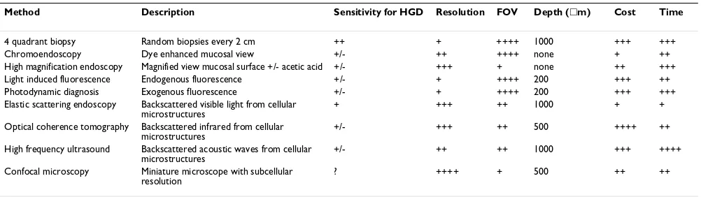
It is likely that this discussion will become a thing of the past. Currently our methodology is grossly inadequate. Ongoing research into the diagnosis and therapy will rad-ically alter our approach to this subject. For example, considerable progress is already being made into methods for predicting which patients are at high risk. The method-ologies currently being evaluated include serum markers, genetic susceptibility profiles and molecular markers using non-endoscopic brushings. In addition, endoscopic methods are being evaluated to target biopsies using tech-nologies such as vital dyes (e.g. methylene blue spraying) in conjunction with zoom endoscopy and optical biopsies or virtual biopsy techniques such as elastic scattering spec-troscopy and fluorescence (autofluorescence or drug induced fluorescence) [226], (Table 10).
At the current time the gold standard treatment is an oesophagectomy, which has an associated morbidity and mortality of between 5 and 10%. However, with the devel-opment of endoscopic treatments this will obviate the need for surgery especially in elderly patients with comor-bidity. These treatments include ablation therapies such as photodynamic therapy and endoscopic mucosal resec-tion of visible lesions [227-229]. Chemoprevenresec-tion strat-egies such as profound acid suppression [230] and COX2 inhibitors [231] are also being discussed. If these or alter-native therapeutic agents could significantly reduce the cancer risk in the population at risk then surveillance may become a thing of the past.
Competing interests
None declared.Authors' contributions
AW wrote the section on the Background, the section on the malignant risk and the section on management of uncomplicated Barrett's. CPJC wrote the section on the epidemiology and edited the chapter. PL co-wrote the
sec-tion on Molecular biology. RCF co-wrote the secsec-tion on molecular biology and wrote the section on surveillance. All authors read and approved the final manuscript.
References
1. Barrett NR: Chronic peptic ulcer of the oesophagus and "oesophagitis".Br J Surg 1950, 38:175-182.
2. Allison PR, Johnstone AS: The oesophagus lined with gastric mucous membrane.Thorax 1953, 8:87-101.
3. Moersch R, Ellis FH, McDonald JR: Pathologic changes occurring in severe reflux oesophagitis. Surg Gynecol Obstet 1959,
108:476-484.
4. Hayward J: The lower end of the oesophagus. Thorax 1961,
16:36-41.
5. Bremner CG, Lynch VP, Ellis FH: Barrett's oesophagus: congeni-tal or acquired? An experimencongeni-tal study of oesophageal mucosal regeneration in the dog.Surgery 1970, 68:209-216. 6. Paull A, Trier JS, Dalton MD, Camp RC, Loeb P, Goyal RK: The
his-tological spectrum of Barrett's oesophagus.N Engl J Med 1976,
295:476-480.
7. Naef AP, Savary M, Ozzello L: Columnar-lined lower oesopha-gus: an acquired lesion with malignant predisposition. Report on 140 cases of Barrett's oesophagus with 12 adenocarcinomas.J Thorac Cardiovasc Surg 1975, 70:826-835. 8. Haggitt RC, Tryzelaar J, Ellis FH, Colcher H: Adenocarcinoma
complicating columnar epithelium-lined (Barrett's) oesophagus.Am J Clin Pathol 1978, 70:1-5.
9. Skinner DB, Walther BC, Riddell RH, Schmidt H, Iascone C, DeMeester TR: Barrett's oesophagus: comparison of benign and malignant cases.Ann Surg 1983, 198:554-565.
10. Reid BJ, Haggitt RC, Rubin LE, Rabinovitch PS: Barrett's oesopha-gus: correlation between flow cytometry and histology in detection of patients at risk for adenocarcinoma. Gastroenter-ology 1987, 93:1-11.
11. Spechler SJ, Goyal RK: The columnar-lined oesophagus, intesti-nal metaplasia and Norman Barrett. Gastroenterology 1996,
110:614-621.
12. Schnell TG, Sontag SJ, Chejfec G: Adenocarcinoma arising in tongues or short segments of Barrett's oesophagus.Dig Dis Sci 1992, 37:137-143.
13. Cameron AJ, Lomboy CT, Pera M, Carpenter HA: Adenocarci-noma of the oesophagogastric junction and Barrett's oesophagus.Gastroenterology 1995, 109:1541-1546.
14. Clark GW, Smyrk TC, Burdiles P, Hoeft SF, Peters JH, Kiyabu M, Hinder RA, Bremner CG, DeMeester TR: Is Barrett's metaplasia the source of adenocarcinomas of the cardia?Arch Surg 1994,
129:609-614.
15. Clark GWB, Ireland AP, Peters JH, Chandrasoma P, DeMeester TR, Bremner CG: Short-segment Barrett's oesophagus: a preva-lent complication of gastroesophageal reflux disease with malignant potential.J Gastrointest Surg 1997, 1:113-122.
Table 10: Comparison of endoscopic surveillance methods for Barrett's oesophagus
Method Description Sensitivity for HGD Resolution FOV Depth (䊐m) Cost Time
4 quadrant biopsy Random biopsies every 2 cm ++ + ++++ 1000 +++ +++
Chromoendoscopy Dye enhanced mucosal view +/- ++ ++++ none + ++
High magnification endoscopy Magnified view mucosal surface +/- acetic acid +/- +++ + none ++ +++ Light induced fluorescence Endogenous fluorescence +/- + ++++ 200 +++ ++ Photodynamic diagnosis Exogenous fluorescence +/- + ++++ 200 +++ +++ Elastic scattering endoscopy Backscattered visible light from cellular
microstructures
+ +++ ++ 1000 + +
Optical coherence tomography Backscattered infrared from cellular microstructures
+/- +++ ++ 500 ++++ ++
High frequency ultrasound Backscattered acoustic waves from cellular microstructures
+/- ++ ++ 1000 +++ ++++
Confocal microscopy Miniature microscope with subcellular resolution
16. Spechler SJ, Zeroogian JM, Antonioli DA, Wang HH, Goyal RK: Prev-alence of metaplasia at the gastro-oesophageal junction. Lan-cet 1994, 344:1533-1536.
17. Trudgill NJ, Suvarna SK, Kapur KC, Riley SA: Intestinal metaplasia at the squamocolumnar junction in patients attending for diagnostic gastroscopy.Gut 1997, 41:585-589.
18. Nandurkar S, Talley NJ, Martin CJ, Ng TH, Adams S: Short segment Barrett's oesophagus: prevalence, diagnosis and associations.Gut 1997, 40:710-715.
19. Weston AP, Krmpotich P, Makdisi WF, Cherian R, Dixon A, McGre-gor DH, Banerjee SK: Short segments Barrett's oesophagus; clinical and histological features associated endoscopic find-ings, and association with gastric intestinal metaplasia.Am J Gastroenterol 1996, 91:981-986.
20. Winters C Jr, Spurling TJ, Chobanian SJ, Curtis DJ, Esposito RL, Hacker JF 3rd, Johnson DA, Cruess DF, Cotelingam JD, Gurney MS, Cattau EL Jr: Barrett's oesophagus – a prevalent occult com-plication of gastro-oesophageal reflux.Gastroenterology 1987,
92:118-124.
21. Stein HJ, Hoeft S, DeMeester TR: Reflux and motility pattern in Barrett's oesophagus.Dis Oesophagus 1992, 5:21-28.
22. Attwood SEA, Ball CS, Barlow AP, Jenkinson L, Norris TL, Watson A:
Role of intragastric and intraoesophageal alkalinisation in the genesis of complications in Barrett's columnar lined lower oesophagus.Gut 1993, 34:11-15.
23. Kauer WK, Peters JH, DeMeester TR, Ireland AP, Bremner CG, Hagen JA: Mixed reflux of gastric and duodenal juices more harmful to the oesophagus than gastric juice alone. The need for surgical therapy re-emphasised.Ann Surg 1995, 222:523-533. discussion 531–33
24. Ball CS, Watson A: Acid sensitivity in reflux oesophagitis with and without complications.Gut 1988, 29:729.
25. Cameron A, Zinmeister A, Ballard D, Carney J: Prevalence of columnar-lined (Barrett's) oesophagus. Comparison of pop-ulation-based clinical and autopsy findings. Gastroenterology 1990, 99:1918-1922.
26. Conio M, Cameron AJ, Romero Y, Branch CD, Schleck CD, Burgart LJ, Zinsmeister AR, Melton LJ 3rd, Locke GR 3rd: Secular trends in the epidemiology and outcome of Barrett's oesophagus in Olmsted Country, Minnesota.Gut 2001, 48:304-309.
27. Bartlesman JFWM, Hameeteman W, Tytgat GNJ: Barrett's oesophagus.Eur J Cancer Prev 1992, 1:323-325.
28. Solaymani-Dodaran M, Coupland C, Logan RFA: Risk of oesopha-geal cancer in Barrett's oesophagus and in gastro-oesopha-geal reflux.Gut 2003, 52:A20.
29. Cameron AJ, Lomboy CT: Barrett's oesophagus: age prevalence and extent of columnar epithelium. Gastroenterology 1992,
103:1241-1245.
30. Caygill CPJ, Reed PI, Johnston BJ, Hill MJ, Ali MH, Levi S: A single centre's 20 years' experience of columnar-lined (Barrett's) oesophagus diagnosis. Eur J Gastroenterol Heptol 1999,
11:1355-1358.
31. Watson A, Reed PI, Caygill CPJ, Epstein O, Winslet MC, Pounder RE:
Changing incidence of columnar-lined (Barrett's) oesopha-gus (CLO) in the UK.Gastroenterology 1999, 116(suppl 2):A351. 32. Todd JA, Johnston DA, Dillon JF: Incidence of Barrett's
oesopha-gus and oesophagitis at upper GI endoscopy. Gut 2000,
46(suppl II):A94.
33. Watson A, Caygill CPJ: The frequency of development of aden-ocarcinoma in Barrett's oesophagus – implications for surveillance.Gastroenterology 2002, 122:A350.
34. Atkinson M, Iftikhar SY, James PD, Robertson CS, Steele RJC: The early diagnosis of oesophageal adenocarcinoma by endo-scopic screening.Eur J Cancer Prev 1992, 1:327-330.
35. Reed PI: The changing pattern of adenocarcinoma of the oesophagogastric junction. In The oesophagogastric Junction Edited by: Giuli R, Galmiche J-P, Jamieson GG, Scarpignato C. Montrouge, John Libby; 1988:1131-1140.
36. Veith M, Masoud B, Meining A, Stolte M: Helicobacter pylori infec-tion: protection against Barrett's mucosa and neoplasia? Digestion 2000, 62:225-231.
37. Sharma P: Helicobactor pylori: a debated factor in gastro-esophageal reflux disease.Dig Dis 2001, 19:127-133.
38. Koop H: Gastroesophageal reflux disease and Barrett's oesophagus.Endoscopy 2002, 34:97-103.
39. Iftikhar SY, James PD, Steele RJC, Hardcastle JD, Atkinson M: Length of Barrett's oesophagus: an important factor in the develop-ment of dysplasia and adenocarcinoma. Gut 1992,
33:1155-1158.
40. Van der Burgh A, Dees J, Hop WCJ, van Blankenstein M: Oesopha-geal cancer is an uncommon cause of death in patients with Barrett's oesophagus.Gut 1996, 39:5-8.
41. Cox MA, Nwokolo CU, Loft DE: Screening for Barrett's oesophagus is worthwhile.Gut 1997, 41(suppl III):E20. 42. Caygill CPJ, Reed PI, McIntyre A, Hill MJ: The UK National
Bar-rett's Oesophagus Registry: a study between two centres.Eur J Cancer Prev 1998, 7:161-164.
43. Caygill C, Reed P, Watson A, Hill M: UK National Barrett's Oesophagus Registry: An Update.Gut 2000, 47(suppl III):A68. 44. Macdonald CE, Wicks AC, Playford RJ: Final results from 10 year cohort of patients undergoing surveillance for Barrett's oesophagus: observational study.BMJ 2000, 321:1252-1255. 45. Csendes A, Smok G, Quiroz J, Burdiles P, Rojas J, Castro C,
Hen-riquez A: Clinical, endoscopic, and functional studies in 408 patients with Barrett's oesophagus, compared to 174 cases of intestinal metaplasia of the cardia.Am J Gastroenterol 2002,
97:554-560.
46. Cotton JP, Lopez M, McLeod S, Todd JA, Johnston DA, Setmarr PW, Dillon DF: Gender differences in the epidemiology of GORD. Gut 2003, 52(suppl I):A46 [Abstract 168].
47. van Blankenstein M, Caygill CPJ, Johnston BJ: The prevalence of Barrett's oesophagus (BO) in a U.K. centre over 15 years.Gut 2002, 50(suppl II):A123.
48. Caygill CPJ, Johnston DA, Lopez M, Johnston BJ, Watson A, Reed PI, Hill MJ: Lifestyle Factors and Barrett's Oesophagus. Am J Gastroenterol 2002, 97:1328-1331.
49. Gray MR, Donnelly RJ, Kingsnorth JN: The role of smoking and alcohol in metaplasia and cancer risk in Barrett's columnar lined epithelium.Gut 1993, 34:727-731.
50. Logan RFA, Riddick A: Barrett's oesophagus – are smoking and drinking alcohol risk factors?Gut 1990, 31:A603.
51. Reed PI, Watson A: UK National Barrett's oesophagus Registry.Eur J Cancer Prev 1996, 5:207.
52. Caygill CPJ, Reed PI, Hill MJ, Watson A: An initial comparison of nine centres registering patients with the UK national Bar-rett's oesophagus Registry (UKBOR).Eur J Cancer Prev 1999,
8:539-542.
53. Caygill CPJ, Reed PI, Hill MJ, Watson A: The UK National Bar-rett's Oesophagus Registry: a progress report.Eur J Cancer Prev 1999, 8:354.
54. The health of the nation: One year on.Her Majesty'sStationary Office 1993:50.
55. Gatenby PAC, Caygill CPJ, Charlett A, Fitzgerald R, Watson A:
Length of Barrett's oesophagus segment: Demographic associations and cancer risk.Gut 2003, 52(suppl I):A41. 56. Blot WJ, Devesa SS, Kneller RW, Fraumeni JF Jr: Rising incidence
of adenocarcinoma of the oesophagus and gastric cardia. JAMA 1991, 265:1287-1289.
57. Watson A: Barrett's oesophagus – 50 years on.Br J Surg 2000,
87:529-531.
58. Cheng KK, Sharp L, McKinney PA, Logan RF, Chilvers CE, Cook-Mozaffari P, Ahmed A, Day NE: A case-control study of oesopha-geal adenocarcinoma in women; a preventable disease.Br J Cancer 2000, 83:127-132.
59. Lagergren J, Bergstrom R, Lindgren A, Nyren O: The role of tobacco, snuff and alcohol use in the aetiology of cancer of the oesophagus and gastric cardia.Int J Cancer 2000, 85:340-346. 60. Romero Y, Cameron AJ, Locke GR 3rd, Schaid DJ, Slezak JM, Branch CD, Melton LJ 3rd: Familial aggregation of gastroesophageal reflux in patients with Barrett's oesophagus and oesophageal adenocarcinoma.Gastroenterology 1997, 113:1449-1456. 61. Lagergren J, Bergstrom R, Lindgren A, Nyren O: Symptomatic
gas-troesophageal reflux as a risk factor for oesophageal adenocarcinoma.N Engl J Med 1999, 340:825-832.
62. Weston AP, Badr AS, Hassanein RS: Prospective multivariate analysis of factors predictive of complete regression of Bar-rett's oesophagus.Am J Gastroenterol 1999, 94:3420-3426. 63. Avidan B, Sonnenberg A, Schnell TG, Chejfec G, Metz A, Sontag SJ: