R E S E A R C H
Open Access
En bloc pelvic resection for advanced
ovarian cancer preceded by central ligation
of vessels supplying the tumor bed: a
description of surgical technique and a
feasibility study
Jacek Jan Sznurkowski
Abstract
Background:The resection of all visible malignancies increases the likelihood for long-term survival in epithelial ovarian cancer. The complete extinguishment of pelvic disease is possible using en bloc pelvic resection. The no-touch isolation technique aims to reduce cancer cells flowing from the primary tumor site to the liver and other organs by ligating blood and lymphatic vessels first. objectives are to present the operative details and to establish the feasibility of the modified technique of en bloc pelvic resection, which begins with the central ligation of vessels supplying the tumor bed.
Methods:Twenty patients with pelvic tumor extensively infiltrating into adjacent pelvic organs were uniformly operated on. The surgical plan commenced with incisions along the lateral peritoneal reflections immediately medial to the white line of Toldt followed by a retroperitoneal central ligation of ovarian and mesenteric vessels and the ovarian lymphovascular flow. Then, the routine steps of en bloc pelvic resection were performed. Data on treatment were assessed.
Results:In all cases, no gross residual disease was achieved. The median durations of the surgical procedure and the hospital stay were 320 min (range: 205–430 min) and 12 days (range: 7–44 days), respectively. The complications were as follows: wound infection (n= 1), anastomosis dehiscence (n= 1), total parenteral nutrition (n= 4), and death (n= 1, PE).
The median follow-up time period was 19 months (range: 8–31 months). No patient experienced a recurrence of
pelvic disease.
Conclusions:Performing a central ligation of vessels supplying the tumor bed prior to an en bloc pelvic resection is feasible with acceptable morbidity and mortality rates.
Keywords:Ovarian cancer, En bloc pelvic resection, Complete pelvic peritonectomy, No-touch isolation, Surgery, Cytoreduction
Correspondence:[email protected]
Department of Surgical Oncology, The Medical University of Gdańsk, ul. Smoluchowskiego 17, 80-214 Gdańsk, Poland
Background
Epithelial ovarian carcinoma (EOC) is the leading cause of death due to gynecologic malignancy, and more than 60 % of women with newly diagnosed EOC present with advanced stage disease [1]. Current standard treatment for advanced ovarian cancer consists of a primary debulking surgery (PDS) followed by platinum-based chemotherapy [2, 3]. Several studies support a greater survival rate in tients who were optimally cytoreduced compared with pa-tients who were suboptimally cytoreduced [4–7].
The possibility of excising all visible lesions (R= 0, microscopic) is crucial for deciding whether to perform an extensive cytoreductive surgery.
En bloc pelvic resection is effective for achieving ma-ximal cytoreduction while eliminating the pelvic disease in advanced primary EOC patients with extensive pelvic organ involvement including viscera [8].
Lymphovascular ligation before tumor manipulation during cancer resection is termed the “no-touch isola-tion” technique [9]. This technique aims to reduce the intraoperative dissemination of cancer cells and was pro-posed by Barnes [10] to decrease the incidence of liver metastases by diminishing the intraoperative dissemin-ation of colorectal cancer cells.
Animal studies have confirmed this reduction [5], and recent data in humans suggest a trend towards reduced tumor cell dissemination with the no-touch isolation method [11, 12]. The benefit of this method in terms of improved patient survival, however, remains unproven [9]. To achieve cytoreduction to microscopic residual disease in disseminated EOC cases with peritoneal and visceral metastases consistent with the requirements of cancer no-touch isolation, a modification of the en bloc pelvic resec-tion procedure was introduced.
This modification aims initially to reach the retroperi-toneal space where the lymphatic and vascular systems supplying the cancer tissue are located. Next, a central vascular ligation is performed. Finally, the intraperitoneal cancer compartment (including the infiltrated portion of bowel) is surrounded from the outside (healthy tissues) by separating the involved peritoneum from the bladder and the pelvic wall and then resected in one block. A colorec-tal anastomosis is created.
This paper aims to present a new surgical approach for advanced EOC and to share the initial experience of applying this approach.
Methods
Population
From May 2013 to October 2014, a modified en bloc pelvic resection was performed on 20 patients with advanced EOC at the Department of Gynecologic Oncology, Oncology Center, in Red Cross Hospital in
Gdynia, Poland. The study was approved by the local Ethics Committee (NKBBN/263/2012).
Inclusion criteria are as follows: age younger than 80 years old, ASA performance status 0 to 2 [13], resectable disease evaluated by computed tomography (CT) scan and/or positron emission tomography (PET), no significant co-morbidities, and provided informed written consent. Exclusion criteria are as follows: other malignant patholo-gies, an extra-abdominal metastasis, a complete intestinal obstruction, or active infections.
The treatment plan consisted of performing a central vascular ligation prior to an en block pelvic resection. This treatment was a component of primary surgery aiming to remove all visible disease using different types of perito-nectomy with related resections defined by Sugarbaker (Table 1) [14]. Each patient was operated on by JJS, who designed the surgical plan. The observation period ended 1st October 2015.
Surgical plan
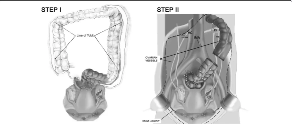
(1) Mobilization of the left and right colons The peritoneum is incised along the lateral peritoneal reflections immediately medial to the white line of Toldt from the left and right paracolic sulci cephalad to the splenic and hepatic flexures of the colon. The mesentery of the left and right colons is divided from its retroperitoneal attachments (Fig.1a).
Both ureters and the plane over Gerota’s fascia are identified to avoid compromising the kidneys. (2) Dissection and central ligation of ovarian vessels
The left and right ovarian vessels are dissected and ligated at the level of the left renal vein and the vena cava, respectively (Fig.1b).
(3) Central lymphovascular ligation with retroperitoneal selective lymph node dissection (if necessary) The lymphovascular flow is ligated by several clips. Enlarged, suspicious lymph nodes are excised at the level of the left renal vein (LRV) down towards the
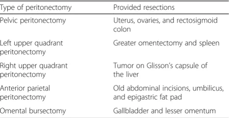
Table 1The visceral resections and parietal peritonectomy procedures
Type of peritonectomy Provided resections
Pelvic peritonectomy Uterus, ovaries, and rectosigmoid colon
Tumor on Glisson’s capsule of the liver
Anterior parietal peritonectomy
Old abdominal incisions, umbilicus, and epigastric fat pad
pelvis in one block. Where possible, a nerve-sparing technique is used.
(4) Central vascular ligation of the tumor-bearing segment of the colon
The inferior mesenteric artery (IMA) is ligated on the aorta. The artery’s individual branches are then resected as they arise from this vascular trunk (Fig.1b). The Y-configuration of the left colic and sigmoidal vessels is converted to a V-configuration to preserve the intermediate arcade. The inferior mesenteric vein (IMV) is divided as it courses around the duodenum. (5) Division of the colon*
The colon is dissected 5 cm beyond the boundary of the cancer. A previous central vascular ligation and the mobilization of the left colon allowed for packing all the viscera, including the proximal colon, into the upper abdomen.
(6) Dissection of the mesorectum 2–3 cm below the retroperitoneal flexure
(7) Stripping of the vesical peritoneum with the underlying fatty tissues from the surface of the bladder
The point of tissue transection is precisely located between the bladder musculature and its adherent fatty tissue with peritoneum. The inferior limit of the dissection is the anterior wall of the vagina. (8) Stripping of the parietal peritoneum
The peritoneal incision around the pelvis is connected to the peritoneal incisions in the right and left paracolic sulci. The parietal pelvic peritoneum is transected from the muscles and fascia down towards the intraperitoneal space. The
inferior limits of the dissection are marked by the boundaries formed by the broad ligament (the mesoovarium, the mesosalpinx, and the mesometrum), the mesorectum, and the mesocolon. (9) Extraperitoneal cutting of the broad ligament
The round ligaments are divided as they enter the internal inguinal ring.
(10) Central ligation of the uterine arteries
Uterine arteries are ligated laterally to the ureter at their origin, the anterior division of the internal iliac artery (marginal ligation). This ligation enables the excision of the entire parametrium in one block with the main specimen.
(11) Dissection of the uterus at the level of the fornices of the vagina
The lower aspect of the uterus is dissected from the vagina, and the vaginal cuff is closed.
(12) Dissection of the rectum
The rectovaginal septum is exposed. The perirectal fat is divided 2–3 cm beneath the peritoneal reflection so that the portion of the tumor
occupying the cul-de-sac is removed intact with the specimen. The rectal musculature is skeletonized using electrosurgery so that a stapler/purse-string can be used to close the rectal stump. Finally, the rectum is divided, and the cancer specimen is removed.
(13) Creation of a colorectal anastomosis
The proximal colon is anastomosed to the rectal remnant with a stapling device.
stapler anvil in the distal descending colon.
The body of the circular stapler and anvil are mated, and the stapler is activated to complete the low colorectal anastomosis.
Side-to-end: The purse-string clamp is applied at the inferior level of the resection. A straight needle is passed through the clamp to form purse-string sutures, which are used to secure the staple anvil in the rectal stump.
The lumen of the colon is visualized and progressively widened using three dilators of increasing diameters (25, 28, and 31 mm). The EEA instrument is inserted through the end of the colon, and the trochar penetrates the sidewall of the bowel. The body of the circular stapler and anvil are mated, and the stapler is activated. The circular stapler is then removed, and the distal end of the colon is closed below the colorectal anastomosis with a linear stapler TA 55.
(14)Leak testing
Of colorectal anastomosis
To evaluate the stapled colorectal anastomosis, the prox-imal and distal tissue rings are examined for completeness.
Air leak testing is conducted, and air is insufflated into the rectum with a water-filled pelvis to check for an air-tight circle of staples. Two hands should easily pass be-neath the sigmoid colon to ensure no tension at the stapled anastomosis. A rectal examination is conducted to check for staple-line bleeding at the anastomosis.
Of the bladder
The integrity of the bladder is analyzed with an intraoper-ative methylene-blue test (IMBT).
*The division of the colon is delayed, while a greater omentectomy is conducted first. This order of opera-tions enables an assessment of the “oncological cleanli-ness” of the entire colon to select the optimal place for cutting. The splenic flexure and the transverse colon are frequently involved, which involvement is hidden by the metastatic omentum.
Results
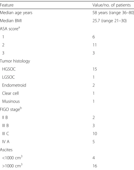
The median age of patients was 58 years (range: 36– 80 years). The median body mass index (BMI) was 25.7 (range: 21–30). High-grade serous ovarian cancer (HGSOC) was the predominating histological type and was detected in 15 of 20 (75 %) cases. The most common FIGO stage was IIIC confirmed in 50 % of patients. Most cases presented with ascites exceeding 1000 ml (80 %). Detailed data of the pre-treatment and disease-related characteristics are shown in Table 2.
In all cases, no gross residual disease was achieved. In all cases, histopathology results also confirmed bowel
infiltration of the cancer cells with the involvement of mesenteric lymph nodes in 11 cases (55 %). Operative details are shown in Table 3. Samples of the removed tumor bed and the oncological clearness of the pelvis after surgery are shown in Fig. 2. The median duration of the procedure and hospital stay was 320 min (range: 205–430 min) and 12 days (range: 7–44 days), respectively. The median estimated blood loss (EBL) was 600 cm3 (range: 400–1000 cm3). Nineteen patients received blood transfusions (99 %). The median number of red blood cell (RBC) transfusion units was 4 (range: 0 to 6 units). Six of 20 patients (30 %) required postoperative intensive care. The median number of days spent by these patients in the intensive care unit (ICU) was 4 (range: 1–6 days). Four pa-tients received total parenteral nutrition (20 %).
One patient experienced a complication associated with en bloc resection, namely an anastomosis site leak. This patient accordingly underwent further surgery during the same admission. The anastomosis site leak was man-aged with a diversion colostomy and pelvic cavity drain-age. The colostomy was reversed after chemotherapy was complete. There were no surgery-related deaths; however, one woman died 12 days post-surgery. Her death was due to pulmonary embolismus, though all 20 patients received combined pharmacologic and mechanical methods of
Table 2Patients characteristics
Feature Value/no. of patients
Median age years 58 years (range 36–80)
Median BMI 25.7 (range 21–30)
ASA scorea
American Society of Anesthesiologists Classification of Physical Status
b
prophylaxis of venous thromboembolism (VTE) during hospital stay.
All surviving patients (n= 19) received platinum-based chemotherapy. The median number of days between the primary surgery and the first cycle of chemotherapy was 48 days (range: 20–147 days). The median follow-up time period was 19 months (range: 8–31 months). No patient experienced a recurrence of disease during the time of observation. Detailed data on the course of treat-ment for particular cases are shown in Table 4.
Discussion
The conventional technique procedure prioritizes the mobilization of the tumor-bearing segment followed by central vascular ligation and the ligation of other portions of the vasculature. Conversely, the no-touch isolation technique prioritizes central vascular ligation, followed by the mobilization of the tumor-bearing segment [9].
In advanced EOC, multi-visceral and peritoneal metasta-ses create a tumor-bearing segment supplied by the extra-peritoneal vascular system.
It is well-known that a complete peritonectomy and visceral resections are required to achieve a complete clearance of pan pelvic disease in these patients. The preponderance of data suggests that colon resection to achieve optimal cytoreduction positively impacts sur-vival rates [15].
To achieve cytoreduction of microscopic residual dis-ease in disseminated EOC cases consistent with the re-quirements of cancer no-touch isolation, a modified en bloc pelvic resection was proposed. This modified resec-tion aims to initially reach the retroperitoneal space where the central vascular ligation is performed.
In the first step of the operation, the peritoneum is incised along the lateral peritoneal reflections immedi-ately medial to the white line of Toldt from the left and right paracolic sulci cephalad to the splenic and hepatic flexures of the colon. The mesentery of the left and right colons is divided from all its retroperitoneal at-tachments. This dividing not only enables the identifi-cation of the ovarian and mesentery vessels but also elevates the entire colon with metastatic omentum, thus allowing the affliction of the upper abdomen with cancer to be properly assessed.
Fig. 2Intraperitoneal cancer compartment (including the infiltrated part of the bowel) and oncological clearness following the modified en block pelvic resection
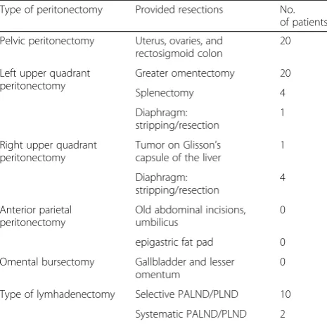
Table 3The visceral resections and parietal peritonectomy procedures conducted during primary debulking surgery (PDS) Type of peritonectomy Provided resections No.
of patients
Pelvic peritonectomy Uterus, ovaries, and rectosigmoid colon
20
Left upper quadrant peritonectomy
Greater omentectomy 20
Splenectomy 4
Diaphragm: stripping/resection
1
Right upper quadrant peritonectomy
Tumor on Glisson’s capsule of the liver
1
Diaphragm: stripping/resection
4
Anterior parietal peritonectomy
Old abdominal incisions, umbilicus
0
epigastric fat pad 0
Omental bursectomy Gallbladder and lesser omentum
0
Type of lymhadenectomy Selective PALND/PLND 10
It has been suggested that the inefficiency of upper ab-domen surgery limits optimal cytoreduction [16]. Hence, this surgical step could reveal all potentially unresectable lesions and is therefore proposed to initiate all aggressive surgical plans to eradicate EOC.
The left and right ovarian vessels are dissected and li-gated at the level of the left renal vein and the vena cava, respectively.
Central vascular ligation of the tumor-bearing segment of the colon is performed. From a practical perspective, this ligation should be proceeded by the opening of the omental bursa to assess the “oncological cleanliness” of the colon.
The surgical plan includes central lymphovascular li-gation and retroperitoneal lymph node dissection (LND). LND is a questionable procedure to use in cases of ad-vanced ovarian cancer. We aimed to excise enlarged, sus-picious nodes during the cytoreduction or to perform a systematic lymphadenectomy in cases with cancer spread macroscopically limited only to the pelvis. Current pro-spective studies such as the LION study (AGO-Ovar) will confirm the appropriateness of using LND in EOC. Not-ably, none of the 12 cases showed postoperative lympho-cele with the lymph node dissection, which result could indicate another positive impact of performing the central lymphovascular ligation prior to excising enlarged, suspi-cious nodes.
The next proposed modification entails an excision of the entire segment of affected peritoneum, including areas without visible lesions. For example, if the peritoneal me-tastases are present in the pelvis, a complete pelvic perito-nectomy is conducted. Recently, radical surgery together with hyperthermic intraperitoneal chemotherapy (HIPEC) as described by Sugarbaker [14] has been widely promoted for use in EOC cases.
Sugarbaker did not excise uninvolved surfaces of the peritoneum because he believed that microscopic disease, if unaddressed, will be eradicated by HIPEC performed immediately after surgery. Existing evidence suggests that primary debulking surgery followed by HIPEC has an un-expectedly negative impact on prognosis in EOC [17]. The results of ongoing randomized control trials will be published not sooner than in the next 3 years. There-fore, the approach of excising the entire segment of in-volved peritoneum, rather than performing experimental perioperative intraperitoneal chemotherapy, seems rea-sonable, particularly given the lack of observed complica-tions related to the excision in our cohort.
Rationale for modification
The impact of no tough isolation technique on intraope-rative cancer dissemination will be difficult to prove. However, the proposed central vascular ligation should, if not decrease intraoperative cancer dissemination, at
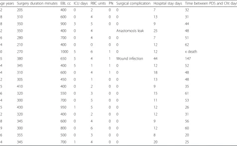
Table 4Modified en block pelvic resection—course of treatment
Age years Surgery duration minutes EBL cc ICU days RBC units PN Surgical complication Hospital stay days Time between PDS and Cht days
62 205 400 0 2 0 0 7 32
58 310 600 0 4 0 0 13 31
38 350 900 3 5 0 0 9 44
62 350 400 0 4 Anastomosis leak 25 48
36 280 700 0 4 0 0 7 51
74 210 400 0 0 0 0 12 62
80 270 1000 5 6 1 0 12 x death
65 380 650 5 4 1 Wound infection 44 147
64 345 400 5 1 1 0 12 52
64 310 600 0 4 1 0 18 48
52 305 450 0 1 0 0 13 48
55 410 400 0 2 0 0 9 35
66 320 550 0 3 0 0 15 61
54 300 700 0 5 0 0 11 53
55 430 950 1 5 0 0 12 26
72 320 400 0 2 0 0 12 31
48 345 600 0 4 0 0 9 56
59 300 800 0 6 0 0 12 60
46 355 500 0 3 0 0 8 20
54 345 700 1 4 0 0 20 25
least decrease complications such as blood loss and lym-phocoele formation. Elevating the entire colon while opening the retroperitoneal space seems to be highly re-liable for predicting optimal cytoreduction. Excising the entire segment of involved peritoneum, including areas without visible lesions, is safe and could be a cost-effective alternative for HIPEC.
Course of treatment
The duration of surgery and hospitalization was com-parable to the respective durations reported in other studies on en block pelvic resection published after 2000 [18–24]. Patients in our cohort had a lower EBL and the highest rate of complete cytoreduction com-pared with other reports [18–24]. We believe that the decrease in blood loss is caused by the introduction of central vascular ligation and by the fact that surgery is starting from the healthy tissues surrounding the tumor compartment. It was proven that the tumor-bearing segment has increased vascularization caused by cancer neoangiogenesis [25]. A surgical operation conducted through an anatomically unchanged (i.e., healthy) com-partment could also be responsible for the increased re-sectability of ovarian cancer tissue.
We observed anastomosis leakage in 1 of 20 cases (5 %). Only in one recent study was the percentage of this complication is higher (8.7 %) [23]. Remaining studies published after 2000 show lower rates of anastomosis leakage, namely 0.8 %, [19] 1.7 % [21], 2.2 % [22], 3 % [24], 3.2 % [20], and 4.5 % [18]. All studies on en block pelvic resection were conducted with a limited number of cases, and the subsequent results of the studies do not reflect the real (average) rate of the anastomosis leakage of colo-rectal anastomosis. A meta-analysis of randomized control trials (RCTs) of colorectal anastomosis surgery published in 2001 revealed that the overall stapled clinical leak rate is 6.3 % [26]. Given these data, our results (5 %) seem fully acceptable.
The single complication observed in our study could be explained by the poor nutritional status of the pa-tient. We routinely analyzed serum albumin levels prior to surgery, yet this method appeared to be ineffective in identifying patients with severe nutritional risk. Since our study, we have incorporated an assessment of nutri-tional status based on the European Society of Parenteral Nutrition (ESPEN) Guidelines on Enteral Nutrition [27] into our qualification process. Currently, all patients with severe nutritional risk receive parenteral nutrition for 10–14 days prior to surgery even if surgery must be consequently delayed.
Recent studies on stapled colorectal anastomosis have indicatedside-to-endanastomosis as safer thanend-to-end anastomosis [28].Side-to-endanastomosis tends to fill the
pelvis, reducing the area of dead space in which a hematoma or collection of hematomas could develop [29]. Indeed, our observed case of leakage was one of two cases in which end-to-end colorectal anastomosis was created, which could thus be additional reason for the complication.
There were no surgery-related deaths; however, one woman died due to pulmonary embolismus 12 days post-surgery. She received combined pharmacologic and mechanical methods of prophylaxis of VTE during her hospital stay. Post mortem, it was found that she had been a mutation carrier of factor V Leiden. This fact was unpredictable; thus, we believe that her death should not affect the appraisal of the described surgical procedure.
All surviving patients received six courses of platinum-based chemotherapy.
The time interval from surgery to the start of chemo-therapy was 48 days (range: 20–147 days). This interval is longer than the average period of 4–5 weeks reported in the literature [30]. This delay has been suggested as impairing a patient’s prognosis [31]. Recently, however, it was demonstrated that the timing of postoperative chemotherapy did not influence the overall survival rate in women without postoperative residual disease [30]. Indeed, none of our optimally debulked patients experi-enced a recurrence of the disease during the time of our observation. The median follow-up occurred only 19 months following surgery (range: 8–31 months). PFS periods reported in the literature range from 14 to 18 months [32]; hence, we believe that an en bloc pelvic resection preceded by a central ligation of the vessels supplying the tumor bed could potentially improve the outcome of ovarian cancer patients.
Prospective clinical trials with a control group are needed to assess the exact oncological benefit of the presented approach.
Summary of proposed modification
peritoneum (not merely the involved portion) from the bladder and the pelvic wall and then resecting in one block. A colorectal anastomosis is created. In our co-hort of 20 ovarian cancer patients, this new approach resulted in low blood loss and a high rate of optimal cytoreduction.
Conclusions
Performing a central ligation of vessels supplying the tumor bed prior to an en bloc pelvic resection is feasible with acceptable morbidity and mortality rates. The im-pact of this modification on intraoperative cancer spread and the decrease in blood loss as well as oncological out-come should be confirmed in future studies.
Competing interests
The author declares no competing interests.
Author’s contributions
JJS conceptualized and designed the study; acquired, analyzed, and interpreted the data; and contributed to the statistical analysis, manuscript preparation, manuscript editing, and manuscript review.
Acknowledgements
Dudziak Mirosław—Head of the Department of Gynecologic Oncology, Oncology Center in Red Cross Hospital in Gdynia, Poland, who supported this trial and assisted in most of the performed operations.
Received: 5 January 2016 Accepted: 21 April 2016
References
1. Siegel R, Naishadham D, Jemal A. Cancer statistics, 2013. CA Cancer J Clin. 2013;63:11–30.
2. Hoskins WJ, Bundy BN, Thigpen JT, Omura GA. The influence of cytoreductive surgery on recurrence-free interval and survival in small-volume stage III epithelial ovarian cancer: a Gynecologic Oncology Group study. Gynecol Oncol. 1992;47:159–66.
3. Ozols RF, Bundy BN, Greer BE, Fowler JM, Clarke-Pearson D, Burger RA, et al. Phase III trial of carboplatin and paclitaxel compared with cisplatin and paclitaxel in patients with optimally resected stage III ovarian cancer: a Gynecologic Oncology Group study. J Clin Oncol. 2003;21:3194–200. 4. Chi DS, Liao JB, Leon LF, Venkatraman ES, Hensley ML, Bhaskaran D, et al.
Identification of prognostic factors in advanced epithelial ovarian carcinoma. Gynecol Oncol. 2001;82:532–7.
5. Winter III WE, Maxwell GL, Tian C, Carlson JW, Ozols RF, Rose PG, et al. Prognostic factors for stage III epithelial ovarian cancer: a Gynecologic Oncology Group study. J Clin Oncol. 2007;25:3621–7.
6. Chi DS, Eisenhauer EL, Lang J, Huh J, Haddad L, Abu-Rustum NR, et al. What is the optimal goal of primary cytoreductive surgery for bulky stage IIIC epithelial ovarian carcinoma (EOC)? Gynecol Oncol. 2006;103:559–64. 7. Bristow RE, Tomacruz RS, Armstrong DK, Trimble EL, Montz FJ. Survival
effect of maximal cytoreductive surgery for advanced ovarian carcinoma during the platinum era: a meta-analysis. J Clin Oncol. 2002;20:1248–59. 8. Chang SJ, Bristow RE. Surgical technique of en bloc pelvic resection for advanced
ovarian cancer. J Gynecol Oncol. 2015;26:155. doi:10.3802/jgo.2015.26.2.155. 9. Atkin G, Chopada A, Mitchell I. Colorectal cancer metastasis: in the surgeon’s
hands? Int Semin Surg Oncol. 2005;2:5.
10. Barnes JP. Physiologic resection of the right colon. Surg Gynecol Obstet. 1952;94:723–6.
11. Gall TM, Jacob J, Frampton AE, Krell J, Kyriakides C, Castellano L, Stebbing J, Jiao LR. Reduced dissemination of circulating tumor cells with no-touch isolation surgical technique in patients with pancreatic cancer. JAMA Surg. 2014;149(5):482–5.
12. Takii Y, Shimada Y, Moriya Y, Nakamura K, Katayama H, Kimura A, et al. A randomized controlled trial of the conventional technique versus the no-touch isolation technique for primary tumor resection in patients with
colorectal cancer: Japan Clinical Oncology Group Study JCOG1006. Jpn J Clin Oncol. 2014;44(1):97–100.
13. Young J, Badgery-Parker T, Dobbins T, Jorgensen M, Gibbs P, Faragher I, Jones I, Currow D. Comparison of ECOG/WHO performance status and ASA score as a measure of functional status. J Pain Symptom Manage. 2015;49(2):258–64. 14. Sugarbaker PH. An overview of peritonectomy, visceral resections, and
perioperative chemotherapy for peritoneal surface malignancy. In: Sugarbaker PH, editor. Cytoreductive surgery & perioperative chemotherapy for peritoneal surface malignancy. Textbook and video atlas. Woodbury: Cine-Med Publishers; 2012.
15. Hoffman MS, Zervose E. Colon resection for ovarian cancer: intraoperative decisions. Gynecol Oncol. 2008;111(2 Suppl):S56–65.
16. Raspagliesi F, Ditto A, Martinelli F, Haeusler E, Lorusso D. Advanced ovarian cancer: omental bursa, lesser omentum, celiac, portal and triad nodes spread as cause of inaccurate evaluation of residual tumor. Gynecol Oncol. 2013;129:92–6. 17. Chiva LM, Gonzalez-Martin A. A critical appraisal of hyperthermic
intraperitoneal chemotherapy (HIPEC) in the treatment of advanced and recurrent ovarian cancer. Gynecol Oncol. 2015;136(1):130–5.
18. Yildirim Y, Ertas IE, Nayki U, Ulug P, Nayki C, Yilmaz I, et al. En-bloc pelvic resection with concomitant rectosigmoid colectomy and immediate anastomosis as part of primary cytoreductive surgery for patients with advanced ovarian cancer. Eur J Gynaecol Oncol. 2014;35(4):400–7. 19. Clayton RD, Obermair A, Hammond IG, Leung YC, McCartney AJ. The
Western Australian experience of the use of en bloc resection of ovarian cancer with concomitant rectosigmoid colectomy. Gynecol Oncol. 2002; 84(1):53–7.
20. Bristow RE, del Carmen MG, Kaufman HS, Montz FJ. Radical oophorectomy with primary stapled colorectal anastomosis for resection of locally advanced epithelial ovarian cancer. J Am Coll Surg. 2003;197(4):565–74. 21. Mourton SM, Temple LK, Abu-Rustum NR, Gemignani ML, Sonoda Y,
Bochner BH, et al. Morbidity of rectosigmoid resection and primary anastomosis in patients undergoing primary cytoreductive surgery for advanced epithelial ovarian cancer. Gynecol Oncol. 2005;99(3):608–14. 22. Park JY, Seo SS, Kang S, Lee KB, Lim SY, Choi HS, et al. The benefits of low
anterior en bloc resection as part of cytoreductive surgery for advanced primary and recurrent epithelial ovarian cancer patients outweigh morbidity concerns. Gynecol Oncol. 2006;103(3):977–84.
23. Richardson DL, Mariani A, Cliby WA. Risk factors for anastomotic leak after recto-sigmoid resection for ovarian cancer. Gynecol Oncol. 2006;103(2):667–72. 24. Peiretti M, Bristow RE, Zapardiel I, Gerardi M, Zanagnolo V, Biffi R, et al.
Rectosigmoid resection at the time of primary cytoreduction for advanced ovarian cancer. A multi-center analysis of surgical and oncological outcomes. Gynecol Oncol. 2012;126(2):220–3.
25. Nishida N, Yano H, Nishida T, Kamura T, Kojiro M. Angiogenesis in cancer. Vasc Health Risk Manag. 2006;2(3):213–9.
26. Matos D, Atallah ÁN, Castro AA, Silva Lustosa SA. Stapled versus handsewn methods for colorectal anastomosis surgery. Cochrane Database Syst Rev. 2001;Issue 3. Art. No.: CD003144. doi:10.1002/14651858.CD003144. 27. Weimann A, Braga M, Harsanyi L, Laviano A, Ljungqvist O, Soeters P, et al.
ESPEN Guidelines on Enteral Nutrition: surgery including organ transplantation. Clin Nutr. 2006;25(2):224–44.
28. Goulder F. Bowel anastomoses: the theory, the practice and the evidence base. World J Gastrointest Surg. 2012;4(9):208–13.
29. Taflampas P, Christodoulakis M, Tsiftsis DD. Anastomotic leakage after low anterior resection for rectal cancer: facts, obscurity, and fiction. Surg Today. 2009;39:183–8.
30. Hofstetter G, Concin N, Braicu I, Chekerov R, Sehouli J, Cadron I, et al. The time interval from surgery to start of chemotherapy significantly impacts prognosis in patients with advanced serous ovarian carcinoma—analysis of patient data in the prospective OVCAD study. Gynecol Oncol. 2013;131(1):15–20. 31. Mahner S, Eulenburg C, Staehle A, Wegscheider K, Reuss A, Pujade-Lauraine E,
et al. Prognostic impact of the time interval between surgery and
chemotherapy in advanced ovarian cancer: analysis of prospective randomized phase III trials. Eur J Cancer. 2013;49(1):142–9. doi:10.1016/j.ejca.2012.07.023. 32. du Bois A, Reuss A, Pujade-Lauraine E, Harter P, Ray-Coquard I, Pfisterer J.