R E S E A R C H
Open Access
Clinical outcomes and prognostic factors
for gastric cancer patients with bone
metastasis
Jota Mikami
1*, Yutaka Kimura
2, Yoichi Makari
1, Junya Fujita
1, Tomoya Kishimoto
1, Genta Sawada
1, Shin Nakahira
1,
Ken Nakata
1, Masaki Tsujie
1and Hiroki Ohzato
1Abstract
Background:Bone metastasis due to gastric cancer is rare, and the clinical features have not been fully evaluated. We investigated the clinical features, treatment outcomes, and prognostic factors in gastric cancer patients with bone metastasis.
Methods:We retrospectively collected data on 34 consecutive patients who were diagnosed radiologically with bone metastasis due to gastric cancer. We estimated the overall survival after the diagnosis of bone metastasis using the Kaplan-Meier product-limit method and evaluated which clinicopathological factors were associated with prognostic factors for survival using univariate and multivariate Cox proportional hazards regression models. Results:The treatment for the primary tumor was surgery in 16 patients (47.1%) and chemotherapy in 18 patients (52.9%). The median serum alkaline phosphatase (ALP) and lactate dehydrogenase (LDH) levels at the time of bone metastasis were 375.5 and 249 IU/L, respectively. Ten patients (29.4%) were diagnosed with bone metastasis and gastric cancer at the same time. The 6-month survival rate after the diagnosis of bone metastasis was 63.8%, and the median survival time was 227.5 days. Multivariate analysis revealed that metachronous metastasis (p= 0.035) and extraosseous metastasis (p= 0.028) were significant risk factors for poor survival.
Conclusions:The prognosis of gastric cancer with bone metastasis was poor, and metachronous metastasis and extraosseous metastasis were shown to be poor prognostic factors. Serum ALP, LDH, and tumor markers are not always high, so aggressive diagnosis using appropriate modalities such as bone scan, MRI, or PET-CT may be necessary in routine practice even in asymptomatic patients.
Keywords:Gastric cancer, Bone metastasis, Prognostic factor
Background
Although the incidence of gastric cancer has decreased in developed countries, it is the second most common cancer worldwide and two thirds of cases are found in developing countries [1]. The main sites of metastasis of gastric cancer are the liver and lungs, and the incidence of bone metastasis due to gastric cancer is only 0.9–2.1% [2], although there may be many gastric cancer patients who have not been diagnosed with metastasis clinically
since the reported frequency of bone metastasis in gas-tric cancer was 13.4–15.9% in an autopsy series [3].
The median survival times of gastric cancer patients with bone metastasis are 3–4 months after the detection of bone metastasis [4, 5]. Since bone metastasis can cause intractable pain leading to poor quality of life, ap-propriate treatment strategies are essential for the af-fected patients [2]. Although the clinical characteristics and poor prognostic factors have been reported, they are not well-defined [4–7]. In this study, we retrospectively examined the clinicopathological features, treatment outcomes, and prognostic factors for survival in gastric cancer patients with bone metastasis.
* Correspondence:johmikami@sakai-hospital.jp
1Department of Surgery, Sakai City Medical Center, 1-1-1 Ebarajicho, Nishi-ku,
Sakai City 593-8304, Osaka, Japan
Full list of author information is available at the end of the article
Methods
This study was approved by the institutional review board at Sakai City Medical Center. We retrospectively collected data on 34 consecutive patients who were radiologically diagnosed with bone metastasis due to gastric cancer be-tween January 2010 and December 2015. All tumors were histologically diagnosed as adenocarcinoma with the stomach, which was recognized as primary tumor. Bone metastases have been treated after the clinical diagnosis by CT, PET-CT, bone scintigraphy, or MRI and after con-firming that there were no other suspicious cancers by en-hanced CT imaging from the chest to the pelvis. Clinicopathological data, such as age at the diagnosis of bone metastasis, gender, the Eastern Cooperative Oncol-ogy Group (ECOG) performance status scale, symptoms at the diagnosis of bone metastasis, tumor localization, differentiation, clinical or pathological stage (according to the 14th edition of the Japanese classification of gastric carcinoma to determine pathological stage [8]) at initial diagnosis, treatment for primary tumor (surgery or chemotherapy), treatment for bone metastasis (chemo-therapy, radio(chemo-therapy, or best supportive care), and the spread of bone metastasis, were determined from patient records. The numerical values of serum alkaline phosphat-ase (ALP), serum lactate dehydrogenphosphat-ase (LDH), carci-noembryonic antigen (CEA), carbohydrate antigen (CA) 19-9, and CA125 were obtained from tests performed at the time of the diagnosis of bone metastasis. When the bone metastasis was observed at the same time as the diagnosis of gastric cancer, we defined it as a synchronous pattern of bone metastasis, while a metachronous pattern of bone metastasis was defined as bone metastasis de-tected at any time after the diagnosis of gastric cancer. For some patients, only a computed tomography (CT) scan was used in the diagnosis because bone metastasis was evident, while many patients were diagnosed using a com-bination of bone scintigraphy, positron emission tomog-raphy (PET)-CT, and magnetic resonance imaging (MRI).
We estimated the overall survival after the diagnosis of bone metastasis using the Kaplan-Meier product-limit method. We also evaluated which clinicopathological factors were associated with prognostic factors for sur-vival using univariate and multivariate Cox proportional hazards regression models. Statistical significance was set at p< 0.05. All statistical analyses were performed using SPSS Statistics software, version 19 (IBM Corp., Armonk, NY, USA).
Results
The median age of the 34 patients at the time the bone metastasis was diagnosed was 66 years (Table 1). There were 26 male patients and 8 females, and 19 patients (55.9%) had undifferentiated adenocarcinoma. The treat-ment for the primary tumor was surgery in 16 patients
(47.1%), and 10 of them had radical resection. Out of 18 patients (52.9%) who used chemotherapy for initial treat-ment, 2 patients had surgery after chemotherapy with S1
Table 1Patient demographics and pathologic features
Factors Patient
(n= 34) Age (years) Median (range) 66 (39–78)
Gender Male 26 (76.5%)
Female 8 (23.5%) ECOG performance status 0–1 16 (47.1%)
2–4 18 (52.9%)
Bone pain Present 11 (32.4%)
Absent 23 (67.6%)
Location Upper 1/3 5 (14.7%)
Middle 1/3 17 (50.0%) Lower 1/3 5 (14.7%) Whole stomach 7 (20.6%) Histologic type Differentiated 15 (44.1%)
Undifferentiated 19 (55.9%)
Stagea I 1 (2.9%)
II 3 (8.8%)
III 8 (23.5%)
IV 22 (64.7%)
Treatment for primary tumor Surgery 16 (47.1%) Chemotherapy 18 (52.9%) ALP (IU/L) Median (range) 375.5 (157–2743) LDH (IU/L) Median (range) 249 (117–1481) CEA Median (range) 8.6 (1.0–3508) CA19-9 Median (range) 53.7 (0.6–1814.0) CA125 Median (range) 20.3 (7.9–1099) Diagnostic modality CT 30 (88.2%)
Bone scan 10 (29.4%)
MRI 9 (26.5%)
PET-CT 1 (2.9%) Pattern of bone metastasis Synchronous 10 (29.4%)
Metachronous 24 (70.6%) Number of bone metastases Single 12 (35.3%) Multiple 22 (64.7%) Extraosseous metastasis Present 26 (76.5%) Absent 8 (23.5%) Treatment of bone metastasis Chemotherapy 26 (76.5%)
Radiotherapy 5 (14.7%) Best supportive care 4 (11.8%)
Abbreviations:ALPserum alkaline phosphatase,LDHlactate dehydrogenase,CEA serum carcinoembryonic antigen,CAcarbohydrate antigen,CTcomputed tomography,MRImagnetic resonance imaging,PETpositron emission tomography a
and cisplatin. Eleven patients (32.4%) had bone pain at the time the bone metastasis was diagnosed. The median serum ALP and LDH levels at the time of bone metasta-sis were 375.5 and 249 IU/L, respectively. To diagnose the bone metastasis, CT scan was used for 30 patients (29.4%), and 15 patients of them underwent bone scin-tigraphy (9 patients), MRI (7 patients), and PET-CT (1 patient) after CT scan (Two patients underwent both bone scintigraphy and and MRI).
Ten patients (29.4%) were diagnosed with bone metas-tasis and gastric cancer at the same time, and 26 patients (76.5%) had at least one other organ affected besides their bones. Of the 24 patients who had metachronous metastasis, the median interval from the diagnosis of gastric cancer to the detection of bone metastasis was 398 days (range, 43–1799, data not shown).
The treatment of the bone metastasis consisted of chemotherapy in 26 patients (76%), radiotherapy in 5 pa-tients (15%), and 4 papa-tients only received the best support-ive care (12%). The most common sites of bone metastases were the thoracic vertebrae (55.9%), pelvic bones (41.2%), lumbar vertebrae (38.2%), and ribs (29.4%) (Table 2). Many patients were treated with chemotherapy, such as S1-based regimens, as first-line chemotherapy, and taxane-based or irinotecan-based regimens as second and subsequent chemotherapies, according to the recommendation of the Japanese Gastric Cancer Treatment Guidelines for patients who have progressive or recurrent gastric cancer [9]. Some patients were treated with radiotherapy for a localized tumor or local symptom relief.
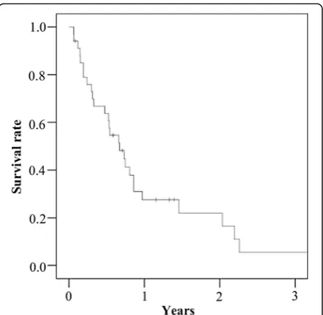
The 6-month survival rate and the median survival time after the diagnosis of bone metastasis were 63.8% and 227.5 days, respectively (Fig. 1). A multivariate ana-lysis revealed that metachronous metastasis (odds ratio 3.6; 95% confidence interval 1.1–11.7; p= 0.035) and extraosseous metastasis (odds ratio 4.1; 95% confidence
interval 1.2–14.9; p= 0.028) were significant risk factors for poor survival (Table 3).
Discussion
In this study, the 6-month survival rate and the median survival time after the diagnosis of bone metastasis were 63.8% and 227.5 days, respectively. In univariate analysis, only pattern of bone metastases (synchronous vs meta-chronous) became an independent prognostic factor. The multivariate analysis was carried out using the pat-tern of bone metastases and variables pointed out to affect prognosis in past reports [2, 7]. As a result, meta-chronous metastasis and extraosseous metastasis were significant risk factors for poor survival.
In the period of this study, there were 622 patients who have been treated for gastric cancer for the first time in our hospital, and 34 of them have been diagnosed with bone metastasis. Most patients develop bone metastasis within 2 years of gastric surgery [3]. In this study, the me-dian interval from the diagnosis of gastric cancer to the detection of bone metastasis was 398 days in the patients who had metachronous metastasis, and the median inter-val from the surgery to the detection of bone metastasis was 562 days. Bone metastases from gastric cancer were not unusual in a multicenter trial [10], and Turkoz et al. suggested that bone metastases should be considered dur-ing the follow-up of gastric cancer patients, even in the early period [3]. Patients may have relatively long-term survival if there is no extraosseous metastasis or local con-trol for metastasis is possible, but it is difficult to diagnose bone metastasis because the majority of affected patients are asymptomatic and evaluations for bone metastases are
Table 2Site of bone metastasis
Site of bone metastasis Patient
(n= 34)
Thoracic vertebrae 19 (55.9%)
Pelvic bones 14 (41.2%)
Lumbar vertebrae 13 (38.2%)
Ribs 10 (29.4%)
Cervical vertebrae 6 (17.6%)
Calvarium 4 (11.2%)
Scapula 3 (8.8%)
Lower extremity 3 (8.8%)
Upper extremity 2 (5.9%)
Clavicle 1 (2.9%)
Sternum 1 (2.9%)
not indicated in routine practice [7]. In our study, there were many cases that were discovered by chance during a routine CT examination. Only 32.4% of patients com-plained about symptoms, such as bone pain. In addition, serum ALP, LDH, or tumor markers were not always high, although there have been several reports that show such serum parameters were useful for diagnosing bone metas-tasis [11–13]. Ahn et al. suggested that an appropriate modality, such as bone scintigraphy, is required to assess bone metastasis at the time of the initial diagnosis and during follow-up observations [5].
Since there were no prospective studies of therapeutic regimens in gastric cancer patients with bone metastasis, the optimal chemotherapy regimens were unknown [2]. In this study, the treatment of metachronous bone metastasis differed depending on the judgment of the attending phys-ician, and there had been various treatments before bone metastasis was recognized, while median survival time (MST) of patients who were treated metachronous bone metastasis with S1-based regimens or irinotecan-based reg-imens was significantly longer than MST of other patients (314 vs 87 days, p= 0.010). The Japanese Gastric Cancer Treatment Guidelines recommend S1-based chemotherapy for progressive or recurrent gastric cancer [9]. On the other hand, since bone metastasis can cause disseminated intravascular coagulation (DIC), the poor general condition of the patient or the presence of thrombocytopenia and severe anemia may make the patient ineligible for chemo-therapy [14]. Hironaka et al. reported that sequential
methotrexate and 5-fluorouracil chemotherapy resulted in a high rate of alleviation of DIC caused by bone metastasis from gastric cancer [15]. In addition, the pain management for patients with bone pain is important and radiation ther-apy may be quite effective [5]. In recent years, it has been reported that the incidence of epidermal growth factor receptor (EGFR) mutations in the bone metastases was high in the lung adenocarcinoma [16, 17]. Thus, EGFR tyrosine kinase inhibitor therapies could be effective for the type of adenocarcinoma. However, there are no reports about genetic mutations in the bone metastases due to gastric cancer. Identifying such mechanisms like gene mu-tations may lead to the development of future treatment.
In our study, the diagnosis of bone metastasis was left to the discretion of the attending physician and various modalities were used, so we could not evaluate which diag-nostic methods were appropriate. In addition, the treat-ments varied depending on when the bone metastasis was detected. Therefore, further investigations are necessary.
Conclusions
The prognosis of gastric cancer with bone metastasis was poor, and metachronous metastasis and extraosseous me-tastasis were shown to be poor prognostic factors. In addition, ALP, LDH, and tumor markers are not always high, so aggressive diagnosis using appropriate modalities such as bone scan, MRI, or PET-CT may be necessary in routine practice even in asymptomatic patients.
Table 3Univariate and multivariate analyses of prognostic factors for survival
Univariate analysis Multivariate analysis
RR (95% CI) pvalue RR (95% CI) pvalue
Age ≥75
<75 1.8 (0.25–13.7) 0.54
Gender Male
Female 1.1 (0.45–2.9) 0.78
ECOG performance status 0–1
2–4 1.4 (0.57–3.4) 0.48 1.1 (0.41–2.8) 0.88
Bone pain Absent
Present 2.0 (0.90–4.5) 0.091 2.7 (0.93–8.1) 0.068 Histologic type Undifferentiated
Differentiated 1.1 (0.53–2.5) 0.73
Stagea I–III
IV 1.0 (0.46–2.2) 0.99
Pattern of bone metastasis Synchronous
Metachronous 2.8 (1.1–7.5) 0.038 3.6 (1.1–11.7) 0.035 Extraosseous metastasis Absent
Present 1.1 (0.47–2.8) 0.77 4.1 (1.2–14.9) 0.028
Abbreviations:CIconfidential interval,ECOGEastern Cooperative Oncology a
Abbreviations
ALP:Alkaline phosphatase; CA: Carbohydrate antigen; CEA: Carcinoembryonic antigen; CT: Computed tomography; DIC: Disseminated intravascular coagulation; ECOG: Eastern Cooperative Oncology Group; LDH: Lactate dehydrogenase; MRI: Magnetic resonance imaging; PET: Positron emission tomography
Acknowledgements
None.
Funding
None.
Availability of data and materials
The datasets analyzed during the current study are available from the corresponding author upon reasonable request.
Authors’contributions
JM performed the data analysis and drafted the manuscripts. YK conceived and designed the study. YM, JF, TK, GS, SN, KN, MT, and HO participated in the study design and coordination. All authors participated in the discussion and approved the final submitted version of the manuscripts.
Competing interests
The authors declare that they have no competing interests.
Consent for publication
Not applicable, but this study has been announced on the home page of our institution instead.
Ethics approval and consent to participate
This study was approved by the institutional review board at Sakai City Medical Center.
Author details
1Department of Surgery, Sakai City Medical Center, 1-1-1 Ebarajicho, Nishi-ku,
Sakai City 593-8304, Osaka, Japan.2Department of Surgery, Kindai University Faculty of Medicine, 377-2 Onohigashi, Sayama City 589-8511, Osaka, Japan.
Received: 8 September 2016 Accepted: 23 December 2016
References
1. World Health Organization. Causes of death. 2004. http://www.who.int/ healthinfo/global_burden_disease/GBD_report_2004update_part2.pdf. Accessed Jul 2014.
2. Nakamura K, Tomioku M, Nabeshima K, Yasuda S. Clinicopathologic features and clinical outcomes of gastric cancer patients with bone metastasis. Tokai j exp clin med. 2014;39:193–8.
3. Turkoz FP, Solak M, Kilickap S, Ulas A, Esbah O, Oksuzoglu B, et al. Bone metastasis from gastric cancer: the incidence, clinicopathological features, and influence on survival. J gastric cancer. 2014;14:164–72.
4. Lee J, Lim T, Uhm JE, Park KW, Park SH, Lee SC, et al. Prognostic model to predict survival following first-line chemotherapy in patients with metastatic gastric adenocarcinoma. Ann oncol. 2007;18:886–91.
5. Ahn JB, Ha TK, Kwon SJ. Bone metastasis in gastric cancer patients. J gastric cancer. 2011;11:38–45.
6. Kim HS, Yi SY, Jun HJ, Lee J, Park JO, Park YS, et al. Clinical outcome of gastric cancer patients with bone marrow metastases. Oncology. 2007;73:192–7. 7. Park HS, Rha SY, Kim HS, Hyung WJ, Park JS, Chung HC, et al. A prognostic
model to predict clinical outcome in gastric cancer patients with bone metastasis. Oncology. 2011;80:142–50.
8. Japanese Gastric Cancer Association. Japanese classification of gastric carcinoma: 3rd English edition. Gastric cancer. 2011;14:101–12.
9. Japanese Gastric Cancer Association. Japanese gastric cancer treatment guidelines 2010 (ver. 3). Gastric cancer. 2011;14:113–23.
10. Silvestris N, Pantano F, Ibrahim T, Gamucci T, De Vita F, Di Palma T, et al. Natural history of malignant bone disease in gastric cancer: final results of a multicenter bone metastasis survey. Plos one. 2013;8:e74402.
11. Choi CW, Lee DS, Chung JK, Lee MC, Kim NK, Choi KW, et al. Evaluation of bone metastases by Tc-99m MDP imaging in patients with stomach cancer. Clin nucl med. 1995;20:310–4.
12. Kobayashi M, Okabayashi T, Sano T, Araki K. Metastatic bone cancer as a recurrence of early gastric cancer—characteristics and possible mechanisms. World j gastroenterol. 2005;11:5587–91.
13. Catalano V, Graziano F, Santini D, D'Emidio S, Baldelli AM, Rossi D, et al. Second-line chemotherapy for patients with advanced gastric cancer: who may benefit? Br j cancer. 2008;99:1402–7.
14. Takashima A, Shirao K, Hirashima Y, Takahari D, Okita NT, Nakajima TE, et al. Sequential chemotherapy with methotrexate and 5-fluorouracil for chemotherapy-naive advanced gastric cancer with disseminated intravascular coagulation at initial diagnosis. J cancer res clin oncol. 2010;136:243–8. 15. Hironaka SI, Boku N, Ohtsu A, Nagashima F, Sano Y, Muto M, et al. Sequential
methotrexate and 5-fluorouracil therapy for gastric cancer patients with bone metastasis. Gastric cancer. 2000;3:19–23.
16. Krawczyk P, Nicos M, Ramlau R, Powrozek T, Wojas-Krawczyk K, Sura S, et al. The incidence of EGFR-activating mutations in bone metastases of lung adenocarcinoma. Pathol oncol res. 2014;20:107–12.
17. Nguyen DX, Massague J. Genetic determinants of cancer metastasis. Nat rev genet. 2007;8:341–52.
• We accept pre-submission inquiries
• Our selector tool helps you to find the most relevant journal
• We provide round the clock customer support
• Convenient online submission
• Thorough peer review
• Inclusion in PubMed and all major indexing services
• Maximum visibility for your research
Submit your manuscript at www.biomedcentral.com/submit


![PE Structure [Deok9] pdf](data:image/gif;base64,R0lGODlhAQABAIAAAP///wAAACH5BAEAAAAALAAAAAABAAEAAAICRAEAOw==)