R E S E A R C H
Open Access
C-reactive protein may be a prognostic factor in
hepatocellular carcinoma with malignant portal
vein invasion
Jong Man Kim
1, Choon Hyuck David Kwon
1, Jae-Won Joh
1*, Justin Sangwook Ko
2, Jae Berm Park
1,
Joon Hyeok Lee
3, Sung Joo Kim
1, Seung Woon Paik
3and Cheol-Keun Park
4Abstract
Background:Hepatocellular carcinoma (HCC) has a high predilection for portal vein invasion, and the prognosis of HCC with malignant portal vein invasion is extremely poor. The objective of this study was to investigate the outcomes and the prognostic factor of recurrence in HCC patients with malignant portal vein invasion.
Methods:We retrospectively reviewed the clinicopathologic data and outcomes of 83 HCC patients with malignant portal vein invasion and 1,056 patients without portal vein invasion who underwent liver resection.
Results:Increased serum alkaline phosphatase (ALP) levels, increased maximum tumor size, and intrahepatic metastasis were predisposing factors for malignant portal vein invasion by multivariate analysis. The median disease-free survival and overall survival of HCC patients with malignant portal vein invasion was 4.5 months and 25 months, respectively. The 1-year, 2-year, and 3-year disease-free survival rates were 30.6%, 26.1%, and 21.2%,
respectively, and the overall survival rates for HCC patients with malignant portal vein invasion were 68.6%, 54.2%, and 41.6%, respectively. The initial detection site was the lung in HCC patients with portal vein invasion and the liver in HCC patients without portal vein invasion. C-reactive protein (CRP) was a significant independent predictor of tumor recurrence in HCC with malignant portal vein invasion after surgery.
Conclusions:Increased ALP levels, increased maximum tumor size, and intrahepatic metastasis were independent predictors of malignant portal vein invasion in HCC. CRP level was closely associated with the predisposing factor of tumor recurrence in HCC patients with malignant portal vein invasion after a surgical resection, and lung metastasis was common.
Keywords:C-reactive protein, Hepatectomy, Hepatocellular carcinoma, Malignant portal vein invasion, Tumor recurrence
Background
Hepatocellular carcinoma (HCC) is a leading cause of cancer death, and its incidence is particularly high in Asian countries, including Korea. Chronic hepatitis B is endemic in Korea, representing the most important risk factor and constituting approximately 70% of all HCC cases [1].
The prognosis of HCC is very poor as a result of intrahepatic metastasis and recurrence, which are closely
associated with portal vein invasion [2,3]. HCC has a tendency to invade the portal vein causing tumor throm-bosis, which has been shown to be an adverse prognostic factor for HCC [4]. The prognosis of HCC with portal vein tumor invasion is extremely poor, with a median survival of 2.7 to 4 months without intervention [5]. However, the characteristics of HCC with malignant portal vein invasion are not well understood.
Although liver transplantation for HCC alters the na-tural history of this disease, liver resection remains one of the main treatment modalities that can provide a good outcome [6].
* Correspondence:jw.joh@samsung.com 1
Department of Surgery, Samsung Medical Center, Sungkyunkwan University School of Medicine, #50 Ilwon-Dong Gangnam-Gu, Seoul 135-710, Korea Full list of author information is available at the end of the article
In this study, we retrospectively compared patients with portal vein invasion to patients without portal vein invasion and analyzed the predisposing factors of tumor recurrence in HCC patients with malignant portal vein invasion who underwent liver resection.
Methods
Patients
From January 2006 to June 2010, 1,139 patients who were newly diagnosed with HCC underwent liver resec-tion at Samsung Medical Center. The diagnoses of HCC with malignant portal vein invasion were confirmed by a histologic examination after surgery. We excluded pa-tients who were younger (age <18 years), had patho-logically proven mixed hepatocellular carcinoma and cholangiocarcinoma, or who were lost to follow-up after liver resection. The demographic, preoperative labora-tory, and pathologic data of all patients were collected from the electronic medical record (EMR) and retro-spectively reviewed. The Child-Pugh classification sys-tem was used to evaluate liver function.
Surgery and pathology
Selection criteria for the liver resection procedure depended on tumor location and extent, liver func-tion, and future liver remnant volume. Child-Pugh class C, severe co-morbidity, and distant metastasis were considered contraindications for hepatectomy. A standard operative technique for the hepatectomy was used for these tumors. Depending on the part of liver to be resected, adequate mobilization was performed. Selective clamping of the portal vein and hepatic ar-tery was performed when feasible; if not, intermittent the Pringle maneuver was performed. The parenchy-mal transection was performed using Cavitron Ultra-sonic Surgical Aspirator (CUSA) under low central venous pressure. A major hepatectomy was defined as a resection of three or more Couinaud segments, and minor hepatectomy was defined as resection of less than three segments. R0 resection status was
de-fined as the presence of microscopic tumor >1 mm from the resection margin.
Postoperative histological assessment and reporting in-cluded the maximal tumor diameter, capsular formation, capsular invasion, portal vein invasion, bile duct inva-sion, microvascular invainva-sion, serosal involvement, in-trahepatic metastasis, multicentric occurrence of HCC, cirrhosis, resection margin, and others. Histologic grade of HCC was assessed according to the Edmonson-Steiner grade system, and groups as well differentiated (grade I), moderate differentiated (grade II), or poorly differentiated (grades III, IV) [7].
Surveillance after surgical resection
After surgery, the patients with HCC with pathologic portal vein invasion were followed up every 2 to 3 months in the postoperative period. A follow-up included physical examination, serum alpha-fetoprotein (AFP), protein in-duced by vitamin K antagonist (PIVKA)-II, liver function test, and chest X-ray. An abdominal computed tomog-raphy (CT) was performed every 3 months or when in-trahepatic recurrence was suspected. Magnetic resonance imaging (MRI) and/or positron emission tomography (PET) scan were performed as CT could not show defini-tively the evidence of recurrence. Detailed information on patients who were identified to have peritoneal recurrence was recorded. Patients with intrahepatic recurrence were treated with radiofrequency ablation (RFA), transarterial chemoembolization (TACE), or sorafenib according to their liver function reserve and the pattern of recurrence. The follow-up time was defined as the length of time from surgery to last follow-up (December 1, 2011) or death. None of the patients were lost to follow-up or died within 30 days after surgery, and 1,139 patients were included in the survival analysis.
Statistical analysis
All data were analyzed using SPSS statistical software (Ver 19.0; SPSS Inc., Chicago, IL, USA). Continuous var-iables are presented as the median and range and were compared by Mann–Whitney U test. Categorical vari-ables were compared by Fisher’s exact test. A multivari-ate analysis was performed to identify the risk factors of the pathological portal vein invasion using logistic re-gression analysis. The disease-free survival rates and overall survival rates were calculated with the Kaplan-Meier method and compared using the log-rank test. Univariate and multivariate analyses were performed to identify the risk factors of HCC recurrence in HCC with pathologic portal vein invasion using Cox regression model. A P value of <0.05 was considered statistically significant.
Results
Demographics of HCC patients with portal vein invasion
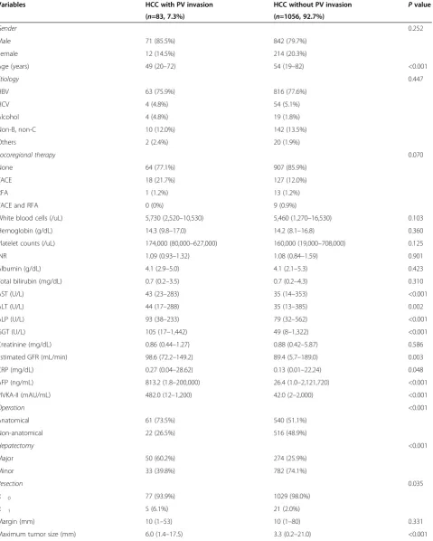
Table 1 Characteristics of HCC patients with and without portal vein invasion
Variables HCC with PV invasion HCC without PV invasion Pvalue
(n=83, 7.3%) (n=1056, 92.7%)
Gender 0.252
Male 71 (85.5%) 842 (79.7%)
Female 12 (14.5%) 214 (20.3%)
Age (years) 49 (20–72) 54 (19–82) <0.001
Etiology 0.447
HBV 63 (75.9%) 816 (77.6%)
HCV 4 (4.8%) 54 (5.1%)
Alcohol 4 (4.8%) 19 (1.8%)
Non-B, non-C 10 (12.0%) 142 (13.5%)
Others 2 (2.4%) 20 (1.9%)
Locoregional therapy 0.070
None 64 (77.1%) 907 (85.9%)
TACE 18 (21.7%) 127 (12.0%)
RFA 1 (1.2%) 13 (1.2%)
TACE and RFA 0 (0%) 9 (0.9%)
White blood cells (/uL) 5,730 (2,520–10,530) 5,460 (1,270–16,530) 0.103
Hemoglobin (g/dL) 14.3 (9.8–17.0) 14.2 (8.1–16.8) 0.360
Platelet counts (/uL) 174,000 (80,000–627,000) 160,000 (19,000–708,000) 0.125
INR 1.09 (0.93–1.32) 1.08 (0.84–1.59) 0.901
Albumin (g/dL) 4.1 (2.9–5.0) 4.1 (2.1–5.3) 0.423
Total bilirubin (mg/dL) 0.7 (0.2–3.5) 0.7 (0.2–4.3) 0.310
AST (U/L) 43 (23–283) 35 (14–353) <0.001
ALT (U/L) 44 (17–288) 35 (13–385) 0.002
ALP (U/L) 93 (38–233) 79 (32–562) <0.001
GGT (U/L) 105 (17–1,442) 49 (8–1,322) <0.001
Creatinine (mg/dL) 0.86 (0.44–1.27) 0.88 (0.42–5.87) 0.586
Estimated GFR (mL/min) 98.6 (72.2–149.2) 89.4 (5.7–189.0) 0.003
CRP (mg/dL) 0.27 (0.04–28.62) 0.13 (0.01–22.24) 0.048
AFP (ng/mL) 813.2 (1.8–200,000) 26.4 (1.0–2,121,720) <0.001
PIVKA-II (mAU/mL) 482.0 (12–1,200) 42.0 (2–2,000) <0.001
Operation <0.001
Anatomical 61 (73.5%) 540 (51.1%)
Non-anatomical 22 (26.5%) 516 (48.9%)
Hepatectomy <0.001
Major 50 (60.2%) 274 (25.9%)
Minor 33 (39.8%) 782 (74.1%)
Resection 0.035
R 0 77 (93.9%) 1029 (98.0%)
R 1 5 (6.1%) 21 (2.0%)
Margin (mm) 10 (1–53) 10 (1–80) 0.331
patients with malignant portal vein invasion did not re-ceive radiation in the preoperative period. There were no differences in gender, cause, and type of locoregional therapy between the two groups. As for laboratory data before the operation, the HCC patients with malignant portal vein invasion had higher serum aspartate amino-transferase (AST), alanine aminoamino-transferase (ALT), alka-line phosphatase (ALP), gamma-glutamyl transferase (GGT), estimated glomerular filtration rate (GFR), and C-reactive protein (CRP) levels compared with HCC pa-tients without portal vein invasion (P <0.05). However, white blood cell (WBC) count, hemoglobin, platelet count, INR, albumin, total bilirubin, direct bilirubin, and creatinine were similar in both groups. The median of AFP and PIVKA-II in HCC patients with malignant por-tal vein invasion was 813.2 ng/mL (range, 1.8–200,000 ng/mL) and 482.0 mAU/mL (range, 12–1,200 mAU/mL), respectively, but the median in HCC patients without por-tal vein invasion was 26.4 ng/mL (range, 1.0–2,121,720 ng/mL) and 42.0 mAU/mL (range, 2–2,000 mAU/mL), respectively (P<0.001 andP<0.001, respectively).
The surgery and pathologic results in HCC patients with malignant portal vein invasion
The proportion of anatomical resection and major hepa-tectomy in HCC patients with malignant portal vein in-vasions was higher than that in HCC patients without portal vein invasion (P <0.001 and P <0.001, respect-ively). Postoperative mortality was not occurred in the patients with malignant portal vein invasion. Patholo-gically proven malignant portal vein invasion consisted of segmental portal vein invasion (n=57) and left or right main portal vein invasion (n=26). Left or right main
portal vein invasion was detected in the preoperative radiology, but segmental portal vein invasion was not di-agnosed in the preoperative radiology. The median tumor size was 6 cm (range, 1.4–17.5 cm) in HCC with malignant portal vein invasion and 3.3 cm (range, 0.2– 21.0 cm) in HCC without portal vein invasion (P<0.001). The proportion of R1 resection in HCC patients with
malignant portal vein invasion was 6.1%, which was higher than that in HCC patients without portal vein in-vasion (P=0.035). None of the studied patients had distant metastasis and there was no patient with R2
re-section in this study. There were 16 cases (19.3%) of grade 3 or 4 HCC with malignant portal vein invasion and 80 (7.6%) cases of HCC without portal vein invasion. The grade in HCC with malignant portal vein invasion was poorer than that in HCC without portal vein inva-sion (P=0.001). Bile duct invainva-sion, intrahepatic metasta-sis, and the absence of capsular formation were more prevalent in HCC with portal vein invasion than in HCC without portal vein invasion (P <0.05). However, no significant difference was found in serosa involvement and multicentric occurrence between the two groups. The median hospitalization in both groups was 9 days and the duration of follow-up in HCC patients with
Table 1 Characteristics of HCC patients with and without portal vein invasion(Continued)
Grade 0.001
1 and 2 67 (80.7%) 976 (92.4%)
3 and 4 16 (19.3%) 80 (7.6%)
Cirrhosis 40 (48.2%) 468 (44.3%) 0.494
Capsule formation <0.001
None 33 (39.8%) 69 (6.6%)
Complete 35 (42.2%) 875 (83.2%)
Partial 15 (18.1%) 108 (10.3%)
Bile duct invasion 7 (8.4%) 24 (2.3%) 0.005
Serosa involvement 2 (2.4%) 12 (1.1%) 0.272
Intrahepatic metastasis 41 (49.4%) 112 (10.6%) <0.001
Multicentric occurrence 1 (1.2%) 57 (5.4%) 0.118
Hospitalization 9 (5–53) 9 (3–102) 0.932
Duration of Follow-up (months) 18 (5–68) 32 (3–82) <0.001
AFP, Alpha-fetoprotein; ALP, Alkaline phosphatase; ALT, Alanine aminotransferase; AST, Aspartate aminotransferase; CRP, C-reactive protein; GFR, Estimated glomerular filtration rate; GGT, Gamma-glutamyl transferase; HBV, Hepatitis B virus; HCV, Hepatitis C virus; INR, International normalized ratio; PIVKA-II, Protein induced by vitamin K antagonist-II; PV, Portal vein; RFA, Radiofrequency ablation; TACE, Transarterial chemoembolization.
Table 2 Risk factors for portal vein invasion in HCC by multivariate analysis
Variables Hazard ratio 95% confidence
interval P
value
ALP 1.017 1.005–1.030 0.007
Maximum tumor size 1.025 1.010–1.040 0.001
Intrahepatic metastasis 6.064 1.553–23.673 0.009
malignant portal vein invasion was 18 months (range, 5-68 months), and that in HCC with portal vein invasion was 32 months (range, 3-82 months). The duration of follow-up in HCC patients with malignant portal vein invasion was shorter than that in HCC patients without portal vein invasion because HCC patients died due to tumor recurrence in the early period after surgery.
Risk factors for malignant portal vein invasion in HCC patients
To identify risk factors for malignant portal vein inva-sion in HCC, we performed a multivariate analysis on all variables that were significantly associated with ma-lignant portal vein invasion on univariate analysis. In-creased serum ALP levels, inIn-creased maximum tumor size, and intrahepatic metastasis were the predisposing factors of malignant portal vein invasion by multivariate analysis (Table 2).
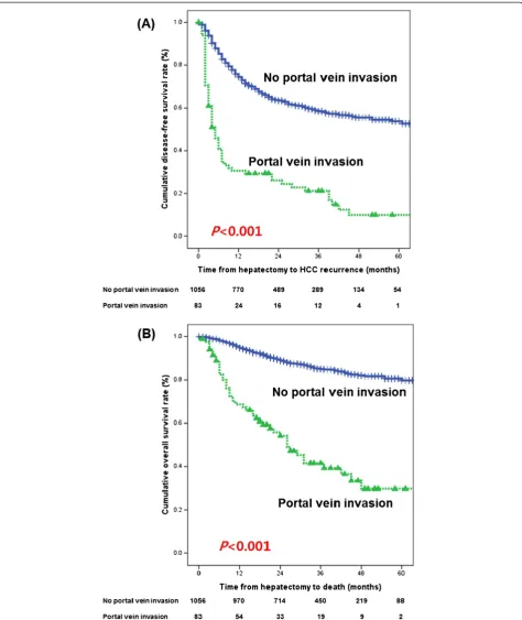
Survival rate after surgery
For the HCC patients with malignant portal vein inva-sion who underwent liver resection, the median disease-free survival was 4.5 months and the overall survival was 25 months. The 1-year, 2-year, and 3-year disease-free survival rates and overall survival rates in HCC patients with malignant portal vein invasion were 30.6%, 26.1%, and 21.2%, and 68.6%, 54.2%, and 41.6%, respectively, but those in HCC patients without portal vein invasion
were 74.4%, 63.5%, and 58.5%, and 95.5%, 89.6%, and 85.1%, respectively (P<0.001 andP<0.001, respectively) (Figure 1).
Recurrence after surgical resection
The incidence of tumor recurrence was 80.7% (67/83) in HCC patients with malignant portal vein invasion and 38.8% (410/1056) in HCC without portal vein invasion. The initial detection site was lung (50.7%) in HCC pa-tients with malignant portal vein invasion and liver Table 3 Initial detection site for recurrence after surgical
resection
Liver 18 (26.9%) 254 (62.0%) <0.001
Lung 34 (50.7%) 95 (23.2%) <0.001
Bone 7 (10.4%) 22 (5.4%) 0.161
Brain 0 (0%) 5 (1.2%) 0.364
Peritoneum 8 (11.9%) 34 (8.3%) 0.351
Table 4 Treatments for HCC recurrence
Recurrence
Liver resection 0 (0%) 18 (4.4%) 0.091
Liver transplantation 3 (4.5%) 7 (1.7%) 0.154
RFA 6 (9.0%) 125 (30.5%) <0.001
TACE 39 (58.2%) 242 (59.0%) 0.894
Sorafenib 12 (17.9%) 67 (16.3%) 0.725
Radiation 9 (13.4%) 42 (10.2%) 0.400
Chemotherapy 5 (7.5%) 23 (5.6%) 0.573
RFA, Radiofrequency ablation; TACE, Transarterial chemoembolization.
Table 5 Risk factors for tumor recurrence in HCC patients with portal vein invasion by univariate analysis
Variables Hazard ratio 95% confidence
interval P
value
Gender - Female 0.657 0.313–1.376 0.265
Age 0.973 0.946–1.000 0.052
AFP 1.000 1.000–1.000 0.005
PIVKA-II 1.001 1.000–1.001 0.128
Locoregional therapy 1.495 0.848–2.634 0.165
White blood cells 0.961 0.835–1.107 0.582
Hemoglobin 1.010 0.853–1.195 0.909
Platelet counts 1.000 0.998–1.003 0.821
INR 6.056 0.254–144.219 0.266
Albumin 0.533 0.290–0.980 0.043
Total bilirubin 1.047 0.654–1.676 0.849
AST 1.005 0.999–1.011 0.103
ALT 1.002 0.996–1.008 0.565
ALP 1.002 0.995–1.008 0.643
Creatinine 0.427 0.091–2.004 0.281
Estimated GFR 1.015 0.997–1.033 0.102
GGT 1.000 0.998–1.001 0.739
CRP 1.133 1.028–1.249 0.012
Operation (non-anatomical) 1.473 0.863–2.515 0.156
Hepatectomy (minor) 0.922 0.367–2.315 0.863
Maximum tumor size 1.003 0.997–1.010 0.282
Grade (3 and 4) 0.627 0.311–1.267 0.194
Cirrhosis 1.426 0.879–2.314 0.150
Capsule formation 1.020 0.353–2.941 0.971
Capsular invasion 0.715 0.439–1.165 0.178
Bile duct invasion 0.752 0.302–1.873 0.540
Serosa involvement 5.863 1.372–25.058 0.017
Intrahepatic metastasis 1.787 1.098–2.910 0.020
Multicentric occurrence 0.048 0.00–195.191 0.474
Margin (mm) 1.006 0.986–1.026 0.566
AFP, Alpha-fetoprotein; ALP, Alkaline phosphate; ALT, Alanine
(62.0%) in HCC patients without portal vein invasion. There was a difference in liver or lung as the initial de-tection site of tumor recurrence between the two groups (P<0.001 andP<0.001, respectively) (Table 3). However, no significant difference was found in the recurrence in bone, brain, and peritoneum between the two groups.
Radiofrequency ablation (RFA) in HCC patients without portal vein invasion was used more often because of high incidence of recurrence in liver, compared with HCC patients with malignant portal vein invasion (P <0.001). No HCC patients with portal vein invasion were treated with liver resection due to recurrence. Liver transplant-ation, TACE, sorafenib, raditransplant-ation, and chemotherapy as treatments for tumor recurrence were similar in both groups (Table 4).
Prognostic factors for recurrence in HCC with malignant portal vein invasion after surgery
Univariate analysis showed that increased AFP level, de-creased serum albumin level, serosa involvement, and in-trahepatic metastasis were closely associated with tumor recurrence after liver resection (Table 5). On multivariate analysis of the prognostic factors for disease-free survival in HCC patients with malignant portal vein invasion, only the CRP level (hazard ratio, 1.133; 95% confidence inter-val, 1.028–1.249; P=0.012) was a significant independent predictor of tumor recurrence after liver resection.
Discussion
The prognosis for patients who have HCC with malignant portal vein invasion remained extremely poor in this study, even though a few long-term survivors were ob-served. Several studies have shown that tumor size, high grade, the presence of fibrous capsule, and platelet counts are strongly associated with portal vein invasion [8-10]. In the present study, increased ALP levels, increased max-imum tumor size, and intrahepatic metastasis were inde-pendent predictors of malignant portal vein invasion in HCC by multivariate logistic regression analysis. There-fore, these factors may be helpful in predicting malignant portal vein invasion before surgery for HCC.
Many studies reported that an increase in the serum CRP level was associated with a shorter survival in pa-tients with various malignancies, including multiple myeloma [11], ovarian cancer [12], colorectal [13], and esophageal cancer [14]. For the serum CRP levels associ-ated with HCC, high CRP was linked to the diffuse type of HCC and elevated preoperative CRP levels in patients with HCC associated with tumor size and portal vein in-vasion, and predicted recurrence after curative resection and a poor surgical outcome [15,16].
The basis for the relationship between elevated CRP and poor prognosis is unclear and there are several pos-sible explanations. An elevated CRP level may reflect a
non-specific inflammatory response to tumor necrosis or local tissue damage, which may be indicative of a fa-vorable environment for the establishment and growth of tumor. The serum level of vascular endothelial growth factor (VEGF), an angiogenic factor, is increased in the presence of raised CRP concentration [17]. Angiogenesis plays an important role in tumor growth and is associated with a poor outcome [18]. This creates a microenvi-ronment that favors tumor angiogenesis, proliferation, growth, and metastases.
Tumor invasion in the remnant liver after surgery is common in HCC patients with malignant portal vein inva-sion [19]. However, our study revealed that lung metasta-sis was common in HCC patients with malignant portal vein invasion after surgery, while liver metastasis was common in HCC patients without portal vein invasion.
Although intrahepatic recurrence predominates, prob-ably because of the early spread of neoplasm and me-tachronous multicentric carcinogenesis, several effective therapeutic modalities can control recurrent disease (such as repeated hepatectomy, TACE, percutaneous ethanol in-jection therapy, microwave coagulation therapy, and RFA) [16,20]. Compared with frequent intrahepatic recurrence, the incidence of distant metastasis is relatively low in HCC [21]. However, extrahepatic metastasis, such as lung, was higher than intrahepatic metastasis in HCC with por-tal vein invasion. The poor prognosis for patients with ex-trahepatic metastasis of HCC occurs because it is an indicator of an aggressive primary HCC [22].
Our study revealed that chest CT must be regularly performed in HCC patients with malignant portal vein invasion after surgical resection. In addition, patients who had preoperatively elevated CRP levels, should be closely monitored after hepatectomy because of a poor prognostic factor of tumor recurrence.
Conclusion
In conclusion, our study reveals that increased ALP levels, increased maximum tumor size, and intrahepatic metasta-sis were independent predictors of malignant portal vein invasion in HCC. CRP levels are closely associated with the predisposing factor of tumor recurrence in HCC pa-tients with malignant portal vein invasion after surgical re-section, and lung metastasis is common.
Competing interests
The authors declare no conflicts of interest.
Authors’contribution
JMK: data collection, analysis, and interpretation, manuscript writing; JWJ and SWP: data design and interpretation; CHDK, JSK: data collection and analysis; JBP, JHL, SJK, and CKP: data collection. All authors read and approved the final manuscript.
Acknowledgements
subject matter or materials discussed in the manuscript. No writing assistance was utilized in the production of this manuscript.
Author details
1Department of Surgery, Samsung Medical Center, Sungkyunkwan University School of Medicine, #50 Ilwon-Dong Gangnam-Gu, Seoul 135-710, Korea. 2Department of Anesthesiology and Pain Medicine, Samsung Medical Center, Sungkyunkwan University School of Medicine, Seoul 135-710, Korea.3Division of Gastroenterology, Department of Medicine, Samsung Medical Center, Sungkyunkwan University School of Medicine, Seoul 135-710, Korea. 4Department of Pathology, Samsung Medical Center, Sungkyunkwan University School of Medicine, Seoul 135-710, Korea.
Received: 13 October 2012 Accepted: 26 March 2013 Published: 23 April 2013
References
1. Song IH, Kim KS:Current status of liver diseases in Korea: hepatocellular carcinoma.Korean J Hepatol2009,Suppl 6:S50–S59.
2. Llovet JM, Burroughs A, Bruix J:Hepatocellular carcinoma.Lancet2003, 362:1907–1917.
3. Matsumata T, Kanematsu T, Takenaka K, Yoshida Y, Nishizaki T, Sugimachi K: Patterns of intrahepatic recurrence after curative resection of hepatocellular carcinoma.Hepatology1989,9:457–460.
4. Jonas S, Bechstein WO, Steinmuller T, Herrmann M, Radke C, Berg T, Settmacher U, Neuhaus P:Vascular invasion and histopathologic grading determine outcome after liver transplantation for hepatocellular carcinoma in cirrhosis.Hepatology2001,33:1080–1086.
5. Villa E, Moles A, Ferretti I, Buttafoco P, Grottola A, Del Buono M, De Santis M, Manenti F:Natural history of inoperable hepatocellular carcinoma: estrogen receptors’status in the tumor is the strongest prognostic factor for survival.Hepatology2000,32:233–238.
6. Forner A, Reig ME, de Lope CR, Bruix J:Current strategy for staging and treatment: the BCLC update and future prospects.Semin Liver Dis2010, 30:61–74.
7. Edmondson HA, Steiner PE:Primary carcinoma of the liver: a study of 100 cases among 48,900 necropsies.Cancer1954,7:462–503.
8. Miyata R, Tanimoto A, Wakabayashi G, Shimazu M, Nakatsuka S, Mukai M, Kitajima M:Accuracy of preoperative prediction of microinvasion of portal vein in hepatocellular carcinoma using superparamagnetic iron oxide-enhanced magnetic resonance imaging and computed tomography during hepatic angiography.J Gastroenterol2006, 41:987–995.
9. Adachi E, Maeda T, Kajiyama K, Kinukawa N, Matsumata T, Sugimachi K, Tsuneyoshi M:Factors correlated with portal venous invasion by hepatocellular carcinoma: univariate and multivariate analyses of 232 resected cases without preoperative treatments.Cancer1996, 77:2022–2031.
10. Hagiwara S, Kudo M, Kawasaki T, Nagashima M, Minami Y, Chung H, Fukunaga T, Kitano M, Nakatani T:Prognostic factors for portal venous invasion in patients with hepatocellular carcinoma.J Gastroenterol2006, 41:1214–1219.
11. Terpos E, Szydlo R, Apperley JF, Hatjiharissi E, Politou M, Meletis J, Viniou N, Yataganas X, Goldman JM, Rahemtulla A:Soluble receptor activator of nuclear factor kappaB ligand-osteoprotegerin ratio predicts survival in multiple myeloma: proposal for a novel prognostic index.Blood2003, 102:1064–1069.
12. Hefler LA, Concin N, Hofstetter G, Marth C, Mustea A, Sehouli J, Zeillinger R, Leipold H, Lass H, Grimm C, Tempfer CB, Reinthaller A:Serum C-reactive protein as independent prognostic variable in patients with ovarian cancer.Clin Cancer Res2008,14:710–714.
13. Nozoe T, Matsumata T, Kitamura M, Sugimachi K:Significance of preoperative elevation of serum C-reactive protein as an indicator for prognosis in colorectal cancer.Am J Surg1998,176:335–338. 14. Shimada H, Nabeya Y, Okazumi S, Matsubara H, Shiratori T, Aoki T, Sugaya
M, Miyazawa Y, Hayashi H, Miyazaki S, Ochiai T:Elevation of preoperative serum C-reactive protein level is related to poor prognosis in esophageal squamous cell carcinoma.J Surg Oncol2003,83:248–252. 15. Lin ZY, Wang LY, Yu ML, Chen SC, Chuang WL, Hsieh MY, Tsai JF, Chang WY:
Role of serum C-reactive protein as a marker of hepatocellular carcinoma in patients with cirrhosis.J Gastroenterol Hepatol2000,15:417–421.
16. Hashimoto K, Ikeda Y, Korenaga D, Tanoue K, Hamatake M, Kawasaki K, Yamaoka T, Iwatani Y, Akazawa K, Takenaka K:The impact of preoperative serum C-reactive protein on the prognosis of patients with
hepatocellular carcinoma.Cancer2005,103:1856–1864.
17. Xavier P, Belo L, Beires J, Rebelo I, Martinez-de-Oliveira J, Lunet N, Barros H: Serum levels of VEGF and TNF-alpha and their association with C-reactive protein in patients with endometriosis.Arch Gynecol Obstet2006, 273:227–231.
18. Fondevila C, Metges JP, Fuster J, Grau JJ, Palacin A, Castells A, Volant A, Pera M:p53 and VEGF expression are independent predictors of tumour recurrence and survival following curative resection of gastric cancer. Br J Cancer2004,90:206–215.
19. Chen XP, Qiu FZ, Wu ZD, Zhang ZW, Huang ZY, Chen YF, Zhang BX, He SQ, Zhang WG:Effects of location and extension of portal vein tumor thrombus on long-term outcomes of surgical treatment for hepatocellular carcinoma.Ann Surg Oncol2006,13:940–946. 20. Minagawa M, Makuuchi M, Takayama T, Kokudo N:Selection criteria for
repeat hepatectomy in patients with recurrent hepatocellular carcinoma. Ann Surg2003,238:703–710.
21. Hong SS, Kim TK, Sung KB, Kim PN, Ha HK, Kim AY, Lee MG:Extrahepatic spread of hepatocellular carcinoma: a pictorial review.Eur Radiol2003, 13:874–882.
22. Uchino K, Tateishi R, Shiina S, Kanda M, Masuzaki R, Kondo Y, Goto T, Omata M, Yoshida H, Koike K:Hepatocellular carcinoma with extrahepatic metastasis: clinical features and prognostic factors.Cancer2011, 117:4475–4483.
doi:10.1186/1477-7819-11-92
Cite this article as:Kimet al.:C-reactive protein may be a prognostic factor in hepatocellular carcinoma with malignant portal vein invasion.
World Journal of Surgical Oncology201311:92.
Submit your next manuscript to BioMed Central and take full advantage of:
• Convenient online submission
• Thorough peer review
• No space constraints or color figure charges
• Immediate publication on acceptance
• Inclusion in PubMed, CAS, Scopus and Google Scholar
• Research which is freely available for redistribution