University of South Carolina
Scholar Commons
Theses and Dissertations
2015
Bacterial Communication and its Role as a Target
for Nanoparticle-Based Antimicrobial Therapy
Kristen Publicover Miller University of South Carolina - Columbia
Follow this and additional works at:https://scholarcommons.sc.edu/etd
Part of theEnvironmental Health Commons
This Open Access Dissertation is brought to you by Scholar Commons. It has been accepted for inclusion in Theses and Dissertations by an authorized administrator of Scholar Commons. For more information, please contactdillarda@mailbox.sc.edu.
Recommended Citation
B
ACTERIALC
OMMUNICATION AND ITS ROLE AS A TARGET FOR NANOPARTICLE -BASED ANTIMICROBIAL THERAPYby
Kristen Publicover Miller
Bachelor of Science Clemson University, 2009
Master of Science Clemson University, 2010
Submitted in Partial Fulfillment of the Requirements
For the Degree of Doctor of Philosophy in
Environmental Health Sciences
The Norman J. Arnold School of Public Health
University of South Carolina
2015
Accepted by:
Alan Decho, Major Professor
Brian Benicewicz, Committee Member
John Ferry, Committee Member
Jamie Lead, Committee Member
Sean Norman, Committee Member
© Copyright by Kristen Publicover Miller, 2015
D
EDICATIONThis dissertation is lovingly dedicated to my parents, Nancy and Jeffery
Publicover. Their love, support, and years of encouragement have been invaluable in the
completion of this degree. I must also dedicate this work to my brother, Daniel
Publicover, for showing me how to find joy in one’s profession and for his love, support,
and humor. Lastly, this dissertation is dedicated to my husband, Philip Miller, who has
A
CKNOWLEDGEMENTSI would like to thank my committee, Dr. Brian Benicewicz, Dr. John Ferry, Dr.
Jamie Lead, and Dr. Sean Norman for their advice and expertise throughout my
dissertation research. I would especially like to thank Dr. Alan Decho, my major advisor
and committee chair, for his support and dedication to my graduate education.
In addition to my committee, I would like to thank the Department of
Environmental Health Sciences for their support and generosity. I would also like to
thank the many students and staff in this department, in the Department of Chemistry and
Biochemistry, and at the University of South Carolina who made my dissertation research
A
BSTRACTThe goal of this dissertation is to establish the environmental and public health
importance of alternative forms of antimicrobial therapy, specifically those that utilize
nanotechnology to combat quorum sensing-controlled bacterial infections. Quorum
sensing (i.e. chemical communication) is an inherent characteristic that is essential to
bacterial pathogenesis and biofilm formation (where most infections occur). A thorough
review of the literature has been conducted to establish an understanding of the state of
nanotechnology research as it relates to combatting bacterial infections. This synthesis,
provided in Chapter 1, demonstrates how the chemical and structural designs of
nanoparticles can be manipulated to specifically target bacterial infections.
Next, an investigation into the development and novel use of nanoparticles
engineered to shut down bacterial quorum sensing is given in Chapter 2. Inhibiting the
quorum sensing process is significant because it does not kill the bacteria, and therefore
does not exacerbate antibiotic resistance. Briefly, the model system demonstrates that
-cyclodextrin functionalized nanoparticles are able to persist in the bacterial cell
environment and quench extracellular bacterial communication molecules, and
effectively silence bacterial communication. The system neutralizes communication
through chelation of common signaling molecules called acyl-homoserine lactones. The
new technology described here provides a seminal step in developing anti-virulence
antimicrobials. Also, this technology utilizes non-toxic nanoparticles that can be
functionalized with biologically-active compounds and tailored to meet specific needs.
This study provides a scaffold and critical stepping stone that will promote more-tailored
future developments in nanoparticle-based antimicrobial therapy.
Chapter 3 provides insight into the environmental importance of bacterial
communication, and the steps taken by bacteria to protect the valuable signal molecules.
Briefly, environmental biofilms consist of extracellular polymeric substances with a high
concentration of nonreducing sugars, such as trehalose. Previous studies have shown that
trehalose is commonly utilized by soil bacteria during periods of drought to maintain
membrane stability and preserve the structure of proteins. The study presented in
Chapter 3 demonstrates that trehalose plays a role in protecting quorum sensing signals
during desiccation through the formation of an extracellular glass. Additionally, the study
provides a survey of the complexity of microbial ecosystems and the role that biofilm
components play in the natural environment. Together, the three chapters of this
dissertation demonstrate the importance of quorum sensing to bacteria and as a target for
T
ABLE OFC
ONTENTSDEDICATION ... iii
ACKNOWLEDGEMENTS ... iv
ABSTRACT ...v
LIST OF TABLES ... ix
LIST OF FIGURES ...x
CHAPTER 1: ENGINEERED NANOPARTICLES TO ATTACK BACTERIA ...1
1.1 NANOPARTICLES AS DRUG CARRIERS: A BRIEF HISTORICAL PERSPECTIVE ...2
1.2 THE ANTIBIOTIC DILEMMA: THE MODE OF DELIVERY COUNTS ...3
1.3 DIRECT AND INDIRECT ANTIMICROBIAL PROPERTIES OF INORGANIC NANOPARTICLES ...15
1.4 LINKING ANTIBIOTICS TO INORGANIC NANOPARTICLES: CHALLENGES IN SURFACE CHEMISTRY DESIGN ...25
1.5 OVERCOMING THE BACTERIAL BARRIERS OF INFECTIONS: NANOPARTICLES AS ANTIMICROBIAL DELIVERY VEHICLES ...45
1.6 CONCLUSION ...53
CHAPTER 2: FUNCTIONALIZED NANOPARTICLES SILENCE BACTERIAL COMMUNICATION ....61
2.1 CHARACTERIZATION OF MONOLAYER NANOPARTICLES ...63
2.2 CHARACTERIZATION OF POLYMER NANOPARTICLES ...64
2.3 NMR DETECTS BINDING OF HSLS AND -CYCLODEXTRIN ...64
2.6 CONCLUSION ...71
2.7 METHODS ...73
CHAPTER 3: EXTRACELLULAR GLASS WITHIN THE EPS MATRIX IS A PROTECTIVE STRATEGY FOR BIOFILMS AGAINST DESICCATION ...92
3.1 RESULTS ...97
3.2 DISCUSSION ...106
3.3 CONCLUSION ...108
3.4 MATERIALS AND METHODS...109
L
IST OFT
ABLESTable 1.1 Description of major endocytosis pathways ...54
Table 2.1 Measured and corrected diffusion coefficients (D) for various mixtures of C8-HSL and cyclodextrin. ...78
Table 2.2 Binding strength of -CD and C6-HSL or C8-HSL as determined by NMR ....78
Table 2.3 Calculated fold change of transcription by V. fischeri after treatment exposures to 125 nM 3OC6- and 0.25 nM C8-HSLs ...79
Table 2.4 Primer sequences used in qPCR ...79
Table 3.1 Composition of sugar monomers within EPS extracted from natural mats ...113
Table 3.2 C6-AHL activity in trehalose as determined by pigmentation response of
C. violaceum CV026. ...114
L
IST OFF
IGURESFigure 1.1 Diagram of cell walls of Gram-positive and Gram-negative bacteria ...54
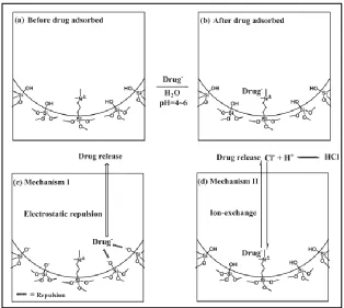
Figure 1.2 Schematic illustration showing the loading and release mechanism of an anionic drug in TA modified MSN ...55
Figure 1.3 (a) Schematic illustration showing the controlled drug release of a pNIPAAm-grafted Au nanocage; (b) Atom-transfer radical polymerization of NIPAAm and acrylic amide monomers ...56
Figure 1.4 Major endocytosis pathways ...57
Figure 1.5 Schematic illustration of multidrug release by MSN-polymer nanocomposite ...57
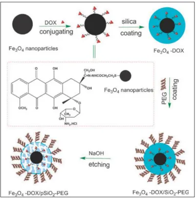
Figure 1.6 Schematic illustration of the synthesis of DOX-associated Fe3O4 nanoparticles coated with a PEG modified porous silica shell ...58
Figure 1.7 Schematic illustration of the synthesis of core-shell drug delivery vehicle ...59
Figure 1.8 Schematic illustration showing the one-pot self-assembly strategy for synthesis of drugs@micelles@MSNs ...59
Figure 1.9 Schematic of biofilm encased in extracellular polymeric secretions ...60
Figure 2.1 N-acyl homoserine lactone ...80
Figure 2.2-cyclodextrin chemical (A) and toroidal (B) structure ...80
Figure 2.3 Schematic representation of monolayer -CD coated fluorescent silicon dioxide nanoparticle ...80
Figure 2.4 1H NMR spectra of the as-synthesized -CD coated silica nanoparticles ...81
Figure 2.5 TGA of (a) dye-labeled monolayer carboxylic acid coated silica nanoparticles; (b) dye-labeled monolayer -CD coated silica nanoparticles ...82
Figure 2.7 TGA of (a) dye-labeled poly(methacrylic acid) grafted silica nanoparticles; (b) dye-labeled poly(-CD) grafted silica nanoparticles ...84
Figure 2.8 Photograph of dye-labeled poly(-CD) grafted silica nanoparticles in
DMSO ...85
Figure 2.9 N-Acyl homoserine lactone molecules synthesized and recognized by
V. fischeri ...85
Figure 2.10 V. fischeri JB10 cultures in marine broth. Bioluminescence induced with 3OC6-HSL and C8-HSL ...86
Figure 2.11 Maximum relative bioluminescence per OD (600 nm) of V. fischeri during exposure to -CD and 2 μM 3OC6-HSL ...87
Figure 2.12 Schematic of nanoparticle-based silencing of bacterial quorum sensing ...88
Figure 2.13 Changes in bioluminescence by V. fischeri during exposures to 2 μM
3OC6-HSL, with either -CD or -CD functionalized Si-NPs ...89
Figure 2.14 Maximum relative bioluminescence per OD (600 nm) of V. fischeri during exposure to 2 μM 3OC6-HSL and -CD or -CD functionalized 15 nm polymer
nanoparticles ...90
Figure 2.15 Mean relative bioluminescence per OD (600 nm) of V. fischeri during exposure to 125 nM 3OC6-HSL and 0.25 nM C8-HSL treated with 250 nM -CD, bare 15 nm Si-NPs, 155 nM -CD functionalized 15 nm Si-NPs, bare 50 nm Si-NPs, or 133 nM -CD functionalized 50 nm Si-NPs ...91
Figure 3.1 Natural microbial mats collected from Salt Pond on San Salvador Island, Bahamasshowing wet- (A) and dry- (B, C) season sections of mat surface ...115
Figure 3.2 SEM of dried surface layer of microbial mat ...116
Figure 3.3 TEM of dry, natural microbial mats showing intact cells surrounded by a dense capsular layer of EPS ...116
Figure 3.4 Vertical cross-section of microbial mat in Salt Pond showing distinct layering of microbial groups ...117
Figure 3.5Abundances of EPS isolated from surface layers of hypersaline mat in Salt Pond, San Salvador Island, Bahamas ...117
Figure 3.7 DSC thermogram recorded upon heating of trehalose anhydrous at rate of 10°C/minute ...119
Figure 3.8 DSC thermogram recorded upon heating of Salt Pond EPS at rate of
10°C/minute ...120
Figure 3.9 TGA thermogram of trehalose dihydrate recorded upon heating at rate of 10°C/minute ...121
Figure 3.10 TGA thermogram of Salt Pond EPS recorded upon heating at rate of
10°C/minute ...122
CHAPTER
1
ENGINEERED NANOPARTICLES TO ATTACK BACTERIA
The seminal realization of the nanotechnology, and its potential to be manipulated at
molecular and atomic scales developed over a half century ago.1 However, largely due to
a lack of technical capability, a time of obtuse contemplation followed. During this initial
lull in nano-based research, the 1950s and 60s saw a surge of attention in the
development of antibiotics – the wonder bullet cited to stop all bacterial disease.
However, this phenomenon was quickly tempered by a rapid emergence of antibiotic
resistance (AR) among pathogens, often in the form of multi-drug resistance (MDR). AR
continues to grow today, and at present, the future utility of traditional antibiotics remains
questionable.
The surge in nanoresearch, coupled with dramatic increases in technical
capabilities, has allowed the disparate fields of nanochemistry and antibiotics to come
together. Traditionally, nanoparticles and nanomaterials have been developed as carriers
to deliver anticancer and other forms of drugs to eukaryote cells. Given this foundation,
the delivery of antibiotics using engineered nanoparticles has become an emerging and
realistic area of research. Overcoming the problem of antibiotic resistance, however,
microorganisms and the physical chemistry of nanoparticles. This understanding is
necessary in order to ultimately target bacteria, and overcome their immense arsenal of
defenses. This review centers on fundamental, RAFT-based and timed-release surface
chemistry, which is currently being developed for nanoparticle delivery of antibiotics to
bacteria. We also integrate approaches being developed to enhance the detection and
quantification of bacteria within biological systems. Concurrent with this chemistry is a
necessary overview of certain bacterial processes, as these directly and indirectly interact
with the chemistry of nanoparticles.
1.1 NANOPARTICLES AS DRUG CARRIERS: A BRIEF HISTORICAL
PERSPECTIVE
Credited for conceiving the idea of a “magic bullet” to selectively target toxic
organisms in the body, Paul Ehrlich inspired many pioneers of the nanoparticle field. In
the 1950s and 60s, Peter Speiser’s group worked on the development of polyacrylic
beads,2,3 then microcapsules,4 and eventually the first nanocapsules.5 Their ultimate goal
was to achieve sustained drug release from nanocapsules in the blood after intravenous
injection.
Since that time, nanoparticles have been used for pharmaceutical and medical
applications, mainly for cancer treatment6 and enhancing the efficacy and targeting of
cancer drugs.7,8 This has allowed for the use of lower concentrations of highly-toxic
drugs in an effort to reduce side effects.9 More recently, and in conjunction with the
important discovery that polyethylene glycol chains on nanoparticles prolong blood
nanoparticles to cross the blood brain barrier, and target deep brain tumors or
infections.11-14 The innovative research of the past 50 years has reinforced the roles of
nanoparticles as drug delivery vehicles and has inspired the current diversity in
nanoparticle research. Today, investigators focus their nanoparticle research on
recognition, sensing, imaging, and delivery in biological systems with a broad range of
core materials.
1.2 THE ANTIBIOTIC DILEMMA: THE MODE OF DELIVERY COUNTS
Antibiotics are molecules produced by microorganisms to inhibit other
microorganisms. Several major classes of antibiotics, based on their chemical structure
and mechanisms of actions, are currently known. Since bacteria produce antibiotics,
inherently they also possess mechanisms to resist their inhibitory actions, which can be
passed on to nearby cells. Bacteria have also demonstrated resistance towards the
naturally antimicrobial compounds used to construct NPs, such as silver and copper. This
section addresses the molecular structure of antibiotics, and provides a basis for
understanding their key activity sites on molecules, and how these relate to their
inhibitory action(s). It sets a stage for why and how NPs can be used to enhance delivery
and activities of antibiotics.
Chemistry of antibiotics: structure and design
Selman Waksman coined the term ‘antibiotic’ to describe any small molecule
made by a microbe intended to antagonize the growth of another microbe.15 Waksman,
antibiotics. Although we have been aware of the healing nature of moldy foods for
thousands of years, it was not until the 19th and 20th centuries that scientists began to
understand that the provenance of antibiotics was related to the antagonism between two
microorganisms. In 1928, British scientist Alexander Fleming witnessed the fungus
Penicillium notatum produce a compound, that he named penicillin, which prevented the
growth of the bacterium Staphylococcus aureus. By 1939, Florey and Chain manipulated
P. notatum to produce large quantities of a stable form of penicillin.16 The cooperation of
scientists from Great Britain and the United States resulted in clinical trials and the mass
production of penicillin during the final, crucial years of World War II. Also during this
period, Waksman and colleagues isolated several antibiotics from filamentous,
soil-dwelling bacteria of the genus Actinomycetes.
Since the groundbreaking work of the 1940s, nearly 25,000 natural, synthetic, and
semisynthetic antimicrobials have been discovered. 20,000 of these are naturally
occurring drugs produced by soil-dwelling fungi and bacteria.17 Although there is an
excess of available bioactive and toxic compounds, less than 1% are clinically useful.
There are two major classes of antibiotics: bactericidal and bacteriostatic. Bactericidal
antibiotics, like the -lactam compounds penicillin and cephalosporin, kill bacteria.
Bacteriostatic antibiotics, like tetracycline and its many derivatives, inhibit the
proliferation of bacteria. Compounds that act on both Gram-positive and Gram-negative
bacteria are classified as broad-spectrum antibiotics. Compounds that act on only a
specific group of bacteria are narrow-spectrum antibiotics. The most studied and relied
to pathogens. For the purpose of this review, we will briefly discuss the pharmacokinetic
and pharmacodynamic properties of the major classes of antibiotics.
Major classes of antibiotics and mechanisms of action
Pharmacokinetics describes the effects of the body on the actions of a drug. The
pharmacokinetics of an antibiotic dictates its absorption, distribution, metabolism, and
elimination by the body. Non-ionized molecules are more lipid soluble than ionized
molecules and can readily diffuse across the cell membrane. Since ionized molecules are
less able to penetrate the lipid membrane; their entry depends on the leakiness of the cell
membrane, as related to electrical resistance. The transmembrane distribution of a weak
electrolyte is influenced by its pKa and the pH gradient across the membrane, where pKa
is the pH at which half of the compound is in its ionized form. Antibiotics rely on passive
transport (paracellular transport or diffusion) or active transport (facilitated diffusion or
drug transporters) to cross cellular barriers. Due to the presence of a carboxyl group
(essentially a proton donor), -lactams are considered weak acids and do not readily cross
the cell membrane (of mammalian or bacterial cells).18
To understand the pharmacokinetic transport of an antibiotic to a bacterial cell, it
is important to briefly consider the fundamental structural differences between
Gram-negative and Gram-positive bacteria (Figure 1.1).
Pharmacodynamics refers to the study of the biochemical and physiological
effects of drugs and their mechanism of action. In the case of antibiotics, the barriers
inflicted by human anatomy, the chemical properties of the antibiotic, and the presence of
efficacy of antimicrobial therapy. The penetration of physical barriers by antibiotics is
directly related to the octanol-water partition coefficient (Kow) of the antibiotic.
Generally, hydrophobic drugs (with high Kows) become concentrated in the lipid
membrane of cells, while hydrophilic drugs typically concentrate in aqueous areas such
as the blood or cytosol. Additionally, the larger and more negatively charged an antibiotic
is, the more difficult it is for that drug to penetrate cell membranes and physical
barriers.20
The extracellular concentration of an antibiotic directly affects the intracellular
concentration in a concentration- and time-dependent fashion,21-24 depending on the drug
and microorganism. An antibiotic intended to target an intracellular infection must cross
the cell membrane and retain activity in the cytosol.25 The equilibrium of free and bound
(to protein, stabilizing agent, serum, etc) antibiotics directly alters active transport, pH
partitioning, and accumulation in the host, and therefore influences its ability to cross the
cell membrane and target an intracellular infection.26 Additionally, the intracellular
activity of antibiotics is largely influenced by the pH of the vacuoles and lysosomes in
which it becomes localized, and the level of bacterial (i.e. growth rate, presence of
persister cells, and small colony variants27-29) and cellular activity.25 Alternatively, the
antibiotic oritavancin (i.e. a semi-synthetic glycopeptide) is unaffected by pH, but shows
decreased intracellular activity, likely due to binding with intra-lysosomal constituents.23
-lactams
To date, -lactam compounds are the most important broad-spectrum antibiotics
fungal-derived drugs include penicillin and cephalosporin. Simply put, -lactams bind
irreversibly to enzymes intended to catalyze the transpeptidation of the peptidoglycan
layer of the bacterial cell wall. -lactams prevent proper reconstruction of a cell wall,
eventually weakening and lysing the bacterial cell.30-31
More specifically, -lactams are a large class of drugs that primarily target the
penicillin-binding proteins (PBPs) required for bacterial cell wall synthesis. This class is
characterized by a highly reactive, four-membered carbonyl lactam ring containing
azetidinone32 and includes penicillins, narrow- and extended-spectrum cephalosporins,
monobactams and carbapenems.33-lactams target peptidoglycan biosynthesis by
blocking the insertion of glycan units into the cell wall to inhibit cell wall development
and by blocking transpeptidation linking and maturation.34 Peptidoglycan, found in both
Gram-negative and Gram-positive bacteria, is an integral part of the cell wall that is
required by bacteria to maintain the structural integrity of the cell and to survive changes
in environmental osmotic pressure.35-lactams form a covalent complex with enzymes
(the PBPs) that generate the mature peptidoglycan molecule. The -lactam antibiotic,
containing a cyclic amide ring, mimics the D-alanyl-D-alanine region of the PBP, forms a
covalent acyl-enzyme complex with the activated serine of the PBP that inhibits the
peptide bond formation reaction catalyzed by PBPs, and is mistakenly incorporated into
the cell wall.30-31, 36 This complex inhibits transpeptidation activity and weakens the
integrity of the cell wall, eventually resulting in cell lysis.36 Specifically, the PBP
inhibition occurs by penicilloylation of the active site, which blocks the hydrolysis of the
bond created with the now open-ringed drug and disables the enzyme.32, 37-38 A review by
such as cell division, autolysin activity, the SOS response, the TCA cycle, Fe-S cluster
synthesis, ROS formation, and envelope and redox-responsive two-component systems.32
Although -lactam antibiotics are highly effective against susceptible bacteria,
they are most useful against extracellular infections within the body. Intracellular
pathogens, such as Listeriamonocytogenes and Salmonella species, are protected from
the immune system by phagocytes and are less accessible to these antibiotics. Reports
show poor accumulation of -lactams in phagocytic and nonphagocytic cells, possibly
due to the acidic conditions of the cytosol and the presence of multidrug efflux pumps in
both human and bacterial cells.39,40 The free carboxylic group on -lactams is essential to
activity, and makes them a weak acid. Due to the acidic nature of the cytosol of
phagocytic and nonphagocyctic cells, -lactams cross the membrane very slowly, if at all.
The efficacy of -lactams is dependent upon time of exposure above MIC, so slow
penetration renders them less effective at targeting intracellular pathogens.41
Resistance to -lactams arises through mutations in PBPs,42 acquisition of new
PBPs with lower affinity for the drug,43 production of -lactamases that inactivate the
drug by hydrolyzing the -lactam ring,33, 42, 44-45 changes in cell wall porins that limit
entry of the drug to the target site, and active efflux of the drug out of the cell by
energy-dependent pumps.34 Bradford46 provides an in-depth review of the characterization and
epidemiology of plasmid and transposon mediated -lactamases. Information on
-lactamase inhibitors, compounds used concurrently with -lactam antibiotics to fight
resistant bacteria, can be found in papers by Drawz & Bonomo and Therrien &
Levesque.33, 47 A detailed overview of bacterial resistance to -lactam antibiotics was
Tetracyclines
Tetracyclines are another group of medically-relevant antibiotics. They are
bacteriostatic against a broad spectrum of Gram-positive and Gram-negative bacteria, and
several protozoans. Tetracyclines act through interrupting protein synthesis by inhibiting
the aminoacyl-tRNA from binding to the 30S ribosomal subunit of bacterial RNA. The
antibacterial activity of tetracyclines is derived from the linear fused tetracyclic nucleus
of six-membered carbocyclic rings, and their strong chelating ability.49
The basic structure of the tetracycline group of antibiotics consists of a linear
fused tetracyclic nucleus with a variety of constituents. These structural features confer
antibacterial activity to the tetracyclines.49-52 Several structure-activity studies have
demonstrated that the rings of tetracycline must contain six members and be purely
carbocyclic to retain antibiotic activity.49 The review by Chopra49 has detailed the mode
of action of tetracycline. Briefly, in Gram-negative bacteria, which have both an outer-
and inner-membrane, tetracycline diffuses across the outer membrane through porin
channels as a positively-charged metal complex. The metal ion-antibiotic complex
accumulates in the periplasm where it dissociates, and the uncharged tetracycline diffuses
through the lipid bilayer of the cytoplasmic (i.e. inner) membrane. At this point,
tetracyclines bind to the small ribosomal subunit and prevent the association of
aminoacyl-tRNA with the ribosome and inhibit protein synthesis. Tetracyclines are
strong chelators and are likely to become chelated within the cytoplasm. Therefore, the
complex that binds to the 16S rRNA likely contains magnesium. Association of
tetracycline and the ribosome is reversible and may explain the bacteriostatic properties
Aminoglycosides
Aminoglycosides (AGAs) were one of the first discovered and clinically used
antibiotics. Hence, they are also one of the most resisted by human pathogens. The
structure of AGAs varies. In general, AGAs consist of an inositol derivative linked to an
amino-sugar and a variety of free hydroxyl and amino groups, potentially containing
further substituents. These free groups interact with the 30S subunit of ribosomal RNA
and interfere with protein translation. More specifically, AGA binds three adenines
present in the A-site of the 30S ribosomal subunit. Binding stabilizes the 30S subunit,
which subsequently allows the noncognate tRNA to bind and initiate a misreading of the
mRNA, followed by the synthesis of faulty proteins.55 It is unknown how the loss of
translational fidelity leads to cell death,56-57 but it has been suggested that the use of
faulty proteins in the inner membrane destabilizes the cell.58-60 Resistance against AGAs
by bacteria is inferred by aminoglycosidic-modifying enzymes (acyltransferases,
phosphotransferases, nucleotidyltransferases, methyltransferases), target modification by
mutation, change of uptake and efflux, and membrane proteases. A recent review by
Becker and Cooper55 goes into specific detail.
Macrolides
The macrolides are a class of natural and semisynthetic bacteriostatic antibiotics
that are often used to treat respiratory, skin, and soft tissue infections. Macrolides contain
a large lactone ring (12-16 atoms) attached to sugars with glycosidic bonds and
substituted by hydroxyl or alkyl groups. Macrolides are subdivided into four classes
Although the precise mechanism is unknown, macrolides bind to the 23s rRNA portion of
the 50S ribosomal subunit to block protein synthesis.61 Kannan & Mankin62 provide a
review of the binding and action of macrolide antibiotics in various microorganisms.
They explain that macrolides bind in the nascent polypeptide exit tunnel of the large
ribosomal subunit close to the peptidyl transferase center (PTC). In this position,
macrolides inhibit the movement of proteins out of the ribosome and prevent translation
elongation.61, 63-64 The macrolide establishes hydrophobic interactions with the rRNA
residues belonging to domains II and V of the 23S rRNA. Crystallographic evidence has
demonstrated that the macrolactone is positioned flat against the wall of the ribosome
with the side chains protruding up towards the PTC active site and down the tunnel
towards the exit. Although highly conserved, the slight variation in rRNA sequences
among bacteria contributes to the spectrum of activity of macrolide antibiotics and
indicates that the binding sites and modes of action may be species-specific.65 A brief
review by Gamerdinger & Deuerling66 summarizes that inhibition of protein translation
in the nascent peptide exit tunnel may be regulated by small metabolites and may play an
important global role in cellular physiology. The inhibitory action of macrolides on the
bacterial ribosomal tunnel is selective. Kannan et al. found that macrolides allow
selective synthesis of full-sized and truncated proteins in bacteria,67 an action that
influences the cellular proteome and may enhance toxicity.
Sulfonamides
Sulfonamides are a structurally-related group of broad spectrum, bacteriostatic,
compounds of this class of drugs contain a 4-aminobenzene sulfonamide backbone that
inhibits the growth and reproduction of bacteria by competing with p-aminobenzoic acid
during folic acid synthesis.68 Sulfonamides are unique polar molecules with amphoteric
properties. At pH 2-3, the amino nitrogen is protonated, while at pH 4.5-11, the amide
nitrogen is deprotonated.69 Application of these drugs began in the early 1940s, but was
quickly phased out due to frequent bacterial resistance. Because of widespread resistance,
sulfonamides are rarely used alone. Currently, combination drugs containing both
sulfonamides and diaminopyrimidines are used to treat both positive and
Gram-negative infections.
A brief review of the mechanisms of action and resistance of sulfonamides are
presented by Skold70. Sulfonamides interrupt the essential folic acid synthesis pathway in
bacteria by mimicking p-aminobenzoic acid (pABA) and inhibiting dihydropteroate
synthase. Often used in combination with sulfonamides, diaminopyrimidine next targets
dihydrofolate synthase (DHPS), an essential enzyme of the folic acid synthesis pathway.
DHPS catalyzes the condensation of p-aminobenzoic acid and
7,8-dihydro-6-hydroxymethlpterin-pyrophosphate (DHPPP) to form dihydropteroic acid (the second to
last step in the folic acid synthesis pathway). At this point, the sulfonamide is bound to
DHPS. Crystallography shows that the sulfonamide is sandwiched between the main
chain of Arg 200 and the side chain of Lys 221 on one side and the side chain of Arg 63
on the other of DHPS. The sulfonamide NH2 donates a hydrogen bond to the carbonyl of
Ser 219 and one sulfonamide oxygen accepts a hydrogen bond from the guanidinium of
acid synthesis pathway and inhibits the growth of bacteria by eventually starving the cell
of folic acid.
Bacteria exhibit resistance to sulfonamides through point mutations that affect
binding of the inhibitor or through the acquisition of drug-insensitive enzymes that
bypass the antibiotic-sensitive step of folic acid synthesis.34
Lipopeptides
Lipopeptides are a class of drugs including both natural and semi-synthetic
antibiotics that target bacterial cell wall synthesis in Gram-negative and Gram-positive
species. Lipopeptides are used in a range of applications as antimicrobial, antitumor,
immunosuppressant, and surfactant agents. The lipopeptides are a structurally diverse
class of antibiotics that contain a short linear peptide or an oligopeptide of either net
negative or positive charge. The peptide portion of the lipopeptide varies in length and
amino acid composition, and is covalently attached to a fatty acid at the N-terminus of the
lipid moiety. In general, lipopeptides target the bacterial cell membrane and are highly
active against multi-drug resistant bacteria. Specifically, lipopeptides form pores in the
bacterial cell membrane, which leads to an imbalance in transmembrane ion flux of K+,
H+, and Ca2+, and cell death.72-73
Lipopeptides, such as the anionic amphomycin and daptomycin, bind directly to
the bacterial cell membrane where they interact with the lipid bilayer via electrostatic and
hydrophobic interactions to cause rapid depolarization of the bacterial membrane
potential.74-77 Upon interaction with the membrane, the peptide adopts a three
interacting directly with the lipid head groups. The formation of a secondary structure is
essential for the interaction of amphiphilic molecules with the bacterial cell membrane.78
The anionic compound daptomycin is active against Gram-positive bacteria, and
requires calcium to lock into an active conformation. The addition of calcium ions leads
to oligomerization of daptomycin and induces the formation of micelle-like structures.
78-79
The lipid tails point inward and the calcium ions help hold the charged side chains of
daptomycin from different monomers together. Calcium is weakly bound (i.e., the system
requires 1000 times more calcium than daptomycin) so the micelle can readily dissociate
at the cell membrane surface.80 The initial binding event between the daptomycin
aggregate and the bacterial membrane may be with the membrane lipid phosphatidyl
glycerol.81 Daptomycin binds strongly to phosphatidyl glycerol head groups in the
bacterial cell membrane.82 The large aggregates alter the membrane shape, leading to
membrane leakage and eventual cell death. The daptomycin aggregates may also interfere
with membrane-associated processes such as cell wall synthesis, cell division, and
energetics.83 A detailed review of daptomycin in clinical microbiology is given by
Humphries and colleagues.83
Glycopeptides and glycolipopeptides
The glycopeptides and glycolipopeptides are a class of natural and semi-synthetic
drugs derived from actinobacteria that target peptidoglycan synthesis in Gram-positive
bacterial cell walls. The glycopeptides and glycolipopeptides act by binding to
peptidoglycan units at the D-alanyl-D-alanine dipeptide terminus to block
peptidoglycan maturation and reduce the mechanical strength of the cell. Some
semi-synthetic glycopeptides have been shown to interact directly with transglycosylase.86
Kahne and colleagues have extensively reviewed the structure and function of
glycopeptides and glycolipopeptides.84
1.3 DIRECT AND INDIRECT ANTIMICROBIAL PROPERTIES OF
INORGANIC NANOPARTICLES
Unique physical-chemical properties of inorganic nanoparticles have been utilized
for hundreds of years. More recently, certain types of inorganic NPs have been found to
exhibit strong antimicrobial properties. However, their applications as antimicrobial
agents are limited by their apparent toxicity to other biological systems (e.g. human
cells). This section examines major types of inorganic NPs, and discusses the chemistry
of their toxic- and/or non-toxic properties and antimicrobial mechanisms of action. This
section also provides an overview of the methods used to determine efficacy of inorganic
nanoparticles (luminescent biosensors, generation of ROS, DNA damage, cell membrane
integrity, electron microscopy, live/dead assays, Raman scattering, SDS-PAGE analysis
of proteins, gene expression, etc.) in a bacterial system. Given the ability of bacteria to
protect themselves from natural stresses and reach a ‘viable but nonculturable’ (VBNC)
state, techniques alternative to traditional culturing are required to judge the antimicrobial
Silver nanoparticles
The antimicrobial nature of silver, and silver compounds, has been utilized for
hundreds of years. The surge of antibiotics in the 1940s reduced the medicinal reliance on
silver, but in recent years there has been a comeback in its uses for wound- and
burn-dressings in a number of forms, including silver nanoparticles. Silver nanoparticles, in
addition to silver ions, metallic silver, silver nitrate, silver sulfadiazine, and silver zeolite,
typically encounter minimal bacterial resistance and possess strong bactericidal
properties.87-90
The antimicrobial effect of silver nanoparticles has been seen in Gram-positive
bacteria, Gram-negative bacteria, and yeast.91-95 Silver nanoparticles are effective at
preventing growth of bacteria on surfaces of agar plates, but are comparatively
less-effective in liquid medium, due to aggregation of colloidal silver in the presence of high
salts and other media components.94, 96-97 These studies suggest that the antimicrobial
ability of silver nanoparticles is dependent upon surface oxidation (of the nanoparticle)
and particle dispersion. The size and shape of silver nanoparticles also play a major role
in their ability to interact with the bacterial surface and release silver ions (Ag+) into
solution. Given their enhanced surface area, smaller, spherical nanoparticles (1-10 nm)
are more effective at attaching to the surface of a cell membrane and disturbing
permeability and respiration than larger nanoparticles.96, 98-99 Thus, reducing the size and
increasing the surface area of nanoparticles provides a greater number of reactive groups,
Ag+ in this case, and is believed to enhance nanoparticle toxicity.100-102 Small
nanoparticles penetrate the bacterial cell membrane and interact with the thiol groups of
and cellular components essential to ATP production,99 and by preventing DNA from
replicating.88, 98
The mechanism of action of silver nanoparticles is not entirely understood, but
current research has demonstrated that silver ions affect essential cellular components
(cell membrane integrity, respiration, and ATP production). To date, there have been very
few observations reported for microbial resistance against silver. Originally discovered in
Salmonella typhimurium,103 and then later in Pseudomonas aeruginosa, Candida
albicans, and the environmental isolate Acinetobacter baumannii, silver resistance has
been the result of both intrinsic and acquired genes.104 The distinction between silver
sensitivity and silver resistance is difficult to determine, however, because silver resistant
phenotypes are not consistent. Silver resistance may be incurred by plasmids, specialized
rapid-efflux pumps, or genetic mutations that are repaired after silver pressure has
diminished. 89, 104-105
Medical devices and wound dressings impregnated with silver nanoparticles have
made a large impact on reducing infection in hospital settings;106 however, the toxicity of
these compounds on humans is not fully understood. Cytotoxicity of silver nanoparticles
on mammalian cells has been observed in vitro, demonstrating that silver nanoparticles
may enhance the generation of reactive oxygen species100, 107 and damage DNA.108-109
The potential for extensive DNA and cell damage is a precursor for carcinogenesis.110
Although the primary condition currently associated with silver is the cosmetic ailment
known as argyria (irreversible bluish-gray discoloration of the skin), further research is
review of the effects of nanoparticles on the cell life cycle is provided by Mahmoudi et
al.111
Titanium dioxide nanoparticles
Titanium dioxide (TiO2) is a naturally-occurring compound that has been used
extensively in cosmetics, sunscreen, and food additives because of its highly refractive
qualities. Additionally, TiO2 nanoparticles have been utilized for their antimicrobial
properties. When photoactivated by UV or visible light, TiO2 catalyzes the cleavage of
water into hydrogen and oxygen, and produces reactive oxygen species (ROS) in
solution.112 Currently, TiO2 nanoparticles, which can be activated by weak UV light, are
used in interior furnishings such as tiles and wallpaper in hospital rooms, air conditioning
and purification units, wastewater/sewage purification systems, and pollution abatement
strategies to reduce bacterial loadings.113
Several studies have been performed to determine the antimicrobial efficiency of
TiO2 nanoparticles. After photoactivation, TiO2 nanoparticles were highly toxic to E. coli
and P. aeruginosa.114-117 The degree of toxicity was directly related to the cell wall of the
microorganism, as E.coli (Gram-negative)was more susceptible than S. aureus and E.
faecalis (Gram-positive), which were more susceptible than the fungi C. albicans and A.
niger.118
The mechanism of action responsible for killing microorganisms exposed to
photoactivated TiO2 is only partially understood. It has been theorized that the ROS
generated through photoactivation are responsible for the antimicrobial efficacy of
membrane-bound proteins,120 in addition to creating single stranded or double stranded breaks in
DNA, rendering it unable to be replicated.121-122 The antimicrobial activity of ROS varies
between environments and experiments; therefore the exact mechanism of action is not
fully understood nor agreed upon.123 Macrophages of mammalian immune systems rely
heavily upon ROS to eliminate pathogens from the body. Therefore, microorganisms are
exposed to ROS often, and have inherent defense mechanisms in place for protection. For
example, Salmonella typhimurium sequesters iron ions to protect their DNA from ROS,
in addition to the expression of a Type III secretion system that prevents the host cell
from reducing hydrogen peroxide (H2O2) to the more-deadly superoxide anion O2-.124,125
Additionally, Salmonella enterica possesses several antioxidant enzymes that detoxify
ROS.126,127 The inherent nature of photocatalysis and the creation of ROS limit the
antimicrobial efficacy of titanium dioxide nanoparticles to aerobically grown organisms
in water, air, or on surfaces.
Gold nanoparticles
Colloidal gold has been used by scientists and medical practitioners for well over
a thousand years. In the present day, due to its exceptional ligand binding ability,
spectroscopic detection, high contrast in electron microscopy, and general stability, gold
nanoparticles are widely used in biological and chemical systems. Extensive reviews on
the syntheses and applications of gold nanoparticles have been published.128-130
Gold nanoparticles are readily taken up by host immune cells, thus enhancing
their ability to deliver drugs to intracellular microbial pathogens.131 The exceptional
delivered to an affected area without releasing high levels of free, toxic drugs into the
broader system. Gold nanoparticles lack the inherent antimicrobial effects noted for silver
and titanium dioxide nanoparticles, therefore the simple presence of gold nanoparticles in
solution with antibiotics is not enough to enhance the efficacy of antibiotics.132-133
However, antibiotics, when conjugated to gold nanoparticles confer an increased and
more targeted local concentration (of antibiotics) and help destroy microorganisms more
efficiently than antibiotics alone, while reducing levels of toxic drugs in the system. For
example, conjugation of ampicillin, streptomycin, and kanamycin to gold nanoparticles
decreased their minimum inhibitory concentrations (MIC) to the bacteria Escherichia
coli, Staphylococcus aureus, and Micrococcus luteus. In addition to enhancing the
efficacy of these drugs, conjugating the antibiotics to gold nanoparticles also made these
drugs more stable and heat tolerant.134
Conjugating antibodies to gold nanoparticles can allow for the targeting of
specific microorganisms. Pissuwan et al. demonstrated that when antibodies were
conjugated to gold nanoparticles, they selectively targeted and destroyed the parasitic
protozoan Toxoplasma gondii after plasmonic heating.135 The in vitro experiment with
antibody-conjugated gold nanoparticles killed approximately 80% of the parasite,
whereas the antibody or gold nanoparticles alone did not kill a significant number of
protozoa. Norman and colleagues used a similar approach to target the a multi-drug
resistant strain of Pseudomonas aeruginosa.136 In their experiment, near-infrared
irradiation was used to photothermally heat gold nanoparticles attached to P. aeruginosa,
resulting in membrane disruption and 78% cell death. Additionally, vancomycin
Enterococci, vancomycin-sensitive strains of Enterococci and other Gram-negative
bacteria.137
Carbon nanotubes
Carbon nanotubes are single-wall (SWNT) or multiwall (MWNT) cylinders of
graphene, valued for their unique size- and structure-dependent properties.138 Due to their
strength and thermal and electrical conductivity, carbon nanotubes have a wide range of
commercial applications. The synthesis and use of commercial carbon nanotubes has
been thoroughly reviewed.139-144 Recently, the biological toxicity of carbon nanotubes has
become a pressing issue as humans and the environment come into more frequent contact
with the nanomaterials.145-148 Studies have defined the major mechanisms of toxicity to
eukaryote cells to be oxidative stress, agglomeration, physical interactions, nutrient
sequestering, shading effects, and metal catalyst residues.149-156 The potential for stress to
humans and the environment indicates that other living organisms, such as bacteria and
fungi may also be susceptible to carbon nanotubes. Currently, the exact mechanism of
toxicity of SWNT towards bacteria is unknown; however, several studies have confirmed
that membrane damage is a common result of SWNT treatment. The role of oxidative
stress is still debated.157
One of the antibacterial first studies, conducted in 2007, showed that direct
contact of carbon nanotubes with E. coli outer membranes caused irrecoverable damage
and cellular lysis.158 Since then, multiple reports have confirmed that SWNT induce
membrane damage and oxidative stress. A major challenge in using carbon nanotubes as
Modified SWNT have also demonstrated exceptional antibacterial activity.
Vecitis and colleagues showed a significant loss in E. coli viability due to both membrane
damage and oxidative stress with an increasing fraction of metallic SWNT.162 They
suggest that the electronic structure of SWNT contributed to oxidative stress, while the
physical structure damaged cell membranes. The difficulty in utilizing SWNT lies in their
ability to nonspecifically damage many cells. However, functionalized SWNT have
demonstrated lower toxicity to human cells than pure SWNT,163-166 while maintaining
antibacterial activity. To this point, a recent study has shown that surface functionalized
SWNT have enhanced functionality against microbes (as demonstrated by loss of cell
wall integrity) with decreased cytotoxicity of human cells.167 The study showed that
aggregation and dispersity of the SWNT were the major contributors to toxicity.
Similarly, SWNT/PLGA composites have been shown to possess antimicrobial activity
against E. coli and Staphylococcus epidermidis upon contact.168 Contact-driven toxicity
may be related to the ability of the tube ends to puncture cell membranes, as short SWNT
had a high density of uncapped tube ends and were found to be more toxic than longer or
capped SWNT. The role of SWNT/PLGA composites is significant in the development of
films for biomedical implants that do not promote bacterial growth. The ability of SWNT
to inhibit biofilm growth, which is often seen on biomedical implants, has been
demonstrated in both Gram-negative169 and Gram-positive bacteria.170
Iron oxide nanoparticles
Iron oxide nanoparticles with a diameter size of 50-100 nm were first applied in
diameter of 10 nm were used in cancer therapy due to the hyperthermia effect.172 For
drug delivery applications, magnetic nanoparticles have been utilized to carry
doxorubicin, human serum albumin, and cottonseed oil to for cancer treatment.173 In
addition, surface functionalization was conducted on magnetic nanoparticles with ligands
and polymers to load drugs.174-175 Recently, magnetic SiO2 nanoparticles were used to kill
pathogenic bacteria.176 As drug delivery vehicles, magnetic nanoparticles possess the
unique advantage of guiding the nanoparticles to a desired location and keeping them
localized at the site using an external magnet.
Porous nanoparticles
The future of nanomedicine and drug delivery rests upon the ability of
nanoparticles to complex a high concentration of drugs, release those drugs in a
controlled and timely manner, control breakdown of the drug-nanoparticle matrix, have
easily manipulated surfaces, and be detectable in vivo.177 Porous nanoparticles, such as
silicon, iron (III) carboxylate, and manganese oxide, are ideal carriers for drugs having
the previously listed capabilities.177-179 Silica nanoparticles have been widely applied as
carriers for delivery of enzymes, antibiotics, and DNA.180 The biocompatibility of silica
nanoparticles makes them an especially ideal carrier for applications associated with the
human body. As an important class of silica materials, mesoporous silicas have attracted
huge interest since their initial synthesis by Mobil Corporation in 1992.181 Mesoporous
silicas have been widely utilized in the fields of catalysis and biomedicine182-183 because
of their uniquely large surface area, controllable particle size and pore size, uniform pore
nanoparticles have the potential to be designed to carry antibiotics to multidrug resistant
infections. Currently, researchers are focusing on utilizing porous nanoparticles to deliver
anticancer drugs into tumors with incredible accuracy184-185. Given the scope of the field
of porous nanoparticles and drug delivery, further details will not be discussed here.
Methods to determine efficacy of inorganic nanoparticles on bacteria
Given the ability of bacteria to protect themselves from natural stresses and reach
a ‘viable but nonculturable’ state, techniques alternative to traditional culturing are
required to judge the antimicrobial properties of inorganic nanoparticles. Bacterial cell
culture techniques are frequently used in conjunction with other methods, such as
luminescent biosensors, detection of reactive oxygen species, cell membrane integrity
determination, and electron microscopy.
For example, the toxicity of silver nanoparticles on the growth rate of E. coli is
often determined by UV spectroscopy (OD 600 nm) of bacterial cultures.97 Many other
studies rely on traditional agar plates to count colony forming units during or after
nanoparticle exposure. As an alternative and additive to these methods, bioluminescent
bacteria such as Vibrio fischeri and genetically modified E. coli have been used in
nanoparticle ecotoxicological studies. The V. fischeri bioluminescent “Flash Assay” is
useful for screening highly turbid nanomaterials that would otherwise confound UV
spectroscopy measurements.186-189 In addition to bacterial biosensors that indicate the
overall toxicity of inorganic nanoparticles, bacterial biosensors have also been developed
to detect the presence of inorganic nanoparticles. Blinova and colleagues presented a
bioluminescence of the genetically modified E. coli increased proportionally with the
increase in available zinc or copper.190 As an alternative to biosensors, the physical
appearance of bacteria during nanoparticle exposure has also been used to determine
toxicity.
Using electron microscopy, the membrane organization of bacterial cells can be
visualized to determine if the nanoparticle treatment induced cellular lysis or cell wall or
cell membrane damage.97, 136, 191 Less powerful microscopic techniques are also used to
determine cell viability after nanoparticle exposure. Bacterial membrane integrity can be
assayed with a fluorescent LIVE/DEAD stain (Syto9 and propidium iodide), and
visualized via confocal scanning laser microscopy.136, 183, 192 In this assay, cells with
intact membranes take up Syto9, while cells with disrupted membranes (and considered
dead) are stained with propidium iodide. Fluorescence stains can also be utilized to detect
the burst of free radicals and reactive oxygen species from bacteria during
nanoparticle-induced death. The oxidation of 2’, 7’-dichlorofluorescin-diacetate (DCFH-DA) can be
used to quantitatively determine the formation of ROS by bacteria under a
microscope.193-195 These alternative techniques to growth studies are important in their
ability to determine cell viability without relying on cell culture techniques.
1.4 LINKING ANTIBIOTICS TO INORGANIC NANOPARTICLES:
CHALLENGES IN SURFACE CHEMISTRY DESIGN
The application of nanoparticles as drug delivery vehicles has attracted great
attention in past decades. Nanoparticles possess unique properties, such as
owing to their unique physicochemical properties when compared to larger particles,
provides the potential for nanoparticles to penetrate and reach areas of bio-membrane
systems where dissolved molecules reach less-effectively. In the present section, we will
explore how nanoparticle-based drug delivery vehicles can improve the solubility,
pharmacokinetics and stability of free drugs.197
Surface functionalization
Surface functionalization of nanoparticles is of great interest because of their
potential applications in chemosensors, coatings, organic light-emitting devices (OLEDs)
and biomedical engineering.198 In the biomedical field, surface functionalization plays a
critical role in tailoring the properties of nanoparticles for enhanced binding capabilities
for therapeutic delivery,199 selective recognition within biological systems,200 and
improved cellular internalization.199
Charged moieties
Positive charges, negative charges, and zwitterionic moieties have been
functionalized onto nanoparticle surfaces. Cationic compounds have been considered
important candidates for antimicrobial agents throughout the past twenty years. Among
them, quaternary ammonium (QA)201 and phosphonium-based compounds202-203 are two
main forms of these agents. QA compounds are the most important and commonly-used
cationic agents used to kill bacteria. Dong and coworkers modified magnetic
nanoparticles with poly(quaternary ammonium) (PQA) to kill E. coli, which retained a
coworkers functionalized glass slides with poly(4-vinyl-N-alkylpyridinium bromide) to
kill airborne bacteria on contact.205 The antimicrobial properties of QA compounds are
likely ascribed to their interactions with bacterial cell membranes, which subsequently
results in disruption of the membranes.206-208 Carmona-Ribeiro reviewed the specific
functions of cationic materials when interacting with bacterial cell membranes and
summarized several general steps for disruption of cell membranes.209 We refer interested
readers to the literature for detailed information in this field.
QA compounds can also be used as drug delivery vehicles to load and release
antibiotics. Lee et al. have demonstrated that MSN can be functionalized with surface
positive charges to deliver an anionic anti-inflammatory drug, sulfasalazine, with
controllable loading, and release by changing pH value.210 The positive charge surface
was synthesized by a condensation reaction between trimethylammonium (TA)-silane
and tetraethoxysilane (TEOS) of MSN. Sulfasalazine was loaded into the nanoparticle
and remained in the framework of MSN under acidic conditions. It was then released by
electrostatic repulsion from the gradually-formed negative surface charges that developed
under neutral conditions (Figure 1.2).
Surface functionalization with negatively-charged compounds has also been
widely investigated for antimicrobial or other biomedical applications. It is reported that
positively charged nanoparticles demonstrate higher internalization in eukaryote cells,
while negatively charged nanoparticles are taken up by diffusion.211 Surface attached
anionic compounds can be employed as drug delivery vehicles to kill bacteria. Riffle and
coworkers modified Fe3O4 nanoparticles with block copolymers PEO-b-PAA.212 The
to conjugate cationic aminoglycoside antibiotics via ionic complexation for therapeutic
applications. The delivery vehicles can also be used to deliver moieties such as metal
ions. Anionic poly(3-sulfopropyl- methacrylate) brushes were prepared on Si/SiO2
surfaces and employed to complex silver ions inside the brushes.213 The surface-attached
silver-containing brushes inhibited the growth of both Gram-negative and Gram-positive
bacteria.
Zwitterionic materials (also called inner salts) with one pair or multiple pairs of
positive and negative charges in their structures have also been anchored on a variety of
surfaces. Surface attached zwitterionic materials were shown to be resistant to bacterial
adhesion and biofilm formation.214 However, most of the applications of these surface
attached zwitterionic moieties are still used in the antifouling field. These anchored
zwitterionic materials are found to highly-resist to protein adsorption. The two main
zwitterionic materials are based on sulfobetaine (SB) and carboxybetaine (CB). Thus, SB
based sulfobetaine methacrylate and CB based carboxybetaine methacrylate materials
have been widely investigated as antifouling materials.214-223
Surface functionalization using different charge moieties can be characterized by
a zeta potential test.224 This test reveals the surface electrical potential, which can be used
to analyze their stability in solution. Generally, nanoparticles have been demonstrated to
exhibit a stable dispersion in solution when the zeta potential is above ±30 mV. It is well
known that the surface charge can inhibit aggregation of nanoparticles, thus surface
modifications that introduce appropriate amounts of charges are an effective method to
Ligands and polymers
Relatively small molecules represent an operational class of materials that have
been widely used to modify the surfaces of nanoparticles. They provide several
advantages, such as low molecular weights, easy coordination onto nanoparticles, and
easy processing conditions. Compared to macromolecules, the relatively smaller size of
these molecules makes surface functionalizations with multiple ligands much easier. In
the last section, charged small-molecule compounds were reviewed for surface
modification. Thus, this section will only cover non-charged small-molecule ligands.
A wide range of small molecule ligands (SMLs) have been coated onto
nanoparticles for applications in biosensing, diagnosis, and drug delivery.225-230 Those
ligands can alter the nanoparticle’s stability, hydrophobic/hydrophilic properties, zeta
potential, cytotoxicity, and the interactions with cells.231-233
Small molecules provide a repulsive layer on particle surfaces, which can enhance
the stability of nanoparticles in suspension and minimize nanoparticle aggregations. Two
factors should be considered while choosing SMLs for nanoparticle stabilization: 1) the
substrate particles, and 2) the dispersion solvent. Generally, silane SMLs are used to
modify SiO2 nanoparticles, thio SMLs are suitable to coat Au-nanoparticles and
phosphate-based SMLs can be employed to functionalize iron oxide and TiO2
nanoparticles. De and coworkers have summarized the surface functionalization of a
variety of nanoparticles with corresponding SMLs.234 In all of the nanoparticle surface
functionalizations, SMLs were bound to surfaces via chemical absorption or physical
organic solvents and water. Choosing appropriate solvents with a polarity close to that of
the dispersion solvent is a necessary step while modifying particles.
Ligand exchange is a significant method to enhance the stability of nanoparticles
in certain solvents. In this process, strongly bound ligands are typically used to replace
weakly bound molecules to pursue firm surface attachment. For example, oleic acid, a
significant and commonly used ligand for stabilizing metal oxide nanoparticles, is a
weakly bound molecule and is generally exchanged with phosphate- and silane-based
ligands235-237 for firmer attachment. Schadler et al. reported using a phosphate-azide
ligand to replace oleic acid on TiO2 andITO surfaces,238 followed by further
functionalization via “click chemistry” on the new ligand.
Generally, oleic acid or oleylamine are added to stabilize magnetic iron oxide
nanoparticles in the preparation process. However, it limits the surface functionalization
of particles and reduces the dispersion of the particles in hydrophilic media. Thus, ligand
exchange is necessary for further applications of magnetic iron oxide nanoparticles.
Bronstein and co-workers used N-(6-aminohexyl)-aminopropyltrimethoxysilane to
replace oleic acid on iron oxide nanoparticles to stabilize the particles.239 Binder and
co-workers employed 1,2-diols bearing -azido or -bromo ligands to replace octylamine or
oleic acid on -Fe2O3 nanoparticles followed by post-functionalization of the new ligand
to obtain fluorescent properties.240 Sun and coworkers replaced oleylamine via ligand
exchange to convert the nanoparticles from hydrophobic to hydrophilic for develop a
stable dispersion in an aqueous environment.241 Hatton et al. replaced oleic acid with
various hydroxyl group containing ligands followed by post-functionalization for
Ligand exchange is also an important tool for surface modification of
nanocrystals. Murray and co-workers used nitrosonium tetrafluoroborate to replace oleic
acid or oleylamine on nanocrystals to stabilize the nanocrystals in various hydrophilic
solvents and made the ligand exchange reversible.243 Talapin et al. used metal
chalcogenide complexes to exchange ligands on nanocrystals, an exchange that resulted
in a hydrophilic property.244-245 In addition, surface functionalization of nanocrystals via
ligand exchange has been widely explored in a variety of environments to obtain new
properties.244-252
Surface coating of polymers on particles impart new properties to the surfaces.
253-254
The coatings can be used to further manipulate the nanoparticle’s stability in
suspensions, the hydrophobic and hydrophilic properties, cytotoxicity, biocompatibility,
and even interactions with cells. It also provides an additional platform to control the
antibiotic loading and release on nanoparticles in drug delivery systems. In this section, a
variety of common surface-modified polymers that were developed in recent years will
be reviewed based on the class of the polymer.
Polymers containing cationic moieties have been coated onto surfaces to kill
bacteria.255-257 The mechanism of the antimicrobial effect of cationic polymers has been
discussed in the previous section. Generally, this class of polymer processes alkyl
pyridinium or quaternary ammonium groups.258 Many quaternary ammonium based
cationic polymers are prepared based on 2-dimethylaminoethyl methacrylate
(DMAEMA).258-261 The surface charge densities (usually greater than 1015 groups/cm2) of
PDMAEMA brushes determine the effect of killing bacteria. The higher charge densities
PDMAEMA-based QA compounds are prepared via the quaternization with alkyl halide, but
viologen-quaternized PDMAEMA demonstrated significantly increased antimicrobial activity
compared to alkyl halide-quaternized QA compounds due to the enhanced cationic
charged densities.262 Other quaternary ammonium based cationic polymer materials have
been developed in recent years. As a renewable material, rosin based polymers containing
multiple quaternary ammonium compounds have been developed to kill bacteria.257, 263-264
Alkyl pyridinium based polymers are usually prepared based on 4-vinyl pyridine.
Quaternized poly (vinylpyridine) brushes were coated on glass surfaces to kill
Gram-positive and negative bacteria with an effective charge density from 1012 to 1016
groups/cm2.265 Klibanov and coworkers have developed surface-immobilized
N-hexyl-poly(vinylpyridine), N-Hexyl,N-methyl-polyethyleneimine (PEI),
N-dodecyl,N-methyl-PEI permanently microbicidal materials.266
Poly(ethylene glycol) (PEG), a hydrophilic polymer, has been used to enhance the
water solubility of materials, and as such, is a significant polymeric material that is
widely-used in bioapplications owing to its unique properties of exceptional
biocompatibility and non-toxicity.267 Surface-coating with PEG can act as antifouling
materials, as it prevents protein absorption and minimizes cell attachment.267-268
Surface-anchored PEGs have also been reported to prevent protein absorption, enhancing
circulation time, improving tumor targeting, and increasing stabilization in salt
solution.269-275
Temperature-responsive poly(N-isopropylacrylamide) (pNIPAAm) is a unique
polymer that alters its conformation predictably in response to different temperature
allowing entrapped antibiotics and biomolecules to be released when the temperature is
raised above the low critical solution temperature (LCST). The polymer swells and closes
the surface pores to inhibit release when the temperature is below the LCST. These
properties can be used to control drug release by adjusting the temperature.276-277
PNIPAAm has a LCST around 32 °C in water, which is perfect for broad applications of
drug delivery278-279 and bio-separations.280-281 Yavuz et al.276 reported the controlled drug
release of pNIPAAm-grafted Au-nanocage by adjusting the near-infrared laser to
generate heat. The monomers NIPAAm and acrylic amide were polymerized by
atom-transfer radical polymerization (ATRP) using a disulfide initiator (Figure 1.3).
PDMAEMA is also a significant temperature-responsive polymer with a LCST around 45
°C in aqueous solutions while at pH 8.5. However, the PDMAEMA polymer is also
pH-responsive due to the presence of multiple amino groups in its structures. Thus, the LCST
is closely-associated with pH with a LCST of >50°C at pH 7.0 and no LCST at lower
(i.e., acidic) pH.282-283
Since pH responsive polymers usually contain ionizable groups in their structures,
they can be protonated and deprotonated under different pH conditions. Generally, there
are two classes of pH responsive polymers, namely acidic- and basic-polyelectrolyte (i.e.,
polyacids and polybases, respectively). Representatives of polyacids are poly(acrylic
acid) (PAA) and poly(methacrylic acid) (PMAA). Both contain multiple carboxylic acids,
which can be used to chemically- or physically-bind small molecules. Using this
approach, they have been coated on nanoparticles to kill bacteria as antibiotic delivery
materials.176, 212, 284-285 The representative of polybase is PDMAEMA. The main