International Journal of Nanomedicine
Supramolecular nanoparticles generated
by the self-assembly of polyrotaxanes
for antitumor drug delivery
Rong Liu1,2,* Yusi Lai1,* Bin He1 Yuan Li1
Gang Wang1
Shuang Chang1 Zhongwei Gu1
1National Engineering Research Center for Biomaterials, Sichuan University, Chengdu, China; 2Dalian Institute of Chemical Physics, Chinese Academy of Sciences, Dalian, China *These authors contributed equally to this paper
Correspondence: Bin He
National Engineering Research Center for Biomaterials, Sichuan University, 29# Wangjiang Road, Chengdu 610064, China
Tel +86 28 8541 2923 Fax +86 28 8541 0653 Email bhe@scu.edu.cn Zhongwei Gu
National Engineering Research Center for Biomaterials, Sichuan University, 29# Wangjiang Road, Chengdu 610064, China
Tel +86 28 8541 0336 Fax +86 28 8541 0653 Email zwgu@scu.edu.cn
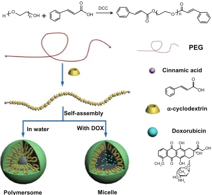
Abstract: A new approach of fabricating supramolecular nanoparticles generated by self-assembly polyrotaxanes for antitumor drug delivery has been reported. Cinnamic-acid-modified poly(ethylene glycol) chains were threaded in α-cyclodextrins to form polyrotaxanes. The poly-rotaxanes self-assembled supramolecular nanoparticles. The morphology of the nanoparticles was changed from nanovesicle to micelle after the antitumor drug, doxorubicin, was loaded. The release profile of the drug-loaded nanoparticles was investigated, and it was found that the sustaining release time could last for 32 hours. The drug-loaded nanoparticles were co-cultured with mouse 4T1 breast cancer cells with a drug concentration of 10 µg/mL; the cell survival rate was 3.3% after a 72-hour incubation. In an in vivo study of breast cancer in a mouse model, the drug-loaded nanoparticles were injected in the tail veins of mice with a dose of 5 mg/kg body weight. The tumor inhibition rate of drug-loaded nanoparticles was 53%, which was better than that of doxorubicin hydrochloride. The cardiac toxicity of doxorubicin was decreased greatly after the encapsulation into supramolecular polyrotaxane nanoparticles.
Keywords: polyrotaxane, self-assembly, nanoparticle, doxorubicin, supermolecular
Introduction
In cancer chemotherapy, nanoparticles exhibit unique advantages in escaping the
capture of the reticuloendothelial system,1 and they also achieve long circulation in
the blood stream to passively target tumor tissues due to their enhanced permeability and retention.2–5 Thus, nanoparticles attract much interest from chemists and materials
scientists for antitumor drug delivery. Nanospheres, micelles, and nanogels have been
extensively reported1,2 as being drug carriers via the fabrication approaches of oil/
water emulsification, amphiphilic self-assembly, and microemulsion polymerization. As the antitumor drugs are physically encapsulated in these nanoparticles, low drug-loading content and encapsulation efficiency are some of the problems faced in drug delivery.6–9 Many strategies have been carried out to improve the drug-loading content.
Mesoporous silicon nanoparticles, graphene, and carbon nanotubes have been used to load drugs due to their high specific surface area and for their ability to adsorb a large
amount of drugs.10–12 The unavoidable drawbacks of these inorganic nanoparticles are
the fact that they are undegradable and they do not circulate for a long time.
Host–guest interaction is an effective approach in fabricating nanoparticles.13–16
Polyrotaxanes are necklace-like inclusion complexes formed by the host–guest interaction between cyclodextrins and polymeric chains via the threading of polymeric chains in the cavity of cyclodextrins.17–20 With the regular stack of cyclodextrins along polymeric chains,
nanoparticles are self-assembled by polyrotaxanes. Moreover, α-cyclodextrin (α-CD)
Dove
press
O R I G I N A L R E S E A R C H
open access to scientific and medical research
Open Access Full Text Article
International Journal of Nanomedicine downloaded from https://www.dovepress.com/ by 118.70.13.36 on 23-Aug-2020
For personal use only.
Number of times this article has been viewed
This article was published in the following Dove Press journal: International Journal of Nanomedicine
is a cyclic oligosaccharide composed of six D-glucose units linked by 1,4-α-glucosidic bonds; it is widely used in pharma-ceutical science for its excellent biocompatibility. Poly(ethylene glycol) (PEG) is regarded as biodegradable material when its molecular weight is lower than 10,000 Dalton, because it is eliminated by the kidney. With this in mind, it is evident that the α-CD/PEG polyrotaxane nanoparticles are attractive drug carriers. Taking the advantages of the hydroxyl functional groups on cyclodextrins into account, cationic segments are immobilized and the polyrotaxanes can be used as nonviral gene vectors. The modified polyrotaxanes can condense DNA efficiently and exhibit excellent gene transfection activity.21,22
The fabrication of nanoparticles via the crystallization of polyrotaxanes has been reported,23–25 and it is evident that
these nanoparticles are compact and cannot trap drugs inside. Herein, we report a new strategy of fabricating polyrotax-ane nanoparticles to encapsulate antitumor drugs. Hydrophobic molecule cinnamic acids were immobilized on the terminal
groups of PEG (Mn = 2000). The cinnamate terminated PEG
was threaded in α-CDs to form polyrotaxanes. The cinnamate
moieties exerted two functions: one was to decrease the number
of α-CDs in PEG chains to avoid the formation of compact
nanoparticles; the other was to provide hydrophobicity in order to trap the hydrophilic antitumor drug inside. Antitumor doxorubicin was used to investigate the release properties of the polyrotaxane nanoparticles. The morphology, in vitro, and in vivo antitumor activities of the drug-loaded polyrotaxane nanoparticles are studied in detail.
Materials and methods
Materials
PEG (Mn = 2000 g/mol) and α-CD were purchased
from Sigma-Aldrich (St Louis, MO) and vacuum-dried
at room temperature for 24 hours before use. N,N′
-dicyclohexylcarbodiimide (DCC), cinnamic acid, and dimethyl
sulfoxide (DMSO)-d6 were purchased from Sigma-Aldrich
and used as received. Doxorubicin hydrochloride (Beijing Zhongshuo Pharmaceutical Technology Development Co, Ltd, Beijing, China) was deprotonated in water, and the pH value was adjusted to 9.6 to obtain doxorubicin.26 Dulbecco’s
modified Eagle’s medium (DMEM), 100 × mycillin, and fetal
bovine serum were purchased from Life Technologies Co, (Carlsbad, CA) and used for the cytotoxicity test. Cell lines NIH/3T3 (mouse embryonic fibroblast cell) and 4T1 (mouse breast cancer cell) were obtained from the Chinese Academy of Science Cell Bank for Type Culture Collection (Shanghai, China). All other chemicals were purchased from Kelong Chemical Co (Chengdu, China) and used as received.
Measurements
1H NMR spectra were performed on a Bruker av-400
spec-trometer (Bruker Corporation, Billerica, MA) using
DMSO-d6 as solvents. Transcranial magnetic stimulation was
used as the internal reference. Ultrasound was performed on ultrasonic cleaner KQ-300DE (Kunshan Ultrasonic Instru-ments Co, Ltd, Kunshan City, China) at a 70% frequency. X-ray diffractometry (XRD) patterns were obtained at room temperature on an X’Pert pro MPD X-ray diffractometer (PANalytical BV, Almelo, the Netherlands), and the powder samples were mounted on a sample holder and scanned from 5° to 50°. Ultraviolet-visible spectra were recorded on Lambda 650 (PerkinElmer Inc, Waltham, MA) in a range from 200 nm to 500 nm. Dynamic light scattering (DLS) experiments were performed on a Malvern Zetasizer Nano ZS (Malvern Instruments Ltd, Malvern, UK) at an angle of
90° at 25°C. Transmission electron microscopy (TEM) was
performed on a Hitachi H600-4 (Hitachi Ltd, Tokyo, Japan). Atomic force microscopy (AFM) imaging was performed using an MFP3D probe (Asylum Research, Santa Barbara, CA) in tapping mode. Cellular uptake was examined by con-focal laser scanning microscopy (Leica TCP SP5). Enzyme-linked immunosorbent assay analysis was performed on enzyme-linked immunosorbent assay instrument Model-550 (Bio-Rad Co, Hercules, CA).
Synthesis of cinnamic-acid-modified
PEG (Cin-PEG)
A total of 20 g of PEG (Mn = 2000) and 4.44 g of cinnamic
acid were dissolved in 200 mL of methylene chloride; 7.98 g of DCC dissolved in 100 mL methylene chloride was slowly dripped into the mixture in an ice bath. After the DCC solu-tion was dripped into the ice bath, the mixture was stirred in the dark at room temperature for 24 hours. The insoluble N,N-dicyclohexylurea was filtered off. The filtrate was concentrated in a rotary evaporator. The modified PEG was precipitated from the concentrate filtrate by the addition of excessive diethyl ether. The product was filtered, washed with water, and dried in a vacuum.
Preparation of polyrotaxanes
To prepare the polyrotaxanes, 1 g of Cin-PEG was dissolved in 20 mL of distilled water, and the solution was added to 10 mL of saturated aqueous solution of α-CDs at room temperature. The mixture was ultrasonically agitated for 10 minutes and then allowed to stand overnight in a refrigerator at 4°C to precipitate white paste. The product was collected by centrifugation, and then was washed with water three times
Dovepress
Liu et al
International Journal of Nanomedicine downloaded from https://www.dovepress.com/ by 118.70.13.36 on 23-Aug-2020
to remove the unthreaded α-CDs and PEG. The white paste precipitate was dried in a vacuum at room temperature to yield the polyrotaxanes. This process was performed in the dark.
Biocompatibility test
Mouse NIH 3T3 fibroblasts were cultured in DMEM at
37°C with 5% CO2. The 3T3 fibroblasts were harvested in
a logarithmic growth phase, seeded on 96 wells at a cellular density of 1 × 10³ cells/well, and incubated for 1 day.
Polyrotaxanes were dissolved and diluted in PBS buffer to give final concentrations ranged from 5.4 × 10⁻³ to 0.54 mg/mL. The
polyrotaxanes and fibroblasts were incubated for 30 hours. The CCK-8 assay at 490 nm was performed and the percentage of cell viability was then determined.
Preparation of drug-loaded polyrotaxane
nanoparticles
Drug-loaded nanoparticles were prepared by concentrate centrifugation. The polyrotaxanes (10 mg) were dissolved in 5 mL water, and stirred for 5 minutes. Doxorubicin (2.5 mg) was dissolved in 1 mL tetrahydrofuran (THF). The solution was dripped into the polyrotaxane solution and stirred for 24 hours. Five milliliters of water was added into the mixture after THF was volatilized. Free doxorubicin and THF were removed using concentrate centrifugation pipe with a dialysis
membrane (MWCO = 3000). The process was repeated three
times. After the centrifugation, the solution was freeze-dried to get the doxorubicin-loaded polyrotaxane nanoparticles.
The doxorubicin loaded nanoparticles were dissolved in 3 mL of DMSO to determine the drug-loading content. The doxorubicin concentration was tested using the UV–VIS spectrophotometer at 485 nm. The drug-loading content was calculated based on the standard curve obtained from doxorubicin in DMSO. The drug-loading content (DLC) and encapsulation efficiency (EE) were calculated according to the following formulae:
DLC (wt%) = (weight of loaded drug/weight of
drug-loaded nanoparticles) × 100% (1)
EE (%) = (weight of loaded drug/weight of
drug in feeding) × 100% (2)
In vitro drug release
The drug-loaded nanoparticles were dispersed and diluted in phosphate-buffered saline (PBS) solution to yield a final concentration of 2 mg/mL. The diluted solution (0.5 mL) was
transferred to dialysis membrane tubes (MWCO = 3000). The
tubes were then immersed in a flask containing 25 mL of PBS
solution (pH = 7.4) and shaken at a speed of 100 rev/min
at 37°C. At specific time intervals, 1 mL of solution was withdrawn from the release medium and replaced with fresh PBS solution.
Cellular uptake
Mouse 4T1 breast cancer cells were cultured in DMEM with
10% fetal bovine serum at 37°C in a 5% CO2 environment.
The 4T1 cells at logarithm phase were seeded on a 6-well culture dish at a cell density of 1 × 10⁴ cells/well. After 1 day,
free doxorubicin hydrochloride and doxorubicin loaded nanoparticles were dispersed and diluted in PBS to give the
final doxorubicin concentration of 10 µg/mL, which were
then added into the plates. After being separately incubated for 3 and 13 hours, the cells were thoroughly washed with PBS three times and visualized under confocal laser scan-ning microscopy (Leica TCP SP5). Doxorubicin was excited at 485 nm with emission at 595 nm.
In vitro inhibition efficacy
The 4T1 cells at logarithm phase were seeded on a 96-well culture dish at a cell density of 1 × 10³ cells/well. After being
incubated for 1 day, free doxorubicin hydrochloride and doxorubicin-loaded nanoparticles were dispersed and diluted in PBS solution to give the final doxorubicin concentration
of 10 µg/mL. The cells were incubated with free
doxoru-bicin hydrochloride and doxorudoxoru-bicin-loaded nanoparticles for 72 hours. The CCK-8 assay at 490 nm was performed at 0, 12, 24, 36, 48, 60, and 72 hours. The percentage of cell viability was then determined. Five parallel samples in each specimen were used for testing.
In vivo antitumor study
Permission to conduct the experiment on animals was obtained from the ethics committee of Sichuan University (Sichuan, China). A total of 48 BALB/c mice (male, body weight: 21–24 g) purchased from the West China Experimental Animal Center of Sichuan University (Sichuan, China) were maintained in a germ-free environment and allowed free access to food and water. All animal experi-ments were performed according to the institutional and National Institutes of Health guidelines for the care and use of research animals.
During the experiment, 5 × 10⁵ 4T1 cells were
subcutane-ously injected into the left flanks of BALB/c mice. After the
inoculated tumor volume reached 100–200 mm3, the mice
were randomly divided into four groups. Each formulation was injected intravenously via the tail vein with a dose of
Dovepress Supramolecular nanoparticles for antitumor drug delivery
International Journal of Nanomedicine downloaded from https://www.dovepress.com/ by 118.70.13.36 on 23-Aug-2020
5 mg of doxorubicin per kg of body weight. Four injections were taken at 4-day intervals. The tumor volume was moni-tored at prescribed time intervals. The tumor volume was calculated using the formula given below:
V [mm3] = 1/2 × LW2 (3)
where L and W represent the long and short diameters of the tumor tissues. On the 19th day after treatment, all mice were sacrificed. The difference in tumor growth was evaluated using the t-test and was considered significant if P, 0.05.
Pathological examination
Sections of the hearts of sacrificed mice treated with doxo-rubicin hydrochloride and doxodoxo-rubicin loaded polyrotaxane nanoparticles were fixed in 10% buffered formaldehyde, embedded in paraffin, and then sectioned. The sections were then stained routinely with hematoxylin-eosin for histologi-cal assessment.
Results and discussion
The structures of Cin-PEG, α-cyclodextrin, and
correspond-ing polyrotaxane were characterized by 1H NMR (Figure 1).
The protons of CH2CH2O in the PEG chain appeared at
δ= 3.6 ppm. The terminal cinnamate groups changed the
chemical environment of the EG (ethylene glycol) unit con-nected to cinnamate and thus led to a signal shift to δ= 3.7 and 4.3 ppm, respectively. The protons in cinnamate were localized from δ= 6 to 8 ppm. The signals of protons in CH = CH were split and appeared at around δ= 7.7 ppm and δ= 6.5 ppm. The signals of protons in the benzene ring appeared in two sites; the peak at δ= 7.4 ppm was attributed to the protons in ortho and para positions. The signal at δ= 7.5 ppm was assigned to the
protons in meta position. The integrity ratio of COOCH2CH2O (Figure 1Ac) to OCH2CH2O (Figure 1Aa) was used to calculate the number of immobilized cinnamic acid. The result showed that both terminal groups of PEG were immobilized with cinnamic acid. The spectrum of polyrotaxane is presented
in Figure 1C. The proton signals of α-cyclodextrin in the
spectrum of polyrotaxane were strong. Moreover, the signal
at δ= 3.5 ppm was noted among the OCH2CH2O protons in
modified PEG, which shifted from δ= 3.6 ppm to δ= 3.5 ppm.
All the protons in PEG and α-CDs were detected in the
spec-trum, which suggested that the polyrotaxane was formed. The mole ratio of EG/CD was calculated from the integrities
of OCH2CH2 in modified PEG and CH (Ca) in α-CDs. The
calculated EG/CD was 2.8, which meant that the average
numbers of α-CD molecules in a Cin-PEG chain were 16 as
the repeated units of EG in PEG (Mn = 2000) were about 44.
The ratio of EG/CD in α-CD/PEG polypseudorotaxane
was 2.0–2.1;27,28 the higher ratio of EG/CD implied that the
terminal cinnamates in the PEG chains reduced the numbers of α-cyclodextrins in PEG chains. It was favorable for weakening the crystallization of polyrotaxane to avoid the formation of compact nanoparticles.
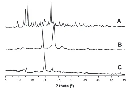
XRD is a powerful tool for confirming the formation of
polyrotaxane. The XRD spectra of α-cyclodextrin, Cin-PEG,
and polyrotaxane are presented in Figure 2. Strong crystal-lization was observed in the spectrum of α-cyclodextrin (Figure 2A). Cin-PEG also exhibited strong crystallinity, and
the prominent peaks of PEG crystal appeared at 2θ= 19.3°
and 23.4° (Figure 2B). In the spectra of polyrotaxane, a very strong peak occurred at 2θ= 19.7° and was presented in the diffraction patterns (Figure 2C). It was the characteristic peak associated with a channel-type crystalline structure by virtue of the polymeric nature of the guest molecules.17–20,29
ppm
HO HO OHbOHc OHf
OH e g
g e
d
d c b
a
O O
O a O
O n − 1 b c g f
f f
f
Ca
CcCe Cb Cd
A
B
C Cf
O O
d e c b a
f
O 6
8 7 6 5 4 3 2 1 0
Figure 1 The 1H NMR spectra of cinnamic-acid-modified PEG (A), α-CD (B), and
polyrotaxanes (C).
Abbreviations: PEG, poly(ethylene glycol); α-CD, α-cyclodextrin.
5 10 15 20 25 30 35 40 45 50
2 theta (°)
A
B
C
Figure 2 The XRD spectra of α-cyclodextrin (A), cinnamic-acid-modified PEG (B),
and polyrotaxane (C).
Abbreviations: XRD, X-ray diffractometry; PEG, poly(ethylene glycol).
Dovepress
Liu et al
International Journal of Nanomedicine downloaded from https://www.dovepress.com/ by 118.70.13.36 on 23-Aug-2020
The XRD result demonstrated that the Cin-PEG was threaded
in α-CDs to form necklace-like polyrotaxanes.
Doxorubicin was trapped in the polyrotaxane nanoparticles. The morphologies of the blank and drug-loaded polyrotaxane nanoparticles were characterized by DLS, TEM, and AFM (Figure 3). The DLS results showed that both the blank and drug-loaded nanoparticles were monodispersed and the mean diameters were 57 and 107 nanometers, respectively. The diameter of the nanoparticles was increased after drug
encapsulation (Figure 3A and B). The TEM photograph of the blank nanoparticles clearly showed that the nanoparticles had a core-shell structure and the shell layer was darker, which meant that this layer was compact (Figure 3C). The AFM image of the blank nanoparticles supported this conclusion. The height of the blank nanoparticles was measured by AFM using the tapping mode. The nanoparticles were sunk inside and the height of the nanoparticles was about 0.8 nanometers, which was nearly the same height as one α-cyclodextrin.30,31 The wall
1.0
0.8
0.6
0.4
0.2
0.0 0.0
800 600 400 200
−200 0
0 20
0.0 0.0
5 nm 4 3 2 1 0 −1 0.2 0.4 0.6 0.8 1.0
2
1
0
−1
−2
0.2
0 50 100 150
nm
Height
µm µm
µm
nm
nm
µm
Height
0.4 0.6 0.8 1.0 40 60 80 100 120 140 0.2 0.4 0.6 0.8 1.0
1.5 1.0 0.5 0.0 −0.5 −1.0 −1.5
0 5 10 15 20 25
N
u
m
b
er
(
%
)
10 100 1000 0 10
5 10 15 20 25
B A
Size (d.nm)
N
u
m
b
er
(
%
)
Size (d.nm)
100 1000
B A
Figure 3 Size distributions and morphologies of blank and drug-loaded polyrotaxane nanoparticles. (A) DLS of blank nanoparticles; (B) DLS of drug-loaded nanoparticles;
(C) TEM photograph of blank nanoparticles; (D) AFM image of blank nanoparticles; (E) TEM photograph of drug-loaded nanoparticles; (F) AFM image of drug-loaded
nanoparticles.
Abbreviations: DLS, dynamic light scattering; TEM, transmission electron microscopy; AFM, atomic force microscopy.
Dovepress Supramolecular nanoparticles for antitumor drug delivery
International Journal of Nanomedicine downloaded from https://www.dovepress.com/ by 118.70.13.36 on 23-Aug-2020
Table 1 The drug-loading content and encapsulation efficiency of
polyrotaxane nanoparticles
Sample EG/CDa Mean diameter
(nm)
DLC (%)
EE (%) Beforeb Afterc
α-CDs/Cin-PEG 2.8 57 ± 8 107 ± 12 18.4 ± 0.7 78.1
Notes:aCalculated from 1H NMR spectrum; bbefore drug-loading; cafter drug-loading.
Abbreviations: EG, ethylene glycol; CD, cyclodextrin; DLC, drug-loading content; EE, encapsulation efficiency; α-CDs, α-cyclodextrins; Cin-PEG, cinnamic-acid-modified poly(ethylene glycol).
0 5 10 15 20 25 30
0 20 40 60 80 100 120
A
cc
u
m
u
la
te
d
r
el
ea
se
(
%
)
Time (h)
A
B
Figure 5 Release profiles of doxorubicin hydrochloride (A) and drug-loaded
polyrotaxane nanoparticles (B).
thickness of the sunk nanoparticles measured by AFM was about 20 nanometers (Figure 3D). Both TEM and AFM images demonstrated that the blank polyrotaxane nanoparticles were hollow with vesicle-like morphology. Interestingly, the mor-phology of drug-loaded nanoparticles changed significantly; it was still core-shell structured, but the core was dark and the shell layer was light in the TEM photograph (Figure 3E). The compact layer was transferred from the shell to the core after the drug was loaded; the shell thickness of the drug-loaded nanoparticles measured from TEM photograph was about 35 nanometers (Figure 3E). The height of the nanoparticles measured by AFM was more than 5 nanometers and no sinking was observed in the nanoparticles (Figure 3F). The results of both TEM and AFM proved that the drug-loaded nanoparticles were no longer nanovesicles but micelle-like nanoparticles.
The hypothesized formation process of the blank and drug-loaded polyrotaxane nanoparticles was illustrated in Figure 4. Cinnamic acid is a hydrophobic molecule, and the polyrotaxanes with Cin-PEG chains are amphiphiles. The amphiphiles formed vesicle-like hollow polymersomes. Once the hydrophobic drug doxorubicin was trapped, the self-assembly of polyrotaxanes was changed. The doxorubicin was centered at the core to gather the hydrophobic segments in the polyrotaxanes, thus, the polyrotaxanes self-assembled micelle-like nanoparticles.
The drug-loading content and encapsulation efficiency of the polyrotaxane nanoparticles were tested and summarized in Table 1. Drug-loading content and encapsulation effi-ciency are important parameters in evaluating the usability of nanoparticles for antitumor drug delivery. A main problem faced by nanodrug carriers was the low drug-loading con-tent; specifically, this property would decrease the curative effect of chemotherapy, and therefore the high dose of drug-loaded nanoparticles had to be used, which increased the risk of the treatment. In most polymeric drug nanocarriers, such as nanospheres and micelles, the drug-loading content
was less than 10%.6–9 In these polyrotaxane nanoparticles,
the drug-loading content and encapsulation efficiency were
18.4% and 78.1%, respectively. The drug-loading content was much higher than that of most polymer micelles.
The release profile of drug-loaded polyrotaxane nano-particles is presented in Figure 5. Hydrophilic doxorubicin hydrochloride was used as a control. The release of doxo-rubicin hydrochloride was very fast in PBS solution and the accumulated release rate was nearly 100% within the first 3 hours. For the drug-loaded nanoparticles, the burst release was about 40% in the first 2 hours. After the burst release, the sustaining release time was more than 30 hours, and the release curve was linear and nearly obeyed the rule of one-order release (Figure 5B). The accumulated release reached about 80% when the release time was 32 hours. The release profile implied that the polyrotaxane nanoparticles were promising for doxorubicin delivery.
PEG
Cinnamic acid
Doxorubicin
Micelle Polymersome
In water With DOX Self-assembly
H OH
O
O O
O O
O OH n OH
+ DCC
n O
O OH OH OH OH
HO O
OO CH3O
NH2
CH3
O
α-cyclodextrin
Figure 4 The illustrated formation of drug-loaded polyrotaxane nanoparticles. Abbreviations: PEG, poly(ethylene glycol); DOX, doxorubicin.
Dovepress
Liu et al
International Journal of Nanomedicine downloaded from https://www.dovepress.com/ by 118.70.13.36 on 23-Aug-2020
The blank polyrotaxane nanoparticles were co-cultured with mouse NIH 3T3 fibroblasts to evaluate the biocompatibility; the result is shown in Figure 6. The incubation time was 24 hours and the nanoparticle concentrations ranged from 5.4 × 10⁻³ mg/mL to 0.54 mg/mL. The cells on the cell
culture plates were used as the control. The cell viability of the nanoparticles in each concentration was around 100%, which demonstrated that the polyrotaxane nanoparticles were non-toxic to NIH 3T3 fibroblasts.
The drug-loaded polyrotaxanes nanoparticles were co-cultured with 4T1 breast cancer cells to evaluate the in vitro inhibition activity (Figure 7). The concentration of
doxoru-bicin was 10 µg/mL. Doxorubicin was hydrophobic, and it
aggregated and precipitated in the cell culture medium, thus it could not be used as a control. Doxorubicin hydrochloride was water-soluble, so doxorubicin hydrochloride was used as the control. The cell viabilities of both control and drug-loaded nanoparticles were decreased with increasing incu-bation time. The cell viabilities of the two groups that were incubated for 24 hours were 27.6% and 31.2%, and those that were incubated for 48 hours exhibited cell viabilities of 2.9% and 6.8%. After 3-day incubation, only 0.3% of cancer cells survived in the doxorubicin hydrochloride group, and the survival rate of cancer cells in the drug-loaded nanopar-ticles was 3.3%. This result revealed that the drug-loaded nanoparticles could inhibit the proliferation of cancer cells efficiently, and that the in vitro antitumor efficiency of drug-loaded nanoparticles was comparable to that of doxorubicin hydrochloride.
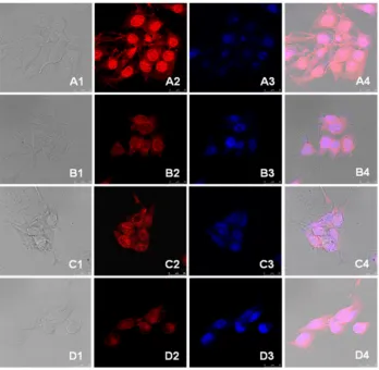
The cellular uptake of the drug-loaded nanoparticles was studied by confocal laser scanning microscopy. Both the drug-loaded nanoparticles and doxorubicin hydrochloride were incubated with 4T1 cells for 3 and 13 hours, respectively. The nuclei of the cells were dyed blue. The photographs
are presented in Figure 8. In the first 3 hours of incubation, a strong red fluorescent effect was observed with doxorubicin hydrochloride in both the cytoplasm and nuclei of the cells. In the cells incubated with drug-loaded nanoparticles, red fluorescent of doxorubicin was also observed in the nuclei, but the red fluorescent in the cytoplasm was stronger than that in nuclei. After being incubated for 13 hours, the red fluorescent distribution was very different. The fluorescent strength in the nuclei of the cells treated with doxorubicin hydrochloride was weakened and the cytoplasm and nuclei were diffuse, while the nucleus membrane was not clear. In the cells treated with drug-loaded nanoparticles, the red fluorescent was strength-ened in the nuclei after 13 hours of incubation.
Doxorubicin hydrochloride, a hydrophilic antitumor drug, was internalized into cells by diffusion, it diffused very quickly both from the medium to the cytoplasm and from the cytoplasm to the nuclei, thus a strong red fluores-cent was observed in both the cytoplasm and the nuclei in the cells treated with doxorubicin hydrochloride for the first 3 hours. With the incubation time increasing, the drug was consumed and the cells were destroyed; thus, the strength of the fluorescent effect in the nuclei was weakened, and the cytoplasm and nuclei of the cells were diffusive. Furthermore, the nucleus membrane was not clear. The internalization of drug-loaded nanoparticles did not occur due to diffusion, so the endocytosis of drug-loaded nanoparticles was slower than that of doxorubicin hydrochloride. As a result, the fluorescent noted in the cells treated with drug-loaded nanoparticles was weak and was mainly localized in the cytoplasm of the cells within the first 3 hours. The doxorubicin was released from the nanoparticles and protonized to hydrophilic due to the acidity in endosomes and/or lysosomes, the protonized doxo-rubicin diffused from cytoplasm to nuclei quickly,32 thus, the
0 20 40 60 80 100 120
0.54 0.27
5.4 x 10⁻² 1.08 x 10²
Cell viability (%
)
Concentration (mg/mL)
5.4 x 10⁻³
Figure 6 The cytotoxicity of blank polyrotaxane nanoparticles.
0 20 40 60 80 100
72 60 48 36 24 12
Cell viability (%
)
Time (h)
Doxorubicin hydrochloride Drug loaded nanoparticles
0
Figure 7 The in vitro inhibition effect of drug-loaded polyrotaxane nanoparticles on 4T1 breast cancer cells.
Note: The concentration of doxorubicin was 10 µg/mL.
Dovepress Supramolecular nanoparticles for antitumor drug delivery
International Journal of Nanomedicine downloaded from https://www.dovepress.com/ by 118.70.13.36 on 23-Aug-2020
fluorescent in the nuclei was stronger after 13-hour incubation than that after 3-hour incubation. These results demonstrated that the trapped doxorubicin could be released from the polyrotaxane nanoparticles efficiently in cytoplasm.
A breast cancer model in mice was used to test the in vivo antitumor efficacy of the drug-loaded polyrotaxane nanoparticles. The concentration of the dose of the drug was 5 mg per kg of body weight, and the drug-loaded nano-particles were administrated by injection into the tail vein of the mice. A total of 12 mice in each group were used as parallel samples; two mice in both the blank nanoparticles and doxorubicin hydrochloride groups died during the in vivo experiments. The tumor volumes were tested, and the statistic results have been presented in Figure 9. The aver-age volumes of tumors administrated with saline and blank
nanoparticles were 1800 and 1550 mm3, respectively. The
volumes of tumors treated with doxorubicin hydrochloride
and drug-loaded nanoparticles were about 950 and 800 mm3.
The statistical results showed a significant difference between the two groups (P, 0.05). Specifically, doxorubicin-loaded
nanoparticles exhibited better tumor growth suppression than nanoparticles loaded with doxorubicin hydrochloride.
The in vivo tumor inhibition rate of the drug-loaded nanoparticles was calculated and the result is pre-sented in Figure 10. The antitumor activity of the drug-loaded Figure 8 Confocal microscopy photographs of doxorubicin and drug-loaded nanoparticles incubated with 4T1 breast cancer cells. (A) doxorubicin hydrochloride and
(B) doxorubicin-loaded nanoparticles incubated for 3 hours; (C) doxorubicin hydrochloride and (D) doxorubicin-loaded nanoparticles incubated for 13 hours.
Note: The photographs from left to right (numbered 1 to 4) are the overlapped photos of bright field and doxorubicin stained nuclei.
10 15 20 25 30
0 400 800 1200 1600 2000
Tumor volume (m
m
3)
Time (days)
Physiological saline Blank nanoparticles Doxorubicin hydrochloride Drug loaded nanoparticles
Figure 9 The in vivo inhibition effect of doxorubicin-loaded polyrotaxane nanoparticles on tumor growth in mice via venous injection.
Dovepress
Liu et al
International Journal of Nanomedicine downloaded from https://www.dovepress.com/ by 118.70.13.36 on 23-Aug-2020
nanoparticles was optimal, with its inhibition rate as high as 53%; conversely, the inhibition rate of doxorubicin hydrochlo-ride was 38%. This result revealed that the in vivo inhibition efficacy of doxorubicin-loaded polyrotaxane nanoparticles was better than that of the doxorubicin hydrochloride-loaded nanoparticles, suggesting that the polyrotaxane nanoparticles were promising carriers of the drug for antitumor drug delivery.
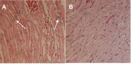
Doxorubicin was seriously toxic to the heart of the mice in the study.33–35 A pathological examination of the drug-loaded
nanoparticles was conducted. The histological photographs of cardiac muscle treated with doxorubicin hydrochloride and drug-loaded nanoparticles are presented in Figure 11. The inflammatory reaction was clearly observed, and the texture of the cardiac muscle was destroyed (Figure 11A) in the sample treated with doxorubicin hydrochloride. In contrast, this phenomenon was not observed in the photographs of the cardiac muscles of mice treated with drug-loaded nanopar-ticles (Figure 11B). This demonstrated that the encapsulation of polyrotaxane nanoparticles could decrease the cytotoxicity of doxorubicin.
Conclusion
A supramolecular nanoparticle generated by the self- assembly
polyrotaxanes composed of α-cyclodextrins and Cin-PEG was
prepared. The morphology of the nanoparticles was changed from nanovesicle to micelle after the antitumor drug doxoru-bicin was loaded. The release profile investigation found that the sustained release could last for 32 hours, and the release replicated one-order release after the burst-release period. The drug-loaded nanoparticles were co-cultured with mouse 4T1 breast cancer cells to evaluate the in vitro antitumor efficacy, and the results showed that the internalization of the drug-loaded nanoparticles was effective, and that the drug could be released in cytoplasm. The results of the in vivo experiment revealed that the drug-loaded polyrotaxane nanoparticles exhibited efficient antitumor activity to breast cancer cells, and this effect was better than that of the doxorubicin hydrochloride. The pathological examination demonstrated that the encapsulation of polyrotaxane nanoparticles could decrease the cardiac toxicity of doxorubicin. The polyrotaxane nanoparticle is a promising carrier for antitumor drug delivery in a wide range of biomedical applications.
Acknowledgments
This research work was supported by the National Basic Research Program of China (National 973 program, No 2011CB606206), the National Science Foundation of China (NSFC, No 31070849, 50830105, 51133004), the Program for New Century Excellent Talents in University, Ministry of Education (MOE, NCET-10-0564), the International Col-laboration Project of Ministry of Science and Technology (MOST, No 2010DFA51550), the Program for Changjiang Scholars, and the Innovative Research Team in University (IRT1163).
Disclosure
The authors report no conflicts of interest in this work.
References
1. Lavasanifar A, Samuel J, Kwon GS. Poly(ethylene oxide)-block-poly(L-amino acid) micelles for drug delivery. Adv Drug Deliv Rev. 2002;54(2):169–190.
2. Li SD, Huang L. Stealth nanoparticles: high density but sheddable PEG is a key for tumor targeting. J Control Release. 2010;145(3):178–181. 3. Ranganathan R, Madanmohan S, Kesavan A, et al. Nanomedicine:
towards development of patient-friendly drug-delivery systems for oncological applications. Int J Nanomedicine. 2012;7:1043–1060. 4. Wang K, Liu L, Zhang T, et al. Oxaliplatin-incorporated micelles
elimi-nate both cancer stem-like and bulk cell populations in colorectal cancer. Int J Nanomed. 2011;6:3207–3218.
5. Cai LL, Liu P, Li X, et al. RGD peptide-mediated chitosan-based polymeric micelles targeting delivery for integrin-overexpressing tumor cells. Int J Nanomedicine. 2011;6:3499–3508.
0 20 40 60
Inhibition rate
(%
)
Drug loaded
nanoparticles hydrochloride Doxorubicin nanoparticles Blank
Figure 10 The inhibition rate of drug-loaded nanoparticles to breast cancer in mouse models.
Figure 11 Histological photographs of mice cardiac muscles administrated with
doxorubicin hydrochloride (A) and drug-loaded polyrotaxane nanoparticles (B).
Note: The arrows show the inflammation.
Dovepress Supramolecular nanoparticles for antitumor drug delivery
International Journal of Nanomedicine downloaded from https://www.dovepress.com/ by 118.70.13.36 on 23-Aug-2020
International Journal of Nanomedicine
Publish your work in this journal
Submit your manuscript here: http://www.dovepress.com/international-journal-of-nanomedicine-journal
The International Journal of Nanomedicine is an international, peer-reviewed journal focusing on the application of nanotechnology in diagnostics, therapeutics, and drug delivery systems throughout the biomedical field. This journal is indexed on PubMed Central, MedLine, CAS, SciSearch®, Current Contents®/Clinical Medicine,
Journal Citation Reports/Science Edition, EMBase, Scopus and the Elsevier Bibliographic databases. The manuscript management system is completely online and includes a very quick and fair peer-review system, which is all easy to use. Visit http://www.dovepress.com/ testimonials.php to read real quotes from published authors. 6. Lee ES, Na K, Bae YH. Doxorubicin loaded pH-sensitive polymeric
micelles for reversal of resistant MCF-7 tumor. J Control Release. 2005;103(3):405–418.
7. Yu D, Peng P, Dharap SS, et al. Antitumor activity of poly(ethylene glycol)-camptothecin conjugate: the inhibition of tumor growth in vivo. J Control Release. 2005;110(1):90–102.
8. Tong R, Cheng J. Paclitaxel-initiated, controlled polymerization of lactide for the formulation of polymeric nanoparticulate delivery vehicles. Angew Chem Int Ed Engl. 2008;47(26):4830–4834. 9. Greenwald RB, Choe YH, McGuire J, Conover CD. Effective drug
delivery by PEGylated drug conjugates. Adv Drug Deliv Rev. 2003; 55(2):217–250.
10. Tang S, Huang X, Chen X, Zheng N. Hollow mesoporous zirconia nanocapsules for drug delivery. Adv Funct Mater. 2010;20(15): 2442–2447.
11. Liu Z, Fan AC, Rakhra K, et al. Supramolecular stacking of doxorubicin on carbon nanotubes for in vivo cancer therapy. Angew Chem Int Ed Engl. 2009;48(41):7668–7672.
12. Liu Z, Robinson JT, Sun XM, Dai HJ. PEGylated nanographene oxide for delivery of water-insoluble cancer drugs. J Am Chem Soc. 2008;130(33):10876–10877.
13. Wang J, Jiang M. Polymeric self-assembly into micelles and hollow spheres with multiscale cavities driven by inclusion complexation. J Am Chem Soc. 2006;128(11):3703–3708.
14. Wang Y, Ma N, Wang Z, Zhang X. Photocontrolled reversible supramolecular assemblies of an azobenzene-containing surfactant with alpha-cyclodextrin. Angew Chem Int Ed Engl. 2007;46(16): 2823–2826.
15. Ohga K, Takashima Y, Takahashi H, Kawaguchi Y, Yamaguchi H, Harada A. Preparation of supramolecular polymers from a cyclodextrin dimer and ditopic guest molecules: control of structure by linker flexibility. Macromolecules. 2005;38(14):5897–5904.
16. Liu Y, Ke CF, Zhang HY, Cui J, Ding F. Complexation-induced transition of nanorod to network aggregates: alternate porphyrin and cyclodextrin arrays. J Am Chem Soc. 2008;130(2):600–605. 17. Rusa CC, Bullions TA, Fox J, Porbeni FE, Wang X, Tonelli AE.
Inclusion compound formation with a new columnar cyclodextrin host. Langmuir. 2002;18(25):10016–10023.
18. Topchieva IN, Tonelli AE, Panova IG, et al. Two-phase channel structures based on alpha-cyclodextrin-polyethylene glycol inclusion complexes. Langmuir. 2004;20(21):9036–9043.
19. Harada A, Kamachi M. Complex formation between poly(ethylene g lycol) and α-cyclodextrin. Macromolecules. 1990;23(10):2821–2823. 20. Li J, Ni X, Zhou Z, Leong KW. Preparation and characterization of
polypseudorotaxanes based on block-selected inclusion complexation between poly(propylene oxide)-poly(ethylene oxide)-poly(propylene oxide) triblock copolymers and alpha-cyclodextrin. J Am Chem Soc. 2003;125(7):1788–1795.
21. Ooya T, Choi HS, Yamashita A, et al. Biocleavable polyrotaxane-plasmid DNA polyplex for enhanced gene delivery. J Am Chem Soc. 2006;128(12):3852–3853.
22. Li J, Yang C, Li HZ, et al. Cationic supramolecules composed of multiple oligoethylenimine-grafted β-cyclodextrins threaded on a polymer chain for efficient gene delivery. Adv Mater. 2006;18(22):2969–2974. 23. Ren L, Ke F, Chen Y, Liang D, Huang J. Supramolecular ABA triblock
copolymer with polyrotaxane as B block and its hierarchical self-assembly. Macromolecules. 2008;41(14):5295–5300.
24. Huang J, Ren L, Chen Y. pH-/temperature-sensitive supramolecular micelles based on cyclodextrin polyrotaxane. Polym Int. 2008;57(5): 714–721.
25. Tsai CC, Leng S, Jeong KU, et al. Supramolecular structure of β-cyclodextrin and poly(ethylene block-poly(propylene oxide)-block-poly(ethylene oxide) inclusion complexes. Macromolecules. 2010;43(22):9454–9461.
26. Shuai X, Ai H, Nasongkla N, Kim S, Gao J. Micellar carriers based on block copolymers of poly(epsilon-caprolactone) and poly(ethylene glycol) for doxorubicin delivery. J Control Release. 2004;98(3): 415–426.
27. Harada A, Li J, Kamachi M. The molecular necklace: a rotaxane containing many threaded α-cyclodextrins. Nature. 1992;356(6367):325–327. 28. Harada A, Li J, Kamachi M. Preparation and properties of inclusion
complexes of polyethylene glycol with .alpha.-cyclodextrin. Macromolecules. 1993;26(21):5698–5703.
29. Uyar T, Hunt MA, Gracz HS, Tonelli AE. Crystalline cyclodextrin inclusion compounds formed with aromatic guests: guest-dependent stoichiometries and hydration-sensitive crystal structures. Cryst Growth Des. 2006;6(5):1113–1119.
30. Miyauchi M, Hoshino T, Yamaguchi H, Kamitori S, Harada A. A [2]rotaxane capped by a cyclodextrin and a guest: Formation of supramolecular [2]rotaxane polymer. J AmChemSoc. 2005;127: 2034–2035.
31. Miyauchi M, Takashima Y, Yamaguchi H, Harada A. Chiral supramolecular polymers formed by host-guest interactions. J Am Chem Soc. 2005;127(9):2984–2989.
32. Liu R, Li D, He B, et al. Anti-tumor drug delivery of pH-sensitive poly(ethylene glycol)-poly(L-histidine-)-poly(L-lactide) nanoparticles. J Control Release. 2011;152(1):49–56.
33. Tan ML, Friedhuber AM, Dunstan DE, Choong PF, Dass CR. The performance of doxorubicin encapsulated in chitosan-dextran sulphate microparticles in an osteosarcoma model. Biomaterials. 2010;31(3): 541–551.
34. Xiao K, Luo J, Li Y, Lee JS, Fung G, Lam KS. PEG-oligocholic acid telodendrimer micelles for the targeted delivery of doxorubicin to B-cell lymphoma. J Control Release. 2011;155(2):272–281.
35. Mukhopadhyay P, Batkai S, Rajesh M, et al. Pharmacological inhibi-tion of CB1 cannabinoid receptor protects against doxorubicin-induced cardiotoxicity. J Am Coll Cardiol. 2007;50(6):528–536.
Dovepress
Dove
press
Liu et al
International Journal of Nanomedicine downloaded from https://www.dovepress.com/ by 118.70.13.36 on 23-Aug-2020