1
Mechanotransduction in Neuronal Cell Development and FunctioningAuthor list: Matteo Chighizola1, Tania Dini1, Cristina Lenardi1, Paolo Milani1, Alessandro Podestà1, Carsten Schulte1*
1Interdisciplinary Centre for Nanostructured Materials and Interfaces (C.I.Ma.I.Na.) and Department of Physics,
Università degli Studi di Milano, via Celoria 16, 20133 - Milan
*Corresponding author:
Carsten Schulte: [email protected]
2
AbstractAlthough many details remain still elusive, it became increasingly evident in recent years that mechanosensing of
microenvironmental biophysical cues and subsequent mechanotransduction are strongly involved in the regulation of
neuronal cell development and functioning. This review gives an overview about the current understanding of brain and
neuronal cell mechanobiology and how it impacts on neurogenesis, neuronal migration, differentiation, and maturation.
Therein; we are focussing particularly on the events in the cell/microenvironment interface and the decisive
extracellular matrix (ECM) parameters (i.e. rigidity and nanometric spatial organisation of adhesion sites) that modulate
integrin adhesion complex-based mechanosensing and mechanotransductive signalling. It will also be outlined how
biomaterial approaches mimicking essential ECM features help to understand these processes and how they can be used
to control and guide neuronal cell behaviour by providing appropriate biophysical cues. In addition, principal
3
1. IntroductionFor a long time research on neuronal cell development and functioning was principally concentrated on the influence of
biochemical factors, but in recent years accumulating evidence made clear that taking a biophysical perspective on these
processes is intriguing and promising for various reasons. One outstanding attribute of neuronal cells is the extreme
polarisation and compartmentalisation that is taking place during neuronal differentiation and maturation. It requires
highly coordinated and dynamic cytoskeletal actions and cell/microenvironment interactions to realise neuronal
migration, neurito- and synaptogenesis, as well as neural network formation and plasticity (Flynn, 2013),(Kerstein et al.,
2015),(Leterrier et al., 2017),(Park and Goda, 2016),(Lilja and Ivaska, 2018). Developing neurons possess growth cones
which are sensory compartments at the tip of neurites, build for the exploration of the microenvironment by their
capacity to integrate chemical and mechanical cues (Chan and Odde, 2008),(Lowery and Van Vactor, 2009),(Myers et
al., 2011),(Vitriol and Zheng, 2012),(Franze et al., 2013),(Kerstein et al., 2015), and specialised to operate in the soft
brain tissue (Chan and Odde, 2008),(Betz et al., 2011),(Kerstein et al., 2015). Also when neurons reached their terminal
differentiation and maturation stage with a complex morphology characterised by an axon and several dendrites,
equipped with numerous fine structures such as synapses and spines, they maintain a remarkable plasticity to enable the
processing of incoming information. This plasticity is highly dependent on integrin-mediated interaction with the
microenvironment (Park and Goda, 2016),(Lilja and Ivaska, 2018).
Biophysical aspects, and in particular integrin-mediated mechanotransductive processes, involved in the
regulation of neuronal cell development and functioning, will be a focus of this review. Special attention will be drawn
to what is happening in the cell/microenvironment interface as these events are, literally and functionally, at the base of
the mechanotransductive signalling and its impact on cellular behaviour. Throughout the review various examples of
bioengineering approaches are indicated that were useful to gain insight into the influence of mechanotransduction on
neuronal differention and/or exploit mechanotransductive mechanisms to guide neuronal cell behaviour in a controlled
manner. Finally, this review will highlight some methods that are used to study biophysical aspects of (neuronal) cells
and their environment. For further reading, the reader will be pointed to reviews that accentuate specific aspects of the
different arguments in more detail.
2. Microenvironmental Cues Influencing Neuronal Cell Development
2.1 Biophysical, structural and compositional peculiarities of the extracellular matrix of the central nervous
system
The extracellular matrix (ECM) of the central nervous system (CNS) has some distinctive and unique features regarding
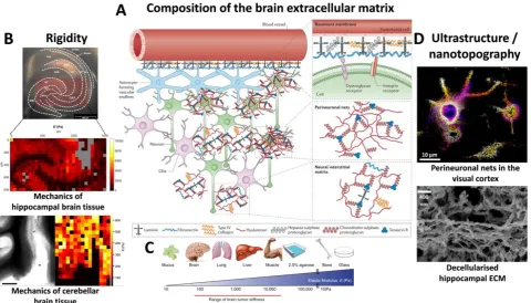
mechanics, structure and composition that differ substantially from the ECM of other organs and tissues (Fig. 1A-D).
Albeit regional mechanical heterogeneity within brain and spinal cord compartments has been reported (Elkin
et al., 2007),(Christ et al., 2010),(Koser et al., 2015),(Antonovaite et al., 2018) (Fig. 1B), the CNS has as a general
softness compared to other tissues (Franze et al., 2013),(Barnes et al., 2017) (Fig. 1C). Interesting observations in this
regard are the stiffening of the human brain tissue due to ageing (Sack et al., 2011) and changes in mechanical
properties in neurodegenerative diseases (e.g., in multiple sclerosis, amyotrophic lateral sclerosis and Alzheimer’s
disease) and brain cancer (e.g., in glioblastoma) (Tyler, 2012),(Barnes et al., 2017),(Tanner, 2018) (Fig. 1C).
The ECM composition of the CNS is furthermore characterised by a high abundance of hyaluronic acid and
4
of the blood-brain-barrier) and proteoglycans (such as e.g. lectican family members) (Fig. 1A) which are highlyintertwined at the nanoscale (Fig. 1D). The non-fibrillar type IV collagen is present in the brain ECM but the amount of
fibrillary proteins (such as e.g. collagen I) is instead relatively low (Ruoslahti, 1996),(Dityatev et al., 2010b),(Lau et al.,
2013).
Apart from the interstitial neural matrix the brain ECM possesses also some special partitions with specific
tasks and locations. In the subventricular zone of the adult brain, heparan sulphate proteoglycan and laminin-rich
structures called fractones can be found close to the brain blood vessels that serve as neural stem cell niches and
neurogenic zones (Kerever et al., 2007),(Mercier, 2016) (Fig. 1A). Another particular brain ECM structure is the
perineuronal net; a specialised scaffold surrounding the neuronal cell body and proximal processes of many neurons in
the CNS, that is essential for the synaptic structure and regulates synaptic plasticity (especially crucial components,
such as tenascin-C and –R, brevican and neurocan) (Pizzorusso et al., 2002),(Geissler et al., 2013),(Sorg et al., 2016)
(Fig. 1A,D).
ECM constituents and in particular integrin/microenvironment interactions are indeed strongly involved in all
steps of neuronal development (Long and Huttner, 2019) which will be further outlined throughout the review, in
particular in paragraph 3.2. Furthermore, alterations in the brain ECM composition and organisation have been found in
neurodegeneration and brain cancer (Bonneh-Barkay and Wiley, 2009),(Lau et al., 2013),(Miyata and Kitagawa,
2017),(Barnes et al., 2017),(Tanner, 2018).
Fig. 1 Compositional, mechanical and structural features of the brain extracellular matrix
5
STORM super-resolution recording of perineuronal nets (chondroitin sulphate proteoglycans stained with Wisteria floribunda agglutinin-Dy749P1). Image courtesy of Xiaowei Zhuang (Harvard University, Cambridge, MA, USA). The lower image shows a scanning electron microscopic recording of the configuration of decellularised hippocampal extracellular matrix. Image from Tajerian et al. (Tajerian et al., 2018).Decellularised brain ECM (an example can be seen in Fig. 1D) that are applied as substrates (either as 2D coating or 3D
hydrogel) foster neurite outgrowth (Medberry et al., 2013) and functional neural network formation (Lam et al., 2019)
better than equivalent decellularised ECM substrates from other tissues. A recent study furthermore shows that
decellularised brain ECM increases the neuronal reprogramming efficiency of mouse embryonic fibroblasts into
induced neurons, compared to e.g. 2D laminin-coated substrates. The promotive effect was already observable when the
brain-derived ECM was also presented as a 2D coating, but strongly pronounced when it was applied as a 3D hydrogel
environment (Jin et al., 2018). This emphasised the importance of appropriate microenvironmental biophysical and
topographical cues, even in the presence of the same biochemical components.
The ECM building blocks and their interactions/crosslinking define the biophysical configuration of the
intricate meshwork and determine its specific mechanical and structural properties, i.e. rigidity and nanotopography
(Gasiorowski et al., 2013),(Young et al., 2016) (Fig. 1A-D). In the next paragraphs it will be detailed how these two
principal biophysical cues deriving from the in vivo microenvironment, or from engineered biomaterials that are
mimicking these pivotal features, impact on the neuronal cell behaviour and functioning.
2.2 Rigidity
Generally speaking, the interaction of (neural) stem cells (shown for embryonal and induced pluripotent stem cells, as
well as foetal and adult neural stem or progenitor cells) with substrates that possess brain-like rigidity (usually ≤1kPa
elastic modulus, Fig. 1C) favours neuronal viability (Georges et al., 2006) and directs their fate towards neuronal
lineage commitment (Saha et al., 2008),(Leipzig and Shoichet, 2009),(Teixeira et al., 2009),(Banerjee et al.,
2009),(Seidlits et al., 2010),(Keung et al., 2011),(Keung et al., 2012),(Keung et al., 2013),(Franze et al.,
2013),(Mammadov et al., 2013),(Musah et al., 2014),(Sun et al., 2014).
Numerous studies furthermore indicated a promotive effect of softer substrates on neurite outgrowth for a
variety of neuronal cell types and different gel materials used as substrates (Balgude et al., 2001),(Willits and Skornia,
2004),(Kostic et al., 2007),(Jiang et al., 2008), (Teixeira et al., 2009),(Sundararaghavan et al., 2009),(Cheng et al.,
2011),(Man et al., 2011),(Koch et al., 2012),(Hopkins et al., 2013),(Franze et al., 2013),(Kerstein et al., 2015),(Mosley
et al., 2017). However, it should be mentioned that also some conflicting results regarding neuron sensitivity towards
substrate rigidity have been reported; in some studies (e.g. with PC12 cells (Leach et al., 2007), cortical neurons
(Norman and Aranda-Espinoza, 2010) or hippocampal neurons (Koch et al., 2012)) neurite outgrowth was insensitive to
the tested mechanical substrate properties, or in one case for cortical neurons an even stronger outgrowth on stiffer
substrates was noted (Stabenfeldt and LaPlaca, 2011). Furthermore, it has been shown that the mechanosensitivity
varies between different neuronal cell types (Koch et al., 2012). Certainly the plethora of utilised combinations between
substrate materials and ligands/adhesive agents (e.g. (hydro)gels made from agarose, collagen I, polyacrylamide,
polydimethylsiloxane, silk fibroin, polyethylene glycol, or methylcellulose, functionalised with often varying
concentrations of collagen I, laminin, fibronectin, matrigel, or poly-lysine; taking into account only some of the cited
references) complicates a comparison of the results. The contradictory effects could be due to not considered aspects,
such as structural differences in porosity, crosslinking and mesh size, which could have led to changes in topographical
6
2.3 TopographyA widely demonstrated impact of accordingly designed anisotropic micro- or nanotopographical features on neuronal
cell behaviour (shown for various neuronal cell types and (neural) stem cells during neuronal differentiation) is the
alignment of cell polarity and neurite/axon outgrowth (Hoffman-Kim et al., 2010),(Kim et al., 2013),(Simitzi et al.,
2017), e.g. along ridges (Rajnicek et al., 1997),(Johansson et al., 2006),(Ferrari et al., 2010),(Lee et al., 2010),(Ferrari et
al., 2011),(Béduer et al., 2012),(Yang et al., 2013),(Yang et al., 2014),(Baek et al., 2018), electrospun fibres (Xie et al.,
2009),(Lim et al., 2010),(Wang et al., 2010),(Gertz et al., 2010),(Smith Callahan et al., 2013), pillar arrays (M. Park et
al., 2016) or elliptical cones (Simitzi et al., 2015). This contact guidance was attributed to focal adhesion confinement
and alignment (Ferrari et al., 2010),(Ferrari et al., 2011),(Tonazzini et al., 2013),(Yang et al., 2014),(Baek et al., 2018),
favouring in this manner the effective neurite outgrowth in a specific direction. This phenomena is of high biomedical
interest and already exploited to improve nerve guidance conduits utilised to promote the regeneration of peripheral
nerve cells (Hoffman-Kim et al., 2010),(Sarker et al., 2018).
However, the potential impact of appropriate nanotopographical features goes beyond these more
geometrically guiding effects; since they can also influence the neuronal program, i.e. gene/protein expression and
differentiation as specific instructive cues (Xie et al., 2009),(Lee et al., 2010),(Lim et al., 2010),(Yang et al.,
2013),(Smith Callahan et al., 2013),(Yang et al., 2014); similar to the effects described for soft substrates. In fact, even
isotropic and disordered nanotopographies (made by quite different methods and materials, such as e.g. silica nanobeads,
carbon nanotubes, silicon nanowires, assembled zirconia nanoclusters, nanorough glass surfaces generated by
reactive-ion etching or platinum-coated polystyrene nanopattern) have been reported to promote neuritogenesis, neuronal
differentiation and neural network maturation (Migliorini et al., 2011),(Kang et al., 2012),(Fabbro et al.,
2012),(Bugnicourt et al., 2014),(Schulte et al., 2016c),(Schulte et al., 2016b),(Chen et al., 2018),(Baek et al.,
2018),(Schulte et al., 2018). Nanotopographies with a suitable dimensionality have the ability to modulate integrin
adhesion complexes (IAC) in a way that impacts on neuronal cell decision making, programming and fate (Yang et al.,
2013),(Yang et al., 2014),(Schulte et al., 2016c),(Schulte et al., 2016b),(Maffioli et al., 2017),(Chen et al., 2018),(Baek
et al., 2018).
In the last decades, it has been unravelled that mechanotransductive processes are at the basis of these
biophysical cue effects on (neuronal) cell development. The next paragraphs will therefore focus on how the cellular
mechanotransductive machinery actually senses and interprets microenvironmental biophysical features in 3.1 (Fig.
2A-D), highlighting in particular also what is known about the neuronal context in 3.2 (Fig. 3).
3. Mechanosensensing in the Cell/Microenvironment Interface and Neuronal Mechantransductive Processes and
Signalling
The adhesive structures in the cell/microenvironment interface that enable the cell to perceive the biophysical
configuration of its microenvironment and to translate the information into appropriate cellular responses are highly
intricate. We will concentrate on the fundamental mechanotransducers of the cells, i.e. integrin adhesion complexes
(IAC), although various cell surface receptors are known to contribute to mechanosensing and -transduction (such as
GPI-anchored proteins (Kalappurakkal et al., 2019) (e.g. uPAR (Ferraris et al., 2014),(Schulte et al., 2016a)), CD44
7
kinases (Yang et al., 2016)), which often however cooperate in some way with integrins. First we will detail processesof mechanosensing in the cell/microenvironent interface in general (Fig. 2A-C) and the decisive extracellular matrix
parameters that influence them (Fig. 2D).
3.1 Mechanosensing and -transduction
The integrin family consists of heterodimeric transmembrane receptors, always build up by one α- (18 types exist) and
one β-subunit (8 types) that can assemble in 24 different combinations. They possess large extracellular domains and
short cytoplasmic tails (with the exception of α6bβ4 integrin that has a longer β-subunit tail). Despite some redundancy,
the different integrin heterodimers have distinct binding specificities to ligands (such as e.g. the RGD motif) present in
the numerous proteins of the ECM (or in some cases receptors in the membrane of other cells) rendering possible the
versatility of integrin signalling and its broad involvement in many cell biological events. However, there are common
features of integrin-mediated cell adhesion realising mechanosensing and interpretation of the microenvironment by
mechanotransductive processes (Changede and Sheetz, 2017),(Gauthier and Roca-Cusachs, 2018),(Sun et al.,
2019),(Kechagia et al., 2019) (Fig. 2A-D).
Integrin activation; i.e. the transition from low to high ligand affinity state by changing the integrin
conformation from bent and closed to extended and open with separated cytoplasmic tails (Shattil et al., 2010),(Zhu et
al., 2013), can either be induced and/or stabilised by integrin ligand binding itself (outside-in signalling), by
intracellular events (inside-out signalling, often through signals arriving from G-protein coupled receptors) (Sun et al.,
2019),(Kechagia et al., 2019), or by modulation of the membrane tension (Wang and Ha, 2013),(Ferraris et al.,
2014),(Paszek et al., 2014),(Schulte et al., 2016a),(Gauthier and Roca-Cusachs, 2018). In any case, the adaptor proteins
talin and kindlin are recruited to the cytoplasmic tail of the integrin β-subunit which is essential for integrin activation
(Jiang et al., 2003),(Theodosiou et al., 2016). The glycocalyx; a pericellular sugar coat that surrounds the cell membrane
and is attached to proteoglycans, glycolipids and -proteins, is another important player in the cell/microenvironment
interface that influences integrin properties. Its compression in the vicinity of integrin/substrate binding sites leads to
mechanical loading of the integrins through force application towards the cell membrane. The compressed glycocalyx
acts furthermore as a steric kinetic trap that impacts on lateral integrin diffusion and promotes integrin clustering
(Paszek et al., 2014). These initial processes are independent of actomyosin contraction (Choi et al., 2008),(Wang and
Ha, 2013),(Changede et al., 2015) (Fig. 2A).
However, the talin rod can bind to filamentous actin (f-actin) which connects the ECM to the actin
cytoskeleton in the nascent adhesions (Jiang et al., 2003); engaging in this manner also the molecular clutch by linking
the integrins to the retrograde actin flow and its forces generated by actin polymerisation and actomyosin contraction
(Chan and Odde, 2008),(Zhang et al., 2008),(Schulte et al., 2016a) (Fig. 2A). An interesting historic side note in the
context of this review is the fact that the molecular clutch hypothesis was first developed (Mitchison and Kirschner,
1988) and decisively elaborated (Chan and Odde, 2008) studying neuronal growth cones. However, whether this initial
structure disassembles immediately or instead is reinforced and matures by recruitment of further proteins and integrin
clustering depends on the extent of force loading within the molecular clutch (Wang and Ha, 2013),(Oria et al., 2017)
(Fig. 2A-B). At sufficient force loading, different stabilising events can take place. Forces in the low pN range are
sufficient to maintain integrins in their extended conformation (which can happen very quickly in less than a second)
(Strohmeyer et al., 2017),(Li and Springer, 2017)). At forces in the order of tens of pN the ECM/integrin binding can
further strengthen by catch bond formation, shown e.g. for α1β5 and αVβ3 integrin, increasing thus the lifetime of the
8
~5 pN to tens of pN) which leads to unfolding of cryptic binding sites for vinculin, first near the membrane andintegrins and later closer to the actin. Vinculin is recruited to these uncovered sites, leading to its movement towards
f-actin at higher force loading (>5-25 pN). During these events vinculin is activated itself and its tail forms a catch bond
with f-actin (maximally stable at ~8 pN), stabilising thereby additionally the nascent adhesions (del Rio et al.,
2009),(Grashoff et al., 2010),(Ciobanasu et al., 2014),(Yao et al., 2014),(Case et al., 2015),(Elosegui-Artola et al.,
2016),(Huang et al., 2017) (Fig. 2A). This force-dependent reinforcement and the consequential increase in lifetime
allows the recruitment of further essential IAC components (Carisey et al., 2013),(Case et al., 2015). The forming IAC
organise into modular nanometric units (with dimensions of ~80-120 nm containing 20-50 integrins (Changede et al.,
2015)) with a stratified nanoarchitecture composed of 3 layers, i.e. an integrin signalling layer (containing e.g. paxillin,
integrin-linked kinase (ILK), focal adhesion kinase (FAK), p130Cas, and src), a force transduction layer (mainly talin
and vinculin) and an actin regulatory layer (e.g., f-actin, α-actinin, zyxin, or VASP) (Case et al., 2015) (Fig. 2B-C). In
further maturation steps the modules can group into structures with increasing dimensions, i.e. first in focal complexes
then focal adhesions, but their dimension and especially composition are quite versatile depending on the cell biological
context. The recruitment of many adaptor and signalling proteins; such as e.g. paxillin, ILK, FAK, src, p130cas, PAK,
or ERK, transforms the IAC into signalling hubs capable of controlling and influencing cell signalling, decision making
and fate in many ways. Integrin downstream signalling controls, e.g., actin cytoskeletal dynamics through modulation
of RhoGTPase activity (in particular RhoA, Rac1, and Cdc42) impacting in this way also on the localisation of
mechanosensitive transcription factors, such as e.g. YAP/TAZ, or proliferation and differentiation by activation of the
ERK/MAPK pathway (Sun et al., 2019),(Kechagia et al., 2019),(Humphries et al., 2019),(Green and Brown, 2019). The
remodelling of the cytoskeleton can furthermore lead to alterations in the nuclear architecture by its connection via the
LINC (Linker of Nuclear and Cytoskeleton) complex which affects the spatial chromosome organisation and gene
expression by mechanoregulatory transcription factors (Uhler and Shivashankar, 2017).
Whether sufficient force loading within molecular clutch can occur to permit the different steps of
reinforcement and IAC maturation depends decisively on critical mechanical and structural microenvironmental
parameters; i.e. the substrate rigidity and the spatial organisation of integrin adhesion sites (in particular in terms of
ligand spacing, density and distribution, as well as topography) (Fig. 2D), respectively their combination (Gauthier and
Roca-Cusachs, 2018),(Kechagia et al., 2019). In general, the lower the rigidity the (s)lower is the force loading per
integrin. If the rigidity is too low, the integrin/ligand bond lifetime is too short and the initial adhesion is likely to
disassemble before the mentioned force thresholds in the molecular clutch can be achieved. Higher rigidities instead
enable a stronger force transmission along the ECM/integrin/talin/f-actin axis surpassing the force thresholds and
favouring thus IAC reinforcement and maturation processes (Elosegui-Artola et al., 2016) (Fig. 2A). Furthermore, it has
been observed that IAC maturation is not taking place if the ligand spacing distance exceeds a certain threshold (>60-70
nm) on rigid substrates (Arnold et al., 2004). Until very recently, this was attributed to a potential direct measurement of
the ligand spacing by an adaptor protein (with talin as a potential candidate) that works as a molecular ruler, but new
data indicate instead that the force loading within the molecular clutch is actually the principal decisive factor.
Considering both parameters; i.e. rigidity and spatial organisation and distribution of adhesion sites, in combination
there are indeed counter-intuitive effects (Oria et al., 2017). Too large ligand spacing distances on high rigidity
substrates can increase the force load per integrin to an extremely high, i.e. eventually too high, level which causes an
adhesion collapse (Liu et al., 2014),(Oria et al., 2017); most probably due to limitations in the maximal integrin
recruitment impeding sufficient force redistribution. On lower substrate rigidities instead an increase in ligand spacing
9
maturation (Oria et al., 2017). However, a minimal adhesion unit of ~3-6 integrins binding ligands in certain vicinity(i.e. in the high tens of nm) promotes integrin clustering, including also the recruitment of unligated integrins. This
accentuates the importance of the distribution of the adhesion sites in terms of order, and explains why introducing
disorder in the adhesion site distribution (and local differences in ligand spacing) can change again the whole outcome
in regards to whether IAC maturation takes place or not (Jiang et al., 2003),(Huang et al., 2009),(Schvartzman et al.,
2011),(Oria et al., 2017). Recently, it has furthermore been shown that nanotopographical features impact on cell
migration with involvement of actomyosin and Rho activity (J. Park et al., 2016) (Fig. 2A-D).
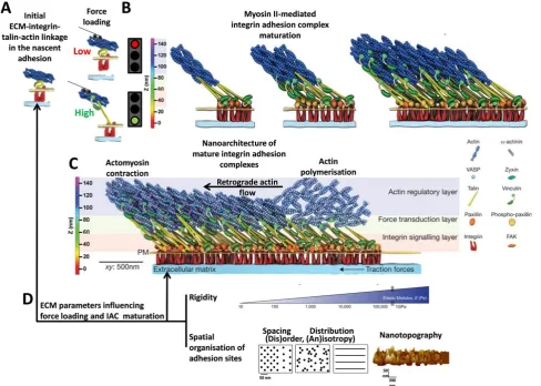
Fig. 2 Integrin-mediated mechanosensing and mechanotransductive sequence with influencing parameters of the
extracellular matrix
(A) The cartoon illustrates the initial ECM-integrin-talin-actin linkage in the nascent adhesions and how the force loading within this molecular clutch determines whether this structure disassembles (in case of too low force loading) or reinforces (in case of sufficient force loading) and (B) matures into integrin adhesion complexes (IAC) by recruitment of further proteins. (C) In this graphic the stratified nanoarchitecture of mature IAC with its different layers is shown. (D) The extent of force loading and IAC maturation is determined by biophysical cues of the extracellular matrix, in particular the rigidity and the spatial organisation of the integrin adhesion sites (in terms of spacing, distribution and nanotopography). Further details on IAC maturation are outlined in paragraph 3.1.
10
Actually these processes in the cell/microenvironment interface are even more complex with various furtherlevels of regulation, such as e.g. mechanisms to modulate the cell surface availability of integrins by endocytosis
(clathrin-dependent or –indepent (CLIC/GEEC) routes), trafficking/recycling, or degradation, as well as talin
competitors/inhibitors or talin cleavage. The interested reader can find further details on certain aspects in recent
reviews of Gauthier and Roca-Cusachs (Gauthier and Roca-Cusachs, 2018), Green and Brown (Green and Brown,
2019), Humphries et al. (Humphries et al., 2019), Sun et al. (Sun et al., 2019) and Kechagia et al. (Kechagia et al.,
2019).
These events in the cell/microenvironment interface contribute essentially to the regulation of many cell
biological behaviours. The next paragraph will outline the current understanding about the involvement of the
mechanotransductive pathway in neuronal cell behaviour (some examples are highlighted in Fig. 3). Considering the
aforementioned local heterogeneity and complexity of biophysical cues in the brain ECM (Fig. 1), these insights are
also highly relevant in. regards to the optimisation of biomaterial approaches that are based on exploitation of
mechanotransductive mechanisms (Fig. 4).
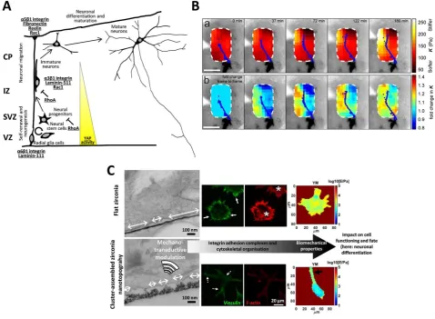
Fig. 3 Mechanotransductive processes in neuronal development and functioning
11
recordings or (b) the stiffness changes over time in the same region. The fluorescently labelled axon was tracked and outlined in blue and documents the directed axon movement towards the softer region (Scale bars = 100 µm). The image has been reproduced from Thompson et al. (Thompson et al., n.d.). (C) The panel demonstrates the modulations along the mechanotransductive sequence induced by the interaction of the neuron-like PC12 cells with ECM-mimicking nanotopographical zirconia substrates produced by the nanofabrication technique supersonic cluster beam deposition, compared to flat zirconia surfaces. In the transmission electron images it can be seen that the cells interact only with the apical part of the nanotopographical asperities which restricts the dimension of the nanometric adhesion sites (indicated by the white arrows) to smaller sizes with respect to the situation on flat zirconia. Also the integrin adhesion complexes (vinculin staining in green recorded by TIRF microscopy) remain of small dimensions (focal contact/point contact size, see white arrows with dashed lines) whereas mature focal adhesions form only on the flat substrate (see white arrows). Consequently, on the nanostructured zirconia no stress fibre formation (epifluorescence of phalloidin staining in red) can be noted, while there are abundant stress fibres on the flat zirconia (examples marked by white asterisks). The cells on the nanotopographical substrate are softer than on the flat surface (quantified by atomic microscopy-based analysis, the colour code indicates the Young’s modulus (YM)) and neurite outgrowth was visible (black arrow). The image was adapted from Schulte et al. (Schulte et al., 2017), Copyright (2017) American Chemical Society.
3.2 Mechanotransductive processes and signalling in neuronal cell development and functioning
Albeit many details remain elusive, it is evident that the ECM configuration and integrin-mediated
mechanosensing/-transduction participate in the control of neuronal cell development and functioning at all stages (Franze et al.,
2013),(Stukel and Willits, 2015),(Park and Goda, 2016),(Barnes et al., 2017),(Lilja and Ivaska, 2018),(Long and
Huttner, 2019),(Xu et al., 2019) (Fig. 3-4).
Integrin/microenvironment interactions and RhoGTPase signalling play an important role during neurogenesis
and the subsequent neuronal long-range migration when the neuronal progenitors move towards the final destination
and terminally differentiate into neurons (Tate et al., 2004),(Fietz et al., 2012),(Long and Huttner, 2019),(Xu et al.,
2019). The regulation of neural progenitor proliferation in the stem cell niche during neurogenesis depends on α6β1
integrin-mediated laminin (predominantly laminin-111) binding which activates MAPK signalling (Campos et al.,
2004),(Flanagan et al., 2006),(Haubst et al., 2006),(Lathia et al., 2007),(Ma et al., 2008),(Shen et al., 2008),(Long and
Huttner, 2019). Interestingly, it has been shown that the mechanosensitive protein YAP sustains the proliferation of
neural progenitors and negatively regulates their neuronal differentiation (shown for the postnatal mouse retina (Zhang
et al., 2012) and chicken neural tube (Cao et al., 2008)). Recently, it was furthermore demonstrated that α3β1
integrin/laminin 511 interaction-dependent YAP activation fosters survival of immature dopaminergic midbrain neurons
in their niche (Zhang et al., 2017) (Fig. 3A).
During mammalian cerebellar cortex development, neuronal progenitors leave the (sub)ventricular zone after
neurogenesis and undergo a multipolar-bipolar transition before they start to migrate along the radial glia cell (RGC)
fibres. The cytoskeletal and morphological changes of this transition are orchestrated by a complex spatiotemporal
regulation of RhoGTPase activity (Konno et al., 2005),(Xu et al., 2019). Later on, the brain ECM glycoprotein reelin is
decisively involved in the control of neuronal cell adhesiveness. It regulates another shift in neuronal migration mode,
by inhibiting the bipolar α3β1 integrin-dependent migration of the immature neurons along the RGC (Anton et al.,
1999),(Hong et al., 2000),(Dulabon et al., 2000),(Schmid et al., 2004) and activating instead the RGC-independent α5β1
integrin/fibronectin binding-dependent terminal translocation during lamination of the developing neocortex (Sekine et
al., 2012). Dynamic and spatiotemporally coordinated RhoGTPase signalling (Xu et al., 2019), actomyosin activity and
traction forces (Solecki et al., 2009),(Jiang et al., 2015) are required to enable neuronal migration. Loss of Rac1 in the
12
axonal guidance and terminal neuronal differentiation (Chen et al., 2007),(Chen et al., 2009). Lowering RhoA activityinstead fosters neuronal lineage commitment (Keung et al., 2011), as well as neurite initiation and outgrowth, in many
neuronal cell models (Yamaguchi et al., 2001),(Dergham et al., 2002),(Da Silva et al., 2003),(Fournier et al.,
2003),(Schulte et al., 2010),(Gu et al., 2013). In line with this, RhoGTPase signalling has been found to be involved in
various biomaterial-induced (either rigidity or topography-based approaches) effects on neuronal differentiation
(Georges et al., 2006),(Saha et al., 2008),(Teixeira et al., 2009),(Seidlits et al., 2010),(Keung et al., 2011),(Yang et al.,
2014),(Schulte et al., 2016c),(Maffioli et al., 2017),(Chen et al., 2018) (Fig. 3A).
Recently, in vivo data obtained in the developing Xenopus brain demonstrated that retinal ganglion cell axons
are sensitive to rapidly changing mechanical properties within the brain tissue that lead to stiffness gradients, causing a
directional growth of the axons towards softer areas (Koser et al., 2016),(Thompson et al., n.d.) (Fig. 3B). The
directional collective migration of Xenopus neural crest cells can instead be induced by stiffening of the underlying
head mesoderm via integrin-dependent mechanosensing (Barriga et al., 2018).
Interesting insights on the processes in the neuron/microenvironment interface and mechanotransductive
signalling were obtained from studies that use biomaterials with topographical surfaces (examples in Fig. 4C).
Nanotopographical features that restrict the maturation of IAC to dimensions beneath focal adhesion size and
consequentially decrease also stress fibre formation (Bugnicourt et al., 2014),(Schulte et al., 2016c),(Baek et al.,
2018),(Baek et al., 2019) and cell rigidity (Schulte et al., 2016c) foster neuronal differentiation (Fig. 3C). These types
of neuron/nanotopography interactions modulated the expression and phosphorylation levels of proteins that are known
to be important components of the IAC and mechanotransductive machinery/signalling sequence (e.g., FAK
phosphorylation and ILK signalling) (Schulte et al., 2016c),(Schulte et al., 2016b),(Maffioli et al., 2017),(Baek et al.,
2018). A study performed with a microtopographical substrates indicated that the ubiquitin E3a ligase; a protein which
targets several integrin signalling/mechanotransduction-related proteins (such as, e.g., src family members) for
degradation and whose deficiency leads to the neurodevelopmental disorder Angelman syndrome, is involved neurite
contact guidance and neuronal topography sensing (Tonazzini et al., 2016). Furthermore, it has been reported that
nanotopography- and soft substrate-promoted neuronal differentiation was accompanied by an increase in Ser127
phosphorylation of YAP and its cytoplasmic retention in a actin cytoskeleton-dependent manner (Musah et al.,
2014),(Sun et al., 2014),(Baek et al., 2018).
Consistent with their function as explorative compartments of developing neurons, neurite growth cones are
particularly influenced and controlled by mechanotransductive processes. The extent of molecular clutch engagement to
the retrograde actin flow-generated forces regulates neurite/axon guidance and pathfinding. Within growth cones
dynamic and small (focal complex size) integrin-mediated interaction sites are formed with microenvironmental cues,
called point contacts. Their spatiotemporal dynamics are tightly governed by a balanced signalling interplay involving
diverse IAC signalling-related components, such as RhoGTPases, src, PAK and FAK (Robles et al., 2005),(Woo and
Gomez, 2006),(Medeiros et al., 2006),(Myers et al., 2011),(Vitriol and Zheng, 2012),(Santiago-Medina et al.,
2013),(Kerstein et al., 2015),(Nichol et al., 2016) (Fig. 4B, on the right). Growth cones are soft structures (with low
elastic modulus around hundreds of Pa and a tension in the range of hundreds of pN (Betz et al., 2011)) and produce
relatively weak forces (compared to other cell types, such as e.g. fibroblasts or epithelial cells). The traction forces are
in the range of tens of pN per um2, executed in particular in the peripheral regions growth cone (such as filopodia and lamellipodial edges) (Hällström et al., 2010),(Betz et al., 2011), (Koch et al., 2012),(Franze et al., 2013),(O’Toole et al.,
13
forces than CNS neurons, though (Koch et al., 2012). The protrusive forces, measured for retinal ganglion cell growthcones, are in the order of 100 pN (Fuhs et al., 2013).
Also later on during neuronal maturation, mechanoregulatory processes are potentially highly relevant in
controlling the spatiotemporal dynamics of synaptogenesis and activity-dependent synapse plasticity. Postsynaptic
terminals of synapses are rich in mechanotransductively active components, such as cell adhesion molecules and
regulators of actin dynamics and organisation (e.g., cofilin, drebrin α-actinin and cortactin) (McGeachie et al.,
2011),(Sheng and Kim, 2011),(Kilinc, 2018). Cofilin, e.g., is strongly recruited to dendritic spines during long-term
potentiation and involved in the remodelling of the synapse structure (Bosch et al., 2014). The configuration and
composition of the ECM and dynamic integrin/ECM (dis)engagement modulates the activity-dependent functional
plasticity of synaptic connectivity and neural circuitry (Chavis and Westbrook, 2001),(Dityatev et al., 2010a),
(McGeachie et al., 2011),(Orlando et al., 2012),(Kerrisk et al., 2013),(Bikbaev et al., 2015),(Park and Goda,
2016),(Kilinc, 2018).
More details about brain ECM or RhoGTPase signalling during neuronal development are outlined in reviews
by Long and Huttner (Long and Huttner, 2019), respectively Xu et al. (Xu et al., 2019). Brain tissue mechanics are
covered in detail by Franze et al. (Franze et al., 2013) and Barnes et al. (Barnes et al., 2017). The involvement of
integrins in synapse formation and plasticity is highlighted in reviews from Park and Goda (Park and Goda, 2016) and
14
Fig. 4 Biomaterial and biophysical approaches to study and/or control mechanotransductive processes thatregulate neuronal cell development and functioning
(A) The scheme illustrates important stages during neuronal cell development. (B) The graphic outlines schematically principal (extra)cellular structures and processes of interest within the integrin adhesion complex-mediated mechanotransductive sequence in neuronal cells (on the right the axon/neurite growth cone is highlighted) and some biophysical methods (AFM: Atomic Force Microscopy, TFM: Traction Force Microscopy) that are used to study them (see also paragraph 4). (C) Examples of different bioengineering approaches are listed that are used for the production of biomaterials which mimic biophysical extracellular matrix (ECM) features, such as hydrogels derived from decellularised brain ECM (Jin et al., 2018), hydrogels made of polymers (in this case polyacrylamide gels with different stiffness ranges, Young’s modulus: left; <14.5 kPa, middle: 14.5-29 kPa, right: >29 kPa) (Hadden et al., 2017), electrospinning, lithographic methods and pattern transfer, as well as reactive-ion etching (RIE) (Chen et al., 2014), carbon nanotubes (Cellot et al., 2011) and assembly of zirconia nanocluster by supersonic cluster beam deposition (Schulte et al., 2017). Various approaches are referenced throughout the review in which these types of substrates are applied and exploited to study and guide neuronal cell mechanotransduction and development. (A-C) Together these mechanobiological approaches can contribute to a better understanding of how mechanotransductive processes impact on neuronal cell development and functioning.
The figure contains adapted elements of images with permission from Jin et al. (Jin et al., 2018), Copyright (2018) Springer Nature; Hadden et al. (Hadden et al., 2017), Copyright (2017) National Academy of Sciences; Chen et al. (Chen et al., 2014), Copyright (2014) Elsevier; as well as elements from Cellot et al. (Cellot et al., 2011), Schulte et al. (Schulte et al., 2016c),(Schulte et al., 2016b), and Maffioli et al. (Maffioli et al., 2017).
4. Biophysical Methods to Study Neuronal Mechanobiology
The aim of research in cellular mechanobiology is to understand how biophysical properties of the cells and the
surrounding microenvironment are intertwined and influence each other mutually to regulate cell morphology and fate
(Fig. 4). In light of this premise, it is fundamental to obtain a precise quantification of the structural and mechanical
properties of the microenvironment, cells or tissues. The exploration of mechanotransductive processes relies
furthermore on the ability to apply accurately controlled physical stimuli to living cells and to measure the forces of
their mechanobiological actions (Iskratsch et al., 2014), in particular also for the understanding of neuronal cell
behaviour (Athamneh and Suter, 2015).
The study of biophysical aspects of cells and their microenvironment and/or the nature of their interaction
requires specific instrumentation (Fig. 4B). This paragraph will give a short overview about some principal
experimental techniques that often were essential to gain the insights into mechanotransductive processes outlined
throughout this review.
Optical/Magnetic Tweezers
Optical and magnetic tweezers were amongst the first tools that enabled cell biologists to quantitatively measure the
weak forces produced by cells, or to apply corresponding forces to the cells (Iskratsch et al., 2014).
The optical tweezers technique consists in using a suitably functionalised µm-sized dielectric bead as a probe,
and exploiting the restoring force generated by the interaction of a laser with the dielectric sphere to control the position
of the bead (Fig. 4B). Measuring the probe displacement in experimental conditions provides a measure of the applied
force. The bead diameter can range from few hundreds of nm to tens of µm depending on the contact region and the
applied pressure required in the experiments. The limitations in bead dimensions are due to the fact that the trapping
force decreases with the bead diameter. The tweezers can be calibrated accurately to know how much force is required
15
pN and the effective force constant of the tweezer is typically in the range of pN/m, providing extreme sensitivity to pNforces (Moffitt et al., 2008),(Neuman and Nagy, 2008),(Capitanio and Pavone, 2013),(Siedlik et al., 2016).
The magnetic tweezers approach relies on the same principle of action as the optical tweezer, but in this case
magnets are used to position or apply forces on and para-ferromagnetic beads. The advantage of the magnetic control is
that many beads can be affected simultaneously (Neuman and Nagy, 2008),(Siedlik et al., 2016),(De Vlaminck and
Dekker, 2012),(Le et al., 2016), and it is also possible to induce twisting to the beads, allowing to test different degrees
of freedom of the system under investigation (Wang et al., 1993),(Strick et al., 2000). The drawback is that the spatial
variation of the field results in a non-uniform force applied to the beads; careful design of the magnetic tweezer
apparatus permits nowadays for nearly constant gradient field over more than hundreds of microns (Neuman and Nagy,
2008),(Siedlik et al., 2016),(De Vlaminck and Dekker, 2012),(Le et al., 2016).
An early use of magnetic tweezers in cell biological research led to one of the seminal works for the
mechanobiology field by Wang et al. in 1993 (Wang et al., 1993), showing the mechanosensitivity of integrins and
mechanotransduction through the actin cytoskeleton. In 1995 Dai and Sheetz (Dai and Sheetz, 1995) characterised the
mechanical properties of the neuronal growth cone by means of optical tweezers, demonstrating the role of the actin
cytoskeleton in affecting the elastic properties of the membrane. Similar experiments were performed later on to
determine the forces exerted by growing filopodia and lamellipodia (Cojoc et al., 2007) or axons (Moore et al.,
2009),(Kilinc et al., 2014) during neuronal differentiation, or to compare forces generated by the growth cones of
neurons from the central nervous system (hippocampal neurons) and the peripheral nervous system (dorsal root ganglia)
(Amin et al., 2013).
Moving the stage in xy-direction or the focus of the laser spot in z-direction in a finely tuned and precisely
controlled manner enables also the application of static or oscillatory forces (in the range of a few pN), in order to study
e.g. the rheological behaviour of cells. With this method, it was recently demonstrated that mechanical stimuli with pN
forces activate calcium channels (Falleroni et al., 2018) and that viscoelastic properties of soma and neurite differ and
that in neurites these properties change in dependency of the substrate rigidity (Grevesse et al., 2015).
Additional details on optical and/or magnetic tweezer-based approaches, focussing on applications relevant for
biophysics and mechanotransduction, can be found in Capitanio and Pavone (Capitanio and Pavone, 2013), Siedlik et al.
(Siedlik et al., 2016) and Le et al. (Le et al., 2016).
Atomic Force Microscopy
AFM belongs to the branch of scanning probe microscopy (SPM). In recent years it has gained increasing importance in
the mechanobiology field as an instrument with versatile application modes that allow high resolution morphological
imaging (i.e., surface topography) and characterisation of mechanical properties (e.g., stiffness or viscoelasticity) of
biological samples, also simultaneously (Haase and Pelling, 2015),(Gavara, 2017),(Alcaraz et al., 2018),(Krieg et al.,
2019).
The probes are attached to an elastic lever (cantilever) that flexes under the interaction forces between the
probe and the surface of the sample. The corresponding cantilever deflections are measured by a quadrant photodiode
that detects the displacement of a laser reflecting from the exterior part of the cantilever. The probe shape and
dimension can be modified according to the needs of the measurement and the information to be obtained. Pyramidal or
narrow conical tips are usually exploited for high resolution imaging application (Krieg et al., 2019). More spherical
(Indrieri et al., 2011),(Puricelli et al., 2015), or even cylindrical (Rico et al., 2007), probes instead are more suitable for
16
contact geometry. This system can be run in different modes depending on which structural and/or mechanicalparameters of the measured samples are to be characterised (Haase and Pelling, 2015),(Gavara, 2017),(Alcaraz et al.,
2018),(Krieg et al., 2019) (Fig. 4B).
During the scan, by keeping the cantilever deflection constant trough a feedback circuit, the surface
morphology of the sample can be reconstructed. This standard imaging application is e.g. widely used to image the
nanotopography of cells (also neuronal cells and their compartments, such as growth cones (Parpura et al.,
1993),(Grzywa et al., 2006),(Xiong et al., 2009)), natural extracellular matrices (Abrams et al., 2000),(Last et al.,
2010),(Gasiorowski et al., 2013), or nanostructured biomaterials (Cellot et al., 2011),(Schulte et al., 2016c),(Schulte et
al., 2016b)). It is also possible to apply forces by pushing the probe into the sample. Measuring the flexure of the
cantilever through the extent of laser dislocation, the upward force acting on the tip can be calculated and mechanical
properties of the sample can be determined. Varying the cantilever geometry allows covering more than six orders of
magnitude in force sensitivity (from tens of pN to tens of µN). The nanomechanical analysis of soft biological samples
(such as e.g. cells and many tissues or extracellular matrices) with AFM is still to-date not straightforward. Attention
must be drawn to the choice of the right experimental conditions for the application of contact mechanics models (such
as Hertz (Hertz, 1882) or Sneddon (Sneddon, 1965)) and also in the data analyses (Puricelli et al., 2015),(Schillers et al.,
2017). In the neuroscience field, by means of these types of AFM-based characterisations it was possible to determine
the mechanical properties (Young’s modulus) of substrates that are able to promote neuronal differentiation processes
(Saha et al., 2008),(Leipzig and Shoichet, 2009),(Teixeira et al., 2009),(Banerjee et al., 2009),(Seidlits et al.,
2010),(Keung et al., 2012),(Keung et al., 2013),(Musah et al., 2014),(Sun et al., 2014), as well as the mechanics of brain
tissue or spinal cord (Elkin et al., 2007),(Christ et al., 2010),(Koser et al., 2016),(Koser et al., 2015),(Antonovaite et al.,
2018) (Fig. 1B and Fig. 3B) and different types of neuronal cells (Lu et al., 2006),(Grzywa et al., 2006),(Spedden et al.,
2012),(Schulte et al., 2016c), including changes due to mechanotransductive processes (Koser et al., 2016),(Schulte et
al., 2016c),(Thompson et al., n.d.) (as detailed also in precedent paragraphs) (Fig. 3B,C).
Instead of applying forces to the cell with the AFM probe to deform the cell in order to analyse the cellular
mechanical properties, the AFM can also be used to measure cellular adhesion forces (e.g., integrin/ligand binding
(Strohmeyer et al., 2017)) by the force spectroscopy technique. There are two principal approaches to apply this
technique. In one case (single-cell force spectroscopy), a cell is attached to a tipless cantilever (using basically the cell
as probe) and brought smoothly into contact with a substrate of interest (Taubenberger et al., 2007),(Helenius et al.,
2008) or even another cell (Puech et al., 2006). In a second approach, an AFM probe is functionalised in a suitable
manner (e.g., with proteins of the ECM) and brought gently into contact with the cell membrane. In both cases after a
sufficient time of contact to enable the desired interaction, the probe is then retracted in order to break the generated
bonds. From the obtained force versus distance curves; typically exhibiting a complex pattern of sudden jumps and
plateaux, it is possible to measure the number of ruptured bonds and the distribution of their strength. Several
configurations have been adopted to study cell adhesion and its mechanisms and biomolecular determinants. Depending
on the probe and type of functionalisation even adhesion forces at the single molecule level can be measured (single
molecule force spectroscopy) (Müller et al., 2009),(Dufrêne et al., 2011).
Further information on AFM methodologies and applications in mechanobiology research is available in more
specific reviews from Haase and Pelling (Haase and Pelling, 2015), Gavara (Gavara, 2017), Alcaraz et al. (Alcaraz et al.,
2018) or Krieg et al. (Krieg et al., 2019).
17
The first cellular reaction to biophysical stimuli in the microenvironment takes place in the interface between cellmembrane (and its embedded receptors) and the ECM.
Traction force microscopy (TFM) aims at measuring the traction forces exerted by the cell towards the
substrate it interacts with. This can be achieved by different approaches, either fluorescent microspheres embedded into
deformable hydrogel substrates or pillar arrays are used (Fig. 4B). When the cells apply forces on these substrates as a
consequence the beads or the pillars are displaced. Tracking the displacement microscopically and knowing the stiffness
of the hydrogel, respectively pillars, permits an estimate of the applied traction forces and to reconstruct a traction force
field. The spatial resolution of TFM is limited by the optical set up and by the relative density of the beads, respectively
pillars. Several models have been proposed to accurately convert the strain field into a stress field (Schwarz and Soiné,
2015). In the neuronal context TFM has been used, e.g., to investigate the forces involved in the growth cones of
neurons (Chan and Odde, 2008),(Betz et al., 2011) or during neuronal migration (Jiang et al., 2015). A combined
TFM/AFM approach was recently applied to study the correlation between cellular rigidity, viscoelasticity, and the
contractile prestress, highlighting the role of the actomyosin machinery (Schierbaum et al., 2019).
Another tool to measure the mechanical forces exerted by the cell are molecular tension probes. The
functioning of these probes is based on an extendable linker (which can be build up by adequate polymers, or
biomacromolecules (oligonucleotide or protein)) flanked by a spectroscopic ruler, consisting of a fluorophore and a
quencher (Fig. 4B). If a sufficient force is applied to the probe, bonds within the linker region sequentially break and
the linker extents leading to a displacement and separation of the fluorophore and quencher. The extent of linker
extension is usually measured by fluorescence resonance energy transfer. Knowing the bond strengths inside the linker
region, the applied force can be quantified (Jurchenko and Salaita, 2015),(Liu et al., 2017). These tension probes can be
utilised as immobilised sensors on a substrate (Liu et al., 2014) or also intracellularly if they are integrated into proteins,
an example is the vinculin tension sensor (Grashoff et al., 2010),(Jurchenko and Salaita, 2015).
5. Conclusion and Outlook
In this review we attempted to outline the crucial involvement of mechanobiological aspects in physiological brain and
neuronal cell development and functioning. In recent years there are furthermore increasing indications that aberrations
in the brain ECM organisation and mechanotransductive processes of neuronal cells strongly contribute to various
neuronal pathologies, such as neurodegenerative diseases (e.g., in multiple sclerosis, amyotrophic lateral sclerosis and
Alzheimer’s disease) and neurodevelopmental disorders (such as, e.g., autism spectrum disorders and schizophrenia)
(Tyler, 2012),(Lau et al., 2013),(Franze et al., 2013),(Park and Goda, 2016) (Lilja and Ivaska, 2018),(Barnes et al.,
2017), or primary brain cancers (e.g., in glioblastoma) (Barnes et al., 2017),(Tanner, 2018). The research in this field is
often still in its infancy, gaining a deeper insight into neuronal cell mechanotransduction by means of biophysical
approaches is therefore essential (Fig. 4) and will help to understand better how abnormal mechanotransductive
processes contribute to the aetiology of brain disorders (Tyler, 2012),(Franze et al., 2013),(Barnes et al., 2017),(Lilja
and Ivaska, 2018),(Tanner, 2018). The biomedical significance of research in this direction is underlined, e.g., by a very
recent study which suggests a glycocalyx/IAC mechanosignalling feedback loop regulated by tension in glioblastoma
multiforme that might be causal for the high recurrence of this tumour after current chemotherapy treatments (Barnes et
al., 2018).
18
This project has received funding from the European Union Horizon 2020 research and innovation programme underthe Marie Sklodowska-Curie grant agreement No. 812772 (Phys2BioMed), and under the FET Open grant agreement
No. 801126 (EDIT).
Conflict of Interest
The authors declare to have no conflict of interest.
REFERENCES
Abrams, G.A., Goodman, S.L., Nealey, P.F., Franco, M., Murphy, C.J., 2000. Nanoscale topography of the basement membrane underlying the corneal epithelium of the rhesus macaque. Cell Tissue Res. 299, 39–46.
Alcaraz, J., Otero, J., Jorba, I., Navajas, D., 2018. Bidirectional mechanobiology between cells and their local extracellular matrix probed by atomic force microscopy. Semin. Cell Dev. Biol., Application of Atomic Force Microscopy in cell biology 73, 71–81. https://doi.org/10.1016/j.semcdb.2017.07.020
Amin, L., Ercolini, E., Ban, J., Torre, V., 2013. Comparison of the force exerted by hippocampal and DRG growth cones. PloS One 8, e73025. https://doi.org/10.1371/journal.pone.0073025
Anton, E.S., Kreidberg, J.A., Rakic, P., 1999. Distinct functions of alpha3 and alpha(v) integrin receptors in neuronal migration and laminar organization of the cerebral cortex. Neuron 22, 277–289.
Antonovaite, N., Beekmans, S.V., Hol, E.M., Wadman, W.J., Iannuzzi, D., 2018. Regional variations in stiffness in live mouse brain tissue determined by depth-controlled indentation mapping. Sci. Rep. 8, 12517. https://doi.org/10.1038/s41598-018-31035-y
Arnold, M., Cavalcanti-Adam, E.A., Glass, R., Blümmel, J., Eck, W., Kantlehner, M., Kessler, H., Spatz, J.P., 2004. Activation of integrin function by nanopatterned adhesive interfaces. Chemphyschem Eur. J. Chem. Phys. Phys. Chem. 5, 383–388. https://doi.org/10.1002/cphc.200301014
Athamneh, A.I.M., Suter, D.M., 2015. Quantifying mechanical force in axonal growth and guidance. Front. Cell. Neurosci. 9. https://doi.org/10.3389/fncel.2015.00359
Baek, J., Cho, S.-Y., Kang, H., Ahn, H., Jung, W.-B., Cho, Y., Lee, E., Cho, S.-W., Jung, H.-T., Im, S.G., 2018. Distinct Mechanosensing of Human Neural Stem Cells on Extremely Limited Anisotropic Cellular Contact. ACS Appl. Mater. Interfaces. https://doi.org/10.1021/acsami.8b10171
Baek, J., Jung, W.-B., Cho, Y., Lee, E., Yun, G.-T., Cho, S.-Y., Jung, H.-T., Im, S.G., 2019. Facile Fabrication of High-Definition Hierarchical Wrinkle Structures for Investigating the Geometry-Sensitive Fate Commitment of Human Neural Stem Cells. ACS Appl. Mater. Interfaces 11, 17247–17255. https://doi.org/10.1021/acsami.9b03479
Balgude, A.P., Yu, X., Szymanski, A., Bellamkonda, R.V., 2001. Agarose gel stiffness determines rate of DRG neurite extension in 3D cultures. Biomaterials 22, 1077–1084.
Banerjee, A., Arha, M., Choudhary, S., Ashton, R.S., Bhatia, S.R., Schaffer, D.V., Kane, R.S., 2009. The influence of hydrogel modulus on the proliferation and differentiation of encapsulated neural stem cells. Biomaterials 30, 4695–4699. https://doi.org/10.1016/j.biomaterials.2009.05.050
Barnes, J.M., Kaushik, S., Bainer, R.O., Sa, J.K., Woods, E.C., Kai, F., Przybyla, L., Lee, M., Lee, H.W., Tung, J.C., Maller, O., Barrett, A.S., Lu, K.V., Lakins, J.N., Hansen, K.C., Obernier, K., Alvarez-Buylla, A., Bergers, G., Phillips, J.J., Nam, D.-H., Bertozzi, C.R., Weaver, V.M., 2018. A tension-mediated glycocalyx–integrin feedback loop promotes mesenchymal-like glioblastoma. Nat. Cell Biol. 20, 1203. https://doi.org/10.1038/s41556-018-0183-3
Barnes, J.M., Przybyla, L., Weaver, V.M., 2017. Tissue mechanics regulate brain development, homeostasis and disease. J. Cell Sci. 130, 71–82. https://doi.org/10.1242/jcs.191742
Barriga, E.H., Franze, K., Charras, G., Mayor, R., 2018. Tissue stiffening coordinates morphogenesis by triggering collective cell migration in vivo. Nature 554, 523–527. https://doi.org/10.1038/nature25742
Bass, M.D., Roach, K.A., Morgan, M.R., Mostafavi-Pour, Z., Schoen, T., Muramatsu, T., Mayer, U., Ballestrem, C., Spatz, J.P., Humphries, M.J., 2007. Syndecan-4-dependent Rac1 regulation determines directional migration in response to the extracellular matrix. J. Cell Biol. 177, 527–538. https://doi.org/10.1083/jcb.200610076
Béduer, A., Vieu, C., Arnauduc, F., Sol, J.-C., Loubinoux, I., Vaysse, L., 2012. Engineering of adult human neural stem cells differentiation through surface micropatterning. Biomaterials 33, 504–514. https://doi.org/10.1016/j.biomaterials.2011.09.073
Betz, T., Koch, D., Lu, Y.-B., Franze, K., Käs, J.A., 2011. Growth cones as soft and weak force generators. Proc. Natl. Acad. Sci. 108, 13420–13425. https://doi.org/10.1073/pnas.1106145108
19
Bonneh-Barkay, D., Wiley, C.A., 2009. Brain extracellular matrix in neurodegeneration. Brain Pathol. Zurich Switz. 19,573–585. https://doi.org/10.1111/j.1750-3639.2008.00195.x
Borghi, F., Scaparra, B., Paternoster, C., Milani, P., Podestà, A., 2018. Electrostatic Double-Layer Interaction at the Surface of Rough Cluster-Assembled Films: The Case of Nanostructured Zirconia. Langmuir 34, 10230– 10242. https://doi.org/10.1021/acs.langmuir.8b01387
Bosch, M., Castro, J., Saneyoshi, T., Matsuno, H., Sur, M., Hayashi, Y., 2014. Structural and molecular remodeling of dendritic spine substructures during long-term potentiation. Neuron 82, 444–459. https://doi.org/10.1016/j.neuron.2014.03.021
Bridgman, P.C., Dave, S., Asnes, C.F., Tullio, A.N., Adelstein, R.S., 2001. Myosin IIB Is Required for Growth Cone Motility. J. Neurosci. 21, 6159–6169. https://doi.org/10.1523/JNEUROSCI.21-16-06159.2001
Bugnicourt, G., Brocard, J., Nicolas, A., Villard, C., 2014. Nanoscale Surface Topography Reshapes Neuronal Growth in Culture. Langmuir 30, 4441–4449. https://doi.org/10.1021/la5001683
Campos, L.S., Leone, D.P., Relvas, J.B., Brakebusch, C., Fässler, R., Suter, U., ffrench-Constant, C., 2004. Beta1 integrins activate a MAPK signalling pathway in neural stem cells that contributes to their maintenance. Dev. Camb. Engl. 131, 3433–3444. https://doi.org/10.1242/dev.01199
Cao, X., Pfaff, S.L., Gage, F.H., 2008. YAP regulates neural progenitor cell number via the TEA domain transcription factor. Genes Dev. 22, 3320–3334. https://doi.org/10.1101/gad.1726608
Capitanio, M., Pavone, F.S., 2013. Interrogating Biology with Force: Single Molecule High-Resolution Measurements with Optical Tweezers. Biophys. J. 105, 1293–1303. https://doi.org/10.1016/j.bpj.2013.08.007
Carisey, A., Tsang, R., Greiner, A.M., Nijenhuis, N., Heath, N., Nazgiewicz, A., Kemkemer, R., Derby, B., Spatz, J., Ballestrem, C., 2013. Vinculin Regulates the Recruitment and Release of Core Focal Adhesion Proteins in a Force-Dependent Manner. Curr. Biol. 23, 271–281. https://doi.org/10.1016/j.cub.2013.01.009
Case, L.B., Baird, M.A., Shtengel, G., Campbell, S.L., Hess, H.F., Davidson, M.W., Waterman, C.M., 2015. Molecular mechanism of vinculin activation and nanoscale spatial organization in focal adhesions. Nat. Cell Biol. 17, 880–892. https://doi.org/10.1038/ncb3180
Case, L.B., Waterman, C.M., 2015. Integration of actin dynamics and cell adhesion by a three-dimensional, mechanosensitive molecular clutch. Nat. Cell Biol. 17, 955–963. https://doi.org/10.1038/ncb3191
Cellot, G., Toma, F.M., Varley, Z.K., Laishram, J., Villari, A., Quintana, M., Cipollone, S., Prato, M., Ballerini, L., 2011. Carbon Nanotube Scaffolds Tune Synaptic Strength in Cultured Neural Circuits: Novel Frontiers in Nanomaterial–Tissue Interactions. J. Neurosci. 31, 12945–12953. https://doi.org/10.1523/JNEUROSCI.1332-11.2011
Chan, C.E., Odde, D.J., 2008. Traction dynamics of filopodia on compliant substrates. Science 322, 1687–1691. https://doi.org/10.1126/science.1163595
Changede, R., Sheetz, M., 2017. Integrin and cadherin clusters: A robust way to organize adhesions for cell mechanics. BioEssays 39, n/a-n/a. https://doi.org/10.1002/bies.201600123
Changede, R., Xu, X., Margadant, F., Sheetz, M.P., 2015. Nascent Integrin Adhesions Form on All Matrix Rigidities after Integrin Activation. Dev. Cell 35, 614–621. https://doi.org/10.1016/j.devcel.2015.11.001
Chavis, P., Westbrook, G., 2001. Integrins mediate functional pre- and postsynaptic maturation at a hippocampal synapse. Nature 411, 317–321. https://doi.org/10.1038/35077101
Chen, L., Liao, G., Waclaw, R.R., Burns, K.A., Linquist, D., Campbell, K., Zheng, Y., Kuan, C.-Y., 2007. Rac1 Controls the Formation of Midline Commissures and the Competency of Tangential Migration in Ventral Telencephalic Neurons. J. Neurosci. 27, 3884–3893. https://doi.org/10.1523/JNEUROSCI.3509-06.2007 Chen, L., Melendez, J., Campbell, K., Kuan, C.-Y., Zheng, Y., 2009. Rac1 deficiency in the forebrain results in neural
progenitor reduction and microcephaly. Dev. Biol. 325, 162–170. https://doi.org/10.1016/j.ydbio.2008.10.023 Chen, W., Han, S., Qian, W., Weng, S., Yang, H., Sun, Y., Villa-Diaz, L.G., Krebsbach, P.H., Fu, J., 2018.
Nanotopography regulates motor neuron differentiation of human pluripotent stem cells. Nanoscale 10, 3556– 3565. https://doi.org/10.1039/c7nr05430k
Chen, W., Shao, Y., Li, X., Zhao, G., Fu, J., 2014. Nanotopographical Surfaces for Stem Cell Fate Control: Engineering Mechanobiology from the Bottom. Nano Today 9, 759–784. https://doi.org/10.1016/j.nantod.2014.12.002 Chen, Y., Lee, H., Tong, H., Schwartz, M., Zhu, C., 2017. Force regulated conformational change of integrin αVβ3.
Matrix Biol. J. Int. Soc. Matrix Biol. 60–61, 70–85. https://doi.org/10.1016/j.matbio.2016.07.002
Cheng, C.-M., LeDuc, P.R., Lin, Y.-W., 2011. Localized bimodal response of neurite extensions and structural proteins in dorsal-root ganglion neurons with controlled polydimethylsiloxane substrate stiffness. J. Biomech. 44, 856– 862. https://doi.org/10.1016/j.jbiomech.2010.12.006
Choi, C.K., Vicente-Manzanares, M., Zareno, J., Whitmore, L.A., Mogilner, A., Horwitz, A.R., 2008. Actin and alpha-actinin orchestrate the assembly and maturation of nascent adhesions in a myosin II motor-independent manner. Nat. Cell Biol. 10, 1039–1050. https://doi.org/10.1038/ncb1763