N
-(Pyrrolidin-1-ylcarbothioyl)benzamide
Aisha A. Al-abbasi,aMohamed Ibrahim Mohamed Tahirb and Mohammad B. Kassima,c*
a
School of Chemical Sciences & Food Technology, Faculty of Science & Technology, Universiti Kebangsaan Malaysia, 43600 Bangi, Selangor, Malaysia,bDepartment of Chemistry, Faculty of Science, Universiti Putra Malaysia, 43400 UPM Serdang, Selangor, Malaysia, andcFuel Cell Institute, Universiti Kebangsaan Malaysia, 43600 Selangor, Malaysia
Correspondence e-mail: mbkassim@ukm.my
Received 5 December 2011; accepted 13 December 2011
Key indicators: single-crystal X-ray study;T= 150 K; mean(C–C) = 0.002 A˚; Rfactor = 0.035;wRfactor = 0.098; data-to-parameter ratio = 15.5.
In the title compound, C12H14N2OS, the pyrrolidine ring
adopts an envelope conformation with the C atom at the 3-position as the flap and makes a dihedral angle of 65.80 (9)
with the benzene ring. In the crystal, N—H O hydrogen bonds join c-glide related molecules into chains extended along [001] that are further connected into (100) layers via
C—H O interactions.
Related literature
For related compounds, their structural parameters and chemical properties, see: Al-abbasi et al. (2010, 2011); Al-abbasi & Kassim (2011); Ngahet al.(2006).
Experimental
Crystal data
C12H14N2OS Mr= 234.31
Monoclinic,P21=c
a= 10.3666 (4) A˚ b= 14.6008 (5) A˚ c= 7.8240 (3) A˚
= 98.446 (4)
V= 1171.40 (8) A˚3 Z= 4
CuKradiation
= 2.29 mm1 T= 150 K
0.130.060.03 mm
Data collection
Oxford Diffraction Gemini area-detector diffractometer Absorption correction: multi-scan
(CrysAlis RED; Oxford Diffraction, 2006) Tmin= 0.870,Tmax= 0.934
8077 measured reflections 2245 independent reflections 1958 reflections withI> 2(I) Rint= 0.024
Refinement
R[F2> 2(F2)] = 0.035 wR(F2) = 0.098 S= 1.03 2245 reflections
145 parameters
H-atom parameters constrained
max= 0.32 e A˚
3
min=0.27 e A˚
3
Table 1
Hydrogen-bond geometry (A˚ ,).
D—H A D—H H A D A D—H A
N1—H1 O1i
0.86 2.05 2.8637 (17) 157
C11—H11A O1ii
0.97 2.52 3.339 (2) 142
Symmetry codes: (i)x;yþ1 2;zþ
1
2; (ii)x;yþ1;z.
Data collection: CrysAlis CCD (Oxford Diffraction, 2006); cell refinement: CrysAlis RED (Oxford Diffraction, 2006); data reduc-tion:CrysAlis RED; program(s) used to solve structure:SHELXS97 (Sheldrick, 2008); program(s) used to refine structure:SHELXL97 (Sheldrick, 2008); molecular graphics:SHELXTL(Sheldrick, 2008); software used to prepare material for publication: SHELXTL, PLATON(Spek, 2009) andpublCIF(Westrip, 2010).
The authors thank Universiti Kebangsaan Malaysia for providing facilities and grants (UKM-OUP-TK-16–73/2011 and UKM-PTS-016–2010) and the Libyan Government for providing a scholarship to AAA.
Supplementary data and figures for this paper are available from the IUCr electronic archives (Reference: GK2440).
References
Al-abbasi, A. A. & Kassim, M. B. (2011).Acta Cryst.E67, o611.
Al-abbasi, A. A., Yamin, B. M. & Kassim, M. B. (2011).Acta Cryst.E67, o1891. Al-abbasi, A. A., Yarmo, M. A. & Kassim, M. B. (2010).Acta Cryst.E66,
o2896.
Ngah, N., Kassim, M. B. & Yamin, B. M. (2006).Acta Cryst.E62, o4501–o4502. Oxford Diffraction (2006). CrysAlis CCD and CrysAlis RED. Oxford
Diffraction, Abingdon, England.
Sheldrick, G. M. (2008).Acta Cryst.A64, 112–122. Spek, A. L. (2009).Acta Cryst.D65, 148–155. Westrip, S. P. (2010).J. Appl. Cryst.43, 920–925. Acta Crystallographica Section E
Structure Reports
Online
supporting information
Acta Cryst. (2012). E68, o201 [doi:10.1107/S1600536811053694]
N-(Pyrrolidin-1-ylcarbothioyl)benzamide
Aisha A. Al-abbasi, Mohamed Ibrahim Mohamed Tahir and Mohammad B. Kassim
S1. Comment
The title compound, I, is a derivative of a previously reported (2S)-1-(benzoylthiocarbamoyl)pyrrolidine-2-carboxylic
acid (II) molecule (Ngah et al. 2006). In the crystal structure of I, the five-membered pyrrolidine ring has an envelope
conformation with a maximum deviation from the mean plane of 0.238 (2)Å at C11. The benzene ring
[C1/C2/C3/C4/C5/C6/C7] and the [(S1/N1/N2/C8/C9/C10] fragment are essentially planar and form dihedral angle of
79.03 (6)°.
The C=S and C=O bond lengths in I [1.6737 (17)Å and 1.2273 (19)Å, respectively] are comparable to those of II [1.662
(5)Å and 1.219 (5)Å]. In addition, the thiourea fragment in both compounds adopted a similar configuration with respect
to the benzoyl and pyrrole fragments.
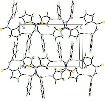
In the crystal, adjacent molecules are linked by N—H···O and C—H···O intermolecular interactions forming a
two-dimensional polymeric structure parallel to (1 0 0) (Figure 2).
S2. Experimental
The title compound was synthesized according to a previously reported compound (Al-abbasi et al., 2010) with some
modifications. A solution of benzoyl chloride (10 mmol) in acetone was added slowly to an equimolar solution of
ammonium thiocyanate in acetone. The reaction mixture was stirred at room temperature before adding piperidine (10
mmol) slowly and left stirring at room temperature for 4 h. The mixture was poured onto a water-ice and then filtered.
The product was recrystallized to give a colourless crystal, suitable for X-ray crystallography (yield 81%; m.p. 407-408
K).
S3. Refinement
The hydrogen atom positions were calculated geometrically and refined in a riding model approximation with C–H bond
Figure 1
The molecular structure of the title compound with displacement ellipsods shown at the 30% probability level.
Figure 2
The crystal packing of the title compound viewed down the c-axis with intermolecular hydrogen bonds drawn as dashed
[image:3.610.130.482.314.651.2]N-(Pyrrolidin-1-ylcarbothioyl)benzamide
Crystal data
C12H14N2OS
Mr = 234.31 Monoclinic, P21/c
Hall symbol: -P 2ybc
a = 10.3666 (4) Å
b = 14.6008 (5) Å
c = 7.8240 (3) Å
β = 98.446 (4)°
V = 1171.40 (8) Å3
Z = 4
F(000) = 496
Dx = 1.329 Mg m−3
Cu Kα radiation, λ = 1.54178 Å Cell parameters from 3639 reflections
θ = 4–71.2°
µ = 2.29 mm−1
T = 150 K Needle, colourless 0.13 × 0.06 × 0.03 mm
Data collection
Oxford Diffraction Gemini area-detector diffractometer
Radiation source: fine-focus sealed tube Graphite monochromator
Detector resolution: 0 pixels mm-1
ω scans
Absorption correction: multi-scan
(CrysAlis RED; Oxford Diffraction, 2006)
Tmin = 0.870, Tmax = 0.934
8077 measured reflections 2245 independent reflections 1958 reflections with I > 2σ(I)
Rint = 0.024
θmax = 71.2°, θmin = 4.3°
h = −12→12
k = −17→17
l = −9→9
Refinement
Refinement on F2 Least-squares matrix: full
R[F2 > 2σ(F2)] = 0.035
wR(F2) = 0.098
S = 1.03 2245 reflections 145 parameters 0 restraints
Primary atom site location: structure-invariant direct methods
Secondary atom site location: difference Fourier map
Hydrogen site location: inferred from neighbouring sites
H-atom parameters constrained
w = 1/[σ2(F
o2) + (0.0577P)2 + 0.4389P] where P = (Fo2 + 2Fc2)/3
(Δ/σ)max < 0.001 Δρmax = 0.32 e Å−3 Δρmin = −0.27 e Å−3
Special details
Experimental. The crystal was placed in the cold stream of an Oxford Cryosystems open-flow nitrogen cryostat (Cosier & Glazer, 1986) with a nominal stability of 0.1 K.
(Cosier, J. & Glazer, A.M., 1986. J. Appl. Cryst., 105, 107.)
Geometry. All e.s.d.'s (except the e.s.d. in the dihedral angle between two l.s. planes) are estimated using the full covariance matrix. The cell e.s.d.'s are taken into account individually in the estimation of e.s.d.'s in distances, angles and torsion angles; correlations between e.s.d.'s in cell parameters are only used when they are defined by crystal symmetry. An approximate (isotropic) treatment of cell e.s.d.'s is used for estimating e.s.d.'s involving l.s. planes.
Refinement. Refinement of F2 against ALL reflections. The weighted R-factor wR and goodness of fit S are based on F2, conventional R-factors R are based on F, with F set to zero for negative F2. The threshold expression of F2 > σ(F2) is used only for calculating R-factors(gt) etc. and is not relevant to the choice of reflections for refinement. R-factors based on F2 are statistically about twice as large as those based on F, and R- factors based on ALL data will be even larger.
Fractional atomic coordinates and isotropic or equivalent isotropic displacement parameters (Å2)
x y z Uiso*/Ueq
O1 0.19040 (11) 0.34576 (8) 0.07829 (14) 0.0257 (3)
N1 0.10339 (13) 0.27023 (9) 0.28912 (17) 0.0225 (3)
H1 0.1201 0.2472 0.3911 0.027*
N2 −0.08955 (12) 0.32949 (10) 0.14768 (16) 0.0221 (3)
C1 0.30423 (16) 0.36102 (11) 0.5288 (2) 0.0258 (4)
H1A 0.2228 0.3603 0.5651 0.031*
C2 0.41496 (17) 0.38288 (13) 0.6445 (2) 0.0313 (4)
H2 0.4074 0.3981 0.7580 0.038*
C3 0.53634 (17) 0.38203 (13) 0.5908 (3) 0.0340 (4)
H3 0.6104 0.3956 0.6690 0.041*
C4 0.54804 (17) 0.36105 (13) 0.4211 (3) 0.0326 (4)
H4 0.6300 0.3601 0.3861 0.039*
C5 0.43831 (16) 0.34158 (12) 0.3035 (2) 0.0288 (4)
H5 0.4459 0.3295 0.1887 0.035*
C6 0.31615 (15) 0.34018 (11) 0.3581 (2) 0.0235 (3)
C7 0.19885 (15) 0.31944 (11) 0.2285 (2) 0.0221 (3)
C8 −0.02146 (15) 0.25496 (11) 0.19383 (19) 0.0215 (3)
C9 −0.22292 (15) 0.32549 (12) 0.0516 (2) 0.0256 (3)
H9A −0.2807 0.2921 0.1162 0.031*
H9B −0.2236 0.2965 −0.0600 0.031*
C10 −0.26313 (17) 0.42580 (13) 0.0310 (2) 0.0309 (4)
H10A −0.3556 0.4330 0.0351 0.037*
H10B −0.2434 0.4503 −0.0775 0.037*
C11 −0.18200 (16) 0.47345 (12) 0.1844 (2) 0.0283 (4)
H11A −0.1726 0.5383 0.1621 0.034*
H11B −0.2210 0.4659 0.2889 0.034*
C12 −0.05187 (16) 0.42453 (11) 0.1987 (2) 0.0257 (4)
H12A 0.0028 0.4511 0.1211 0.031*
H12B −0.0059 0.4268 0.3159 0.031*
Atomic displacement parameters (Å2)
U11 U22 U33 U12 U13 U23
C12 0.0261 (8) 0.0219 (8) 0.0281 (8) −0.0030 (6) 0.0001 (6) 0.0002 (6)
Geometric parameters (Å, º)
S1—C8 1.6737 (16) C4—H4 0.9300
O1—C7 1.2275 (19) C5—C6 1.395 (2)
N1—C7 1.363 (2) C5—H5 0.9300
N1—C8 1.413 (2) C6—C7 1.496 (2)
N1—H1 0.8600 C9—C10 1.525 (2)
N2—C8 1.318 (2) C9—H9A 0.9700
N2—C9 1.4746 (19) C9—H9B 0.9700
N2—C12 1.480 (2) C10—C11 1.528 (2)
C1—C2 1.391 (2) C10—H10A 0.9700
C1—C6 1.392 (2) C10—H10B 0.9700
C1—H1A 0.9300 C11—C12 1.516 (2)
C2—C3 1.384 (3) C11—H11A 0.9700
C2—H2 0.9300 C11—H11B 0.9700
C3—C4 1.385 (3) C12—H12A 0.9700
C3—H3 0.9300 C12—H12B 0.9700
C4—C5 1.384 (2)
C7—N1—C8 123.71 (13) N2—C8—N1 115.24 (14)
C7—N1—H1 118.1 N2—C8—S1 124.55 (12)
C8—N1—H1 118.1 N1—C8—S1 120.18 (12)
C8—N2—C9 122.08 (14) N2—C9—C10 103.72 (13)
C8—N2—C12 126.18 (13) N2—C9—H9A 111.0
C9—N2—C12 111.41 (13) C10—C9—H9A 111.0
C2—C1—C6 119.56 (16) N2—C9—H9B 111.0
C2—C1—H1A 120.2 C10—C9—H9B 111.0
C6—C1—H1A 120.2 H9A—C9—H9B 109.0
C3—C2—C1 120.06 (17) C9—C10—C11 104.10 (13)
C3—C2—H2 120.0 C9—C10—H10A 110.9
C1—C2—H2 120.0 C11—C10—H10A 110.9
C2—C3—C4 120.29 (16) C9—C10—H10B 110.9
C2—C3—H3 119.9 C11—C10—H10B 110.9
C4—C3—H3 119.9 H10A—C10—H10B 109.0
C5—C4—C3 120.25 (17) C12—C11—C10 102.97 (14)
C5—C4—H4 119.9 C12—C11—H11A 111.2
C3—C4—H4 119.9 C10—C11—H11A 111.2
C4—C5—C6 119.58 (17) C12—C11—H11B 111.2
C4—C5—H5 120.2 C10—C11—H11B 111.2
C6—C5—H5 120.2 H11A—C11—H11B 109.1
C1—C6—C5 120.22 (15) N2—C12—C11 103.01 (13)
C1—C6—C7 121.10 (14) N2—C12—H12A 111.2
C5—C6—C7 118.64 (15) C11—C12—H12A 111.2
O1—C7—N1 123.00 (14) N2—C12—H12B 111.2
O1—C7—C6 121.50 (14) C11—C12—H12B 111.2
C6—C1—C2—C3 −1.3 (3) C9—N2—C8—N1 −178.38 (13)
C1—C2—C3—C4 1.2 (3) C12—N2—C8—N1 −5.5 (2)
C2—C3—C4—C5 0.6 (3) C9—N2—C8—S1 −0.6 (2)
C3—C4—C5—C6 −2.1 (3) C12—N2—C8—S1 172.25 (12)
C2—C1—C6—C5 −0.3 (3) C7—N1—C8—N2 −59.7 (2)
C2—C1—C6—C7 −177.91 (15) C7—N1—C8—S1 122.50 (15)
C4—C5—C6—C1 2.0 (3) C8—N2—C9—C10 178.74 (14)
C4—C5—C6—C7 179.69 (15) C12—N2—C9—C10 4.89 (17)
C8—N1—C7—O1 −8.9 (2) N2—C9—C10—C11 −26.57 (16)
C8—N1—C7—C6 170.83 (14) C9—C10—C11—C12 38.33 (17)
C1—C6—C7—O1 142.08 (16) C8—N2—C12—C11 −154.72 (15)
C5—C6—C7—O1 −35.6 (2) C9—N2—C12—C11 18.82 (17)
C1—C6—C7—N1 −37.7 (2) C10—C11—C12—N2 −34.56 (16)
C5—C6—C7—N1 144.65 (16)
Hydrogen-bond geometry (Å, º)
D—H···A D—H H···A D···A D—H···A
N1—H1···O1i 0.86 2.05 2.8637 (17) 157
C11—H11A···O1ii 0.97 2.52 3.339 (2) 142
C12—H12A···O1 0.97 2.54 3.035 (2) 112