5-Pentyl-1
H
-tetrazole
Thorsten Rieth, Dieter Schollmeyer and Heiner Detert*
University Mainz, Duesbergweg 10-14, 55099 Mainz, Germany Correspondence e-mail: detert@uni-mainz.de
Received 9 December 2010; accepted 13 December 2010
Key indicators: single-crystal X-ray study;T= 193 K; mean(C–C) = 0.002 A˚;
Rfactor = 0.040;wRfactor = 0.109; data-to-parameter ratio = 16.3.
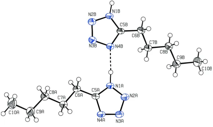
The title compound C6H12N4, is one of a few known tetrazoles with an alkyl chain in the 5-position. The asymmetric unit contains two independent molecules. The molecules are linked by N—H N interactions into chains with graph-set notation D(2) andC2
2
(8) along [010]. The two independent molecules form a layered structure, the layers being composed of interdigitating strands of alternatingly oriented and nearly identical molecules.
Related literature
For synthetic methods see: Mihina & Herbst (1950); Stevenet al. (1993); Detert & Schollmeyer (1999); Sugiono & Detert (2001); Glanget al.(2008); Borchmannet al.(2010). For the properties of tetrazole, see: Huisgen et al. (1960a,b, 1961); Singh (1980); Pernice et al. (1988); Huff et al. (1996). For graph-set notation, see: Bernsteinet al.(1995).
Experimental
Crystal data
C6H12N4
Mr= 140.20
Triclinic,P1 a= 8.7812 (14) A˚ b= 9.6770 (12) A˚ c= 11.614 (2) A˚ = 93.136 (10)
= 112.059 (9)
= 116.389 (7)
V= 789.6 (2) A˚3 Z= 4
CuKradiation = 0.63 mm1
T= 193 K
0.500.400.30 mm
Data collection
Enraf–Nonius CAD-4 diffractometer 3182 measured reflections 2991 independent reflections
2764 reflections withI> 2(I) Rint= 0.070
3 standard reflections every 60 min intensity decay: 2%
Refinement
R[F2> 2(F2)] = 0.040 wR(F2) = 0.109
S= 1.04 2991 reflections
184 parameters
H-atom parameters constrained max= 0.24 e A˚3
min=0.20 e A˚3
Table 1
Hydrogen-bond geometry (A˚ ,).
D—H A D—H H A D A D—H A
N1A—H1A N4B 0.96 1.82 2.7773 (14) 175
N1B—H1B N4Ai
0.95 1.84 2.7779 (14) 170
Symmetry code: (i)x;yþ1;z.
Data collection: CAD-4 Software (Enraf–Nonius, 1989); cell refinement: CAD-4 Software; data reduction: CORINC (Dra¨ger & Gattow, 1971); program(s) used to solve structure:SIR97(Altomare
et al., 1999); program(s) used to refine structure:SHELXL97 (Shel-drick, 2008); molecular graphics:PLATON(Spek, 2009); software used to prepare material for publication:PLATON.
Supplementary data and figures for this paper are available from the IUCr electronic archives (Reference: BX2337).
References
Altomare, A., Burla, M. C., Camalli, M., Cascarano, G. L., Giacovazzo, C., Guagliardi, A., Moliterni, A. G. G., Polidori, G. & Spagna, R. (1999).J. Appl. Cryst.32, 115–119.
Bernstein, J., Davis, R. E., Shimoni, L. & Chang, N.-L. (1995).Angew. Chem. Int. Ed. Engl.34, 1555–1573.
Borchmann, D., Kratochwil, M., Glang, S. & Detert, H. (2010). Proceedings of the 36th German Topical Meeting on Liquid Crystals, pp. 133–138. Detert, H. & Schollmeyer, D. (1999).Synthesis, pp. 999–1004. Dra¨ger, M. & Gattow, G. (1971).Acta Chem. Scand.25, 761–762.
Enraf–Nonius (1989).CAD-4 Software. Enraf–Nonius, Delft, The Nether-lands.
Glang, S., Schmitt, V. & Detert, H. (2008). Proceedings of the 36th German Topical Meeting on Liquid Crystals, pp. 125–128.
Huff, B. E., LeTourneau, M. E., Staszak, M. A. & Ward, J. A. (1996). Tetrahedron Lett.37, 3655–3658.
Huisgen, R., Sauer, J. & Seidel, M. (1960a).Chem. Ber.93, 2885–2891. Huisgen, R., Sturm, H. J. & Markgraf, J. H. (1960b).Chem. Ber.93, 2106–2124. Huisgen, R., Sturm, H. J. & Seidel, M. (1961).Chem. Ber.94, 1555–1562. Mihina, J. S. & Herbst, R. M. (1950).J. Org. Chem.15, 1082–1092.
Pernice, P., Castaing, M., Menassa, P. & Kraus, J. L. (1988).Biophys. Chem.32, 15–20.
Sheldrick, G. M. (2008).Acta Cryst.A64, 112–122.
Singh, H. (1980).Progress in Medicinal Chemistry, edited by G. P. Ellis & G. B. West Vol. 17, pp. 151–184. Amsterdam: Elsevier/North Holland Biomedical Press.
Spek, A. L. (2009).Acta Cryst.D65, 148–155.
Steven, J., Wittenberger, S. J. & Donner, B. G. (1993).J. Org. Chem.58, 4139– 4141.
Sugiono, E. & Detert, H. (2001).Synthesis, pp. 893–896.
Acta Crystallographica Section E Structure Reports
Online
supporting information
Acta Cryst. (2011). E67, o156 [https://doi.org/10.1107/S1600536810052244]
5-Pentyl-1
H
-tetrazole
Thorsten Rieth, Dieter Schollmeyer and Heiner Detert
S1. Comment
The title compound (I), is formed by the addition of triethylammonium azide to capronitrile in refluxing toluene and
acidic work-up. In the crystal, molecules are linked by N— H··· N interactions into chains with graph-set notation D(2) a
C22(8) along [010] (Bernstein et al., 1995), Table 1. Both molecules of the title compound have very similar geometries.
The heterocycles and alkyl chains are coplanar with the molecules A oriented to the opposite site of the molecules B.
These strands form layers via interdigitation of the alkyl chains.
S2. Experimental
The title compound was prepared as follows: Triethyl ammonium chloride (8.95 g, 0.06 mol) and sodium azide (3.90 g,
0.06 mol) were added to a solution of hexanoic acid nitrile (4.36 g, 0.045 mol) in toluene (35 ml) and the mixture was
stirred under reflux for 72 h. The mixture was filtered, the solvent evaporated and the residue dissolved in water.
Hydro-chloric acid (6M, 15 ml) was added and the product was extracted with ether/petroleum ether (1/1, 3*30 ml). The cooled
organic solutions were dried with sodium sulfate. The solvents were evaporated and the residue crystallized upon
standing at ambient temperature within 5 days. Recrystallization from toluene yielded 5-pentyltetrazole in 78% yield as
colorless needles.
S3. Refinement
Hydrogen atoms attached to carbons were placed at calculated positions with C—H = 0.95 Å (aromatic) or 0.98–0.99 Å
(sp3 C-atom). The Hydrogen atoms attached to N1A and N1B were located in diff. Fourier maps. All H atoms were
refined in the riding-model approximation with isotropic displacement parameters (set at 1.2–1.5 times of the Ueq of the
Figure 1
View of compound I. Displacement ellipsoids are drawn at the 50% probability level.
5-pentyl-1H-tetrazole
Crystal data
C6H12N4
Mr = 140.20 Triclinic, P1 Hall symbol: -P 1
a = 8.7812 (14) Å
b = 9.6770 (12) Å
c = 11.614 (2) Å
α = 93.136 (10)°
β = 112.059 (9)°
γ = 116.389 (7)°
V = 789.6 (2) Å3
Z = 4
F(000) = 304
Dx = 1.179 Mg m−3 Melting point: 315 K
Cu Kα radiation, λ = 1.54178 Å Cell parameters from 25 reflections
θ = 65–69°
µ = 0.63 mm−1
T = 193 K Block, colourless 0.50 × 0.40 × 0.30 mm
Data collection
Enraf–Nonius CAD-4 diffractometer
Radiation source: rotating anode Graphite monochromator
ω/2θ scans
3182 measured reflections 2991 independent reflections 2764 reflections with I > 2σ(I)
Rint = 0.070
θmax = 70.0°, θmin = 4.3°
h = −10→9
k = 0→11
l = −14→14
3 standard reflections every 60 min intensity decay: 2%
Refinement
Refinement on F2 Least-squares matrix: full
R[F2 > 2σ(F2)] = 0.040
wR(F2) = 0.109
S = 1.04 2991 reflections
Primary atom site location: structure-invariant direct methods
Secondary atom site location: difference Fourier map
w = 1/[σ2(F
o2) + (0.0611P)2 + 0.1618P] where P = (Fo2 + 2Fc2)/3
(Δ/σ)max < 0.001 Δρmax = 0.24 e Å−3
Δρmin = −0.20 e Å−3
Extinction correction: SHELXL97 (Sheldrick, 2008), Fc*=kFc[1+0.001xFc2λ3/sin(2θ)]-1/4 Extinction coefficient: 0.0088 (12)
Special details
Geometry. All e.s.d.'s (except the e.s.d. in the dihedral angle between two l.s. planes) are estimated using the full covariance matrix. The cell e.s.d.'s are taken into account individually in the estimation of e.s.d.'s in distances, angles and torsion angles; correlations between e.s.d.'s in cell parameters are only used when they are defined by crystal symmetry. An approximate (isotropic) treatment of cell e.s.d.'s is used for estimating e.s.d.'s involving l.s. planes.
Refinement. Refinement of F2 against ALL reflections. The weighted R-factor wR and goodness of fit S are based on F2, conventional R-factors R are based on F, with F set to zero for negative F2. The threshold expression of F2 > σ(F2) is used only for calculating R-factors(gt) etc. and is not relevant to the choice of reflections for refinement. R-factors based on F2 are statistically about twice as large as those based on F, and R- factors based on ALL data will be even larger.
Fractional atomic coordinates and isotropic or equivalent isotropic displacement parameters (Å2)
x y z Uiso*/Ueq
N1A 0.76292 (14) 0.44445 (11) 0.33040 (10) 0.0321 (2)
H1A 0.7628 0.5432 0.3397 0.038*
N2A 0.77322 (17) 0.38035 (13) 0.22933 (10) 0.0392 (3)
N3A 0.77338 (17) 0.25012 (13) 0.24908 (11) 0.0413 (3)
N4A 0.76329 (16) 0.22891 (12) 0.36157 (10) 0.0358 (3)
C5A 0.75588 (16) 0.35102 (13) 0.41070 (11) 0.0286 (3)
C6A 0.74035 (18) 0.38071 (13) 0.53191 (11) 0.0321 (3)
H6A 0.6195 0.3797 0.5105 0.039*
H6B 0.8458 0.4884 0.5875 0.039*
C7A 0.74667 (18) 0.25697 (14) 0.60683 (12) 0.0336 (3)
H7A 0.6359 0.1501 0.5540 0.040*
H7B 0.8633 0.2525 0.6233 0.040*
C8A 0.74449 (18) 0.29643 (15) 0.73469 (12) 0.0360 (3)
H8A 0.6351 0.3123 0.7186 0.043*
H8B 0.8619 0.3983 0.7905 0.043*
C9A 0.7308 (2) 0.16675 (18) 0.80548 (14) 0.0441 (3)
H9A 0.6116 0.0655 0.7507 0.053*
H9B 0.8383 0.1489 0.8197 0.053*
C10A 0.7338 (3) 0.2091 (2) 0.93461 (16) 0.0607 (4)
H10A 0.7183 0.1198 0.9740 0.091*
H10B 0.6298 0.2299 0.9215 0.091*
H10C 0.8555 0.3050 0.9916 0.091*
N1B 0.77122 (14) 0.95284 (11) 0.40731 (9) 0.0305 (2)
H1B 0.7639 1.0469 0.3995 0.037*
N2B 0.74619 (15) 0.88499 (12) 0.50153 (10) 0.0350 (3)
N3B 0.74529 (16) 0.75189 (12) 0.47932 (10) 0.0364 (3)
N4B 0.76875 (15) 0.73261 (12) 0.37130 (10) 0.0336 (2)
C5B 0.78464 (16) 0.85919 (13) 0.32751 (11) 0.0288 (3)
C6B 0.81748 (19) 0.89627 (13) 0.21354 (12) 0.0359 (3)
H6C 0.9558 0.9640 0.2423 0.043*
C7B 0.74053 (18) 0.74860 (13) 0.10847 (11) 0.0326 (3)
H7C 0.8051 0.6880 0.1436 0.039*
H7D 0.6030 0.6784 0.0813 0.039*
C8B 0.77032 (18) 0.79245 (14) −0.00809 (11) 0.0341 (3)
H8C 0.7002 0.8484 −0.0453 0.041*
H8D 0.9071 0.8678 0.0205 0.041*
C9B 0.70495 (19) 0.64908 (15) −0.11205 (12) 0.0380 (3)
H9C 0.5680 0.5737 −0.1411 0.046*
H9D 0.7749 0.5930 −0.0751 0.046*
C10B 0.7361 (2) 0.69521 (18) −0.22777 (13) 0.0501 (4)
H10D 0.6736 0.5983 −0.2974 0.075*
H10E 0.8727 0.7536 −0.2030 0.075*
H10F 0.6822 0.7635 −0.2575 0.075*
Atomic displacement parameters (Å2)
U11 U22 U33 U12 U13 U23
N1A 0.0489 (6) 0.0245 (5) 0.0357 (5) 0.0239 (4) 0.0240 (5) 0.0118 (4)
N2A 0.0627 (7) 0.0336 (6) 0.0384 (6) 0.0314 (5) 0.0295 (5) 0.0145 (4)
N3A 0.0686 (7) 0.0333 (6) 0.0397 (6) 0.0336 (5) 0.0308 (6) 0.0135 (5)
N4A 0.0564 (6) 0.0268 (5) 0.0379 (6) 0.0269 (5) 0.0264 (5) 0.0118 (4)
C5A 0.0357 (6) 0.0209 (5) 0.0337 (6) 0.0163 (5) 0.0171 (5) 0.0077 (4)
C6A 0.0446 (6) 0.0263 (6) 0.0350 (6) 0.0218 (5) 0.0219 (5) 0.0094 (5)
C7A 0.0440 (7) 0.0288 (6) 0.0360 (6) 0.0214 (5) 0.0214 (5) 0.0112 (5)
C8A 0.0426 (7) 0.0348 (6) 0.0348 (6) 0.0211 (5) 0.0193 (5) 0.0100 (5)
C9A 0.0566 (8) 0.0519 (8) 0.0428 (7) 0.0358 (7) 0.0283 (6) 0.0228 (6)
C10A 0.0868 (12) 0.0807 (12) 0.0495 (9) 0.0573 (10) 0.0426 (9) 0.0353 (8)
N1B 0.0454 (6) 0.0229 (5) 0.0326 (5) 0.0217 (4) 0.0203 (4) 0.0098 (4)
N2B 0.0511 (6) 0.0294 (5) 0.0353 (5) 0.0242 (5) 0.0242 (5) 0.0116 (4)
N3B 0.0544 (6) 0.0303 (5) 0.0370 (5) 0.0255 (5) 0.0263 (5) 0.0151 (4)
N4B 0.0520 (6) 0.0262 (5) 0.0362 (5) 0.0250 (5) 0.0256 (5) 0.0133 (4)
C5B 0.0386 (6) 0.0206 (5) 0.0312 (6) 0.0168 (5) 0.0169 (5) 0.0074 (4)
C6B 0.0554 (7) 0.0235 (6) 0.0359 (6) 0.0205 (5) 0.0260 (6) 0.0118 (5)
C7B 0.0440 (7) 0.0240 (6) 0.0350 (6) 0.0174 (5) 0.0220 (5) 0.0094 (5)
C8B 0.0457 (7) 0.0276 (6) 0.0339 (6) 0.0194 (5) 0.0206 (5) 0.0117 (5)
C9B 0.0507 (7) 0.0322 (6) 0.0347 (6) 0.0208 (6) 0.0224 (6) 0.0097 (5)
C10B 0.0734 (10) 0.0466 (8) 0.0388 (7) 0.0305 (7) 0.0324 (7) 0.0150 (6)
Geometric parameters (Å, º)
N1A—C5A 1.3330 (15) N1B—C5B 1.3339 (15)
N1A—N2A 1.3494 (14) N1B—N2B 1.3446 (14)
N1A—H1A 0.9564 N1B—H1B 0.9474
N2A—N3A 1.2937 (14) N2B—N3B 1.2956 (14)
N3A—N4A 1.3628 (15) N3B—N4B 1.3616 (14)
N4A—C5A 1.3223 (14) N4B—C5B 1.3222 (14)
C6A—H6A 0.9900 C6B—H6C 0.9900
C6A—H6B 0.9900 C6B—H6D 0.9900
C7A—C8A 1.5222 (16) C7B—C8B 1.5203 (16)
C7A—H7A 0.9900 C7B—H7C 0.9900
C7A—H7B 0.9900 C7B—H7D 0.9900
C8A—C9A 1.5226 (18) C8B—C9B 1.5167 (17)
C8A—H8A 0.9900 C8B—H8C 0.9900
C8A—H8B 0.9900 C8B—H8D 0.9900
C9A—C10A 1.520 (2) C9B—C10B 1.5212 (17)
C9A—H9A 0.9900 C9B—H9C 0.9900
C9A—H9B 0.9900 C9B—H9D 0.9900
C10A—H10A 0.9800 C10B—H10D 0.9800
C10A—H10B 0.9800 C10B—H10E 0.9800
C10A—H10C 0.9800 C10B—H10F 0.9800
C5A—N1A—N2A 109.67 (9) C5B—N1B—N2B 109.65 (9)
C5A—N1A—H1A 127.6 C5B—N1B—H1B 129.2
N2A—N1A—H1A 122.7 N2B—N1B—H1B 120.8
N3A—N2A—N1A 106.07 (10) N3B—N2B—N1B 106.22 (9)
N2A—N3A—N4A 110.18 (10) N2B—N3B—N4B 110.06 (9)
C5A—N4A—N3A 106.84 (9) C5B—N4B—N3B 106.84 (9)
N4A—C5A—N1A 107.23 (10) N4B—C5B—N1B 107.23 (10)
N4A—C5A—C6A 127.37 (10) N4B—C5B—C6B 127.50 (10)
N1A—C5A—C6A 125.40 (10) N1B—C5B—C6B 125.25 (10)
C5A—C6A—C7A 112.98 (9) C5B—C6B—C7B 113.84 (9)
C5A—C6A—H6A 109.0 C5B—C6B—H6C 108.8
C7A—C6A—H6A 109.0 C7B—C6B—H6C 108.8
C5A—C6A—H6B 109.0 C5B—C6B—H6D 108.8
C7A—C6A—H6B 109.0 C7B—C6B—H6D 108.8
H6A—C6A—H6B 107.8 H6C—C6B—H6D 107.7
C8A—C7A—C6A 111.95 (10) C8B—C7B—C6B 111.84 (10)
C8A—C7A—H7A 109.2 C8B—C7B—H7C 109.2
C6A—C7A—H7A 109.2 C6B—C7B—H7C 109.2
C8A—C7A—H7B 109.2 C8B—C7B—H7D 109.2
C6A—C7A—H7B 109.2 C6B—C7B—H7D 109.2
H7A—C7A—H7B 107.9 H7C—C7B—H7D 107.9
C7A—C8A—C9A 113.17 (11) C9B—C8B—C7B 113.44 (10)
C7A—C8A—H8A 108.9 C9B—C8B—H8C 108.9
C9A—C8A—H8A 108.9 C7B—C8B—H8C 108.9
C7A—C8A—H8B 108.9 C9B—C8B—H8D 108.9
C9A—C8A—H8B 108.9 C7B—C8B—H8D 108.9
H8A—C8A—H8B 107.8 H8C—C8B—H8D 107.7
C10A—C9A—C8A 112.79 (12) C8B—C9B—C10B 112.71 (11)
C10A—C9A—H9A 109.0 C8B—C9B—H9C 109.0
C8A—C9A—H9A 109.0 C10B—C9B—H9C 109.0
C10A—C9A—H9B 109.0 C8B—C9B—H9D 109.0
C8A—C9A—H9B 109.0 C10B—C9B—H9D 109.0
C9A—C10A—H10A 109.5 C9B—C10B—H10D 109.5
C9A—C10A—H10B 109.5 C9B—C10B—H10E 109.5
H10A—C10A—H10B 109.5 H10D—C10B—H10E 109.5
C9A—C10A—H10C 109.5 C9B—C10B—H10F 109.5
H10A—C10A—H10C 109.5 H10D—C10B—H10F 109.5
H10B—C10A—H10C 109.5 H10E—C10B—H10F 109.5
C5A—N1A—N2A—N3A −0.33 (14) C5B—N1B—N2B—N3B 0.27 (13)
N1A—N2A—N3A—N4A 0.02 (14) N1B—N2B—N3B—N4B −0.24 (13)
N2A—N3A—N4A—C5A 0.30 (15) N2B—N3B—N4B—C5B 0.13 (14)
N3A—N4A—C5A—N1A −0.49 (13) N3B—N4B—C5B—N1B 0.04 (13)
N3A—N4A—C5A—C6A 178.87 (11) N3B—N4B—C5B—C6B 178.27 (11)
N2A—N1A—C5A—N4A 0.52 (14) N2B—N1B—C5B—N4B −0.19 (13)
N2A—N1A—C5A—C6A −178.86 (11) N2B—N1B—C5B—C6B −178.47 (11)
N4A—C5A—C6A—C7A 3.98 (18) N4B—C5B—C6B—C7B 28.32 (18)
N1A—C5A—C6A—C7A −176.77 (11) N1B—C5B—C6B—C7B −153.76 (12)
C5A—C6A—C7A—C8A 176.01 (10) C5B—C6B—C7B—C8B 177.66 (10)
C6A—C7A—C8A—C9A 174.26 (11) C6B—C7B—C8B—C9B 176.94 (11)
C7A—C8A—C9A—C10A 178.51 (12) C7B—C8B—C9B—C10B −179.91 (11)
Hydrogen-bond geometry (Å, º)
D—H···A D—H H···A D···A D—H···A
N1A—H1A···N4B 0.96 1.82 2.7773 (14) 175
N1B—H1B···N4Ai 0.95 1.84 2.7779 (14) 170
