Effect of patient positioning on the duration of venous reflux
in duplex ultrasound for venous insufficiency
M Bonfield
1, F Cramp
2and T Robinson
11Vascular Studies Unit, Department of Medical Physics and Clinical Engineering, University Hospitals Bristol NHS Foundation Trust,
Bristol BS2 8HW, UK;2Faculty of Health and Life Sciences, University of the West of England, Bristol BS16 1DD, UK Corresponding author: M Bonfield. Email: Michelle.Bonfield@UHBristol.nhs.uk
Abstract
Introduction:There is currently little research regarding optimum patient position for venous insufficiency assessment although standing is considered the gold standard in many professional guidelines. Some patients are unable to stand for the examination and scanning patients in a standing position is physically challenging for the sonographer. This pilot study aimed to evaluate the effect of varying patient positioning on the duration of venous reflux.
Method:Venous reflux duration was measured in symptomatic participants with suspected venous insufficiency.
Measurements were taken in the standing position (gold standard) and four alternative positions: 258reverse-Trendelenburg (RT) tilt, sitting on the edge of the examination couch, 108RT tilt and 08RT tilt. The mean reflux duration measured in each different position was compared with the gold standard.
Results:Complete measurements were obtained from 16 patients (8 men and 8 women). For an incompetent vein, statistical analysis demonstrated a significant difference only between the standing position and the 08position (U¼19.0; exactP,0.01 [2-tailed]).
Conclusion:Results suggest that several alternative positions could be used for assessing incompetent veins as long as the patient is not lying supine with 08tilt. This would offer much greater flexibility, which may be of benefit to both patients and sonographers.
Keywords: Vascular, duplex ultrasound, research, diagnostic imaging, ultrasound
Ultrasound2012;20: 92 – 97. DOI: 10.1258/ult.2012.011055
Venous pathology
Venous flow from the lower limbs has to compete against gravity and most veins in the legs contain one-way valves to facilitate this. When the valves fail, they allow prolonged retrograde flow and it is this backward motion of blood that is documented during venous reflux testing with ultra-sound. Reflux lasting longer than 0.5 seconds is considered the accepted threshold for defining abnormal venous reflux.1 Prolonged venous reflux causes the blood to pool in the leg veins which leads to venous hypertension. This condition is called venous incompetence or venous insuffi-ciency. Chronic venous insufficiency can lead to varicose veins, venous eczema, lipodermatosclerosis and ulceration.2 Venous disorders are not generally life-threatening but they are associated with high levels of morbidity and are at great cost to the National Health Service (NHS).
Rationale for research
When sonographically assessing venous incompetence, the standing position is widely recommended in various
reference texts and consensus documents used in sonogra-phy.3 – 6Not all patients, however, are able to stand for all or part of the examination and scanning patients in a stand-ing position is physically challengstand-ing for the sonographer. There is a high prevalence of work-related upper limb dis-orders in the field of sonography. The result is sonographers scanning in pain, episodes of long-term leave of absence and an increase in the number of sonographers retiring early on grounds of ill health.7 This comes at great cost to both the NHS and the individual.
A comprehensive literature search using keywords relat-ing to venous ultrasound and patient positionrelat-ing was performed across five key databases: Allied and Complementary Medicine (AMED), Cumulative Index to Nursing and Allied Health ( CINAHL), Embase, Medline and Pubmed. The search yielded 10 studies of which several incorporated assessing patients for lower limb venous ultrasound in different positions but for which patient positioning was not the prime focus. Only three of the 10 studies looked at patient positioning directly. Studies by Mahmutyazicioglu et al.8 and Musil9 included
only normal subjects and are therefore less relevant to the target patient group. In addition, both of these studies and the study by Cairnduff et al.10 examined patients only in
the standing position and one other position.
Study aim
The aim of the study was to evaluate the effect of varying patient positioning on the duration of venous reflux in the assessment of lower limb venous insufficiency with ultra-sound. The evaluation compared reflux duration measured in four different patient positions with that measured in the standing position (gold standard):
†
Supine with examination couch at 08 tilt;†
Supine with examination couch at 108RT tilt;†
Supine with examination couch at 258RT tilt;†
Sitting on the edge of the examination couch.Methodology
Sampling and recruitment
Approval (10/H0106/31) was gained for the study from the North Somerset and South Bristol Research Ethics Committee and the University of the West of England Research Ethics Sub-Committee of the School of Health and Life Sciences, Bristol, UK. The sample set was a convenience sample appropriate for a pilot investigation. Participants were selected from a known population: patients presenting with symptoms of primary varicose veins in the distribution of the long saphenous vein, over a data collection period of six months. All participants demonstrated significant reflux of .0.5 seconds (the point at which venous reflux is con-sidered significant and not due to a normal element of venous reflux occurring at valve closure)1 in the long
saphenous vein in the standing position. The additional inclusion criterion was that the patient was able to stand unassisted for 15 minutes. Exclusion criteria were those patients who had limited mobility and those who had pre-viously undergone interventions on the long saphenous vein and were presenting with recurrent varicose veins. Recurrent pathology usually indicates tortuous vessels which would present difficulties in locating a suitable and reproducible site for cursor placement. Prior to commencing data collection, participants provided written informed consent.
Data collection methods
Data collection took place on symptomatic patients as part of routine lower limb venous insufficiency assessments at the Vascular Studies Unit in the Bristol Royal Infirmary. Ultrasound was performed using a Logiq 9 (GE Medical Systems, Wauwatosa, WI, USA) equipped with a 10-MHz linear transducer. The equipment setting was the standard superficial venous pre-set which was adjusted as required for each patient for optimum imaging. Manual distal com-pression was used to augment flow and three repeated measurements of reflux duration were taken at the follow-ing sites (Figure 1):
†
The long saphenous vein 5 cm distal to the SFJ (sapheno-femoral junction);†
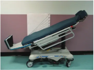
The femoral vein 5 cm distal to the SFJ. [image:2.595.337.531.573.718.2]Five centimetres is the length of the ultrasound transducer which facilitated the speed and ease of location of the measurement site hence minimizing the additional examin-ation time for the patient. An adjustable examinexamin-ation couch with foot rest and reverse-Trendelenburg (RT) tilt option (5051 Eye Stretcher Chair, Stryker Medical, Newbury, UK) was used for the four alternative positions. The couch was manually adjusted to each degree of tilt using a large hand-drawn protractor that was permanently fixed to the wall (Figure 2). Placement of the protractor was undertaken using a spirit level to ensure that the 08position was accu-rately placed. With no adjustment settings made, the Figure 1 Measurement site 5 cm distal to the saphenofemoral junction
couch was at 08tilt and the maximum degree of RT tilt the couch could obtain was 258. The 08, 258RT and sitting pos-itions could therefore all be obtained without aligning the couch with the protractor each time. Only the 108 RT pos-ition required alignment with the protractor on each occasion.
The gold standard position was tested first as this formed the basis for the clinical diagnosis. The patient was asked to stand facing the sonographer. An examination couch was positioned behind the patient and its height adjusted to the level of the patient’s mid-thigh to provide partial support. The side bars of the examination couch were raised and the patient was asked to hold on to the bar for increased stability. The patient was instructed to shift their weight onto the limb that was not being examined and par-tially externally rotate the symptomatic limb keeping the foot on the floor but non-weight bearing. This facilitated placement of the transducer to image the required anatom-ical sites. The display monitor of the ultrasound machine was adjusted in order that the Doppler trace occupied one-third of the screen and the image occupied two-thirds of the screen to make accurate measurements of reflux dur-ation while providing the optimal image of the vessel to ensure accurate placement of the cursor. The Doppler trace time was adjusted to ensure that the total duration of venous reflux could be captured (Figure 3). The velocity scale was also adjusted to capture the total flow augmenta-tion to aid the investigator in achieving reproducible aug-mentation for each measurement.
The venous reflux measurements were then repeated with the patient positioned in each of the alternative positions. A set of three measurements was recorded with the patient positioned in the five different positions. The order of the
alternative positions was decided randomly using the pre-prepared envelopes technique to avoid carry-over effects (Figure 4). The random sequences for the pre-prepared envelopes were generated using the website www.randomi-ser.org. This was undertaken by an independent person not involved in the data collection process. The random allo-cation envelope for each participant was opened only after the gold standard test had been completed in order to avoid any operator bias. The venous reflux measurements were undertaken by a single clinical vascular scientist (Michelle Bonfield) and on a single ultrasound machine to minimize interoperator and intermachine variability. Where possible, assessments were conducted in the morning as it has been suggested that venous function may deteriorate with activity during the day.11Room
temp-erature was maintained at approximately 248C via an air-conditioning system.
Statistical analysis
[image:3.595.133.459.460.726.2]The arithmetic mean and standard deviation of each set of three repeated measurements were calculated for each patient and each position. As the data were not normally distributed, a Kruskal –Wallis Test was undertaken to deter-mine if there was a significant difference between the mean data for the different positions. Where a significant differ-ence was identified, further post hoc testing was justified using the Mann-Whitney U test pairing the data from the gold standard position (standing) with that for the four alternative positions. For all statistical analysis a two-tailed significance was used as it was unknown if the reflux dur-ation measurements obtained in the four alternative pos-itions would be greater or lesser than those obtained in
the gold standard position. As the direction of any effect was unknown it was important to test for the possibility of the relationship in both directions.
Results
[image:4.595.121.481.59.300.2]From 56 referrals for lower limb venous ultrasound during the six-month data collection period, 37 patients did not meet the inclusion criteria. Of these, 14 had undergone pre-vious surgical intervention to the long saphenous vein, 19 did not demonstrate venous reflux .0.5 seconds in the long saphenous vein in the standing position and four were unable to stand unassisted for 15 minutes. Nineteen patients met the inclusion criteria but three declined partici-pation in the study. Complete measurements were obtained from 16 patients. The mean age of the participants was 42.8 years (standard deviation of 18.1, range 22– 88). Eight par-ticipants were men and eight were women. Eight left and eight right limbs were included in the study. Thirteen of the 16 patients were seen in morning appointments between 9:30 and 11:00. All participants demonstrated sig-nificant reflux of .0.5 seconds in the superficial venous system rather than the deep venous system. The mean reflux data from the long saphenous vein are presented in Table 1.
The shortest mean reflux duration in the long saphenous vein (672 ms) was obtained in the 08 position and the longest mean reflux duration (3565 ms) in the 258 RT pos-ition. There was a large standard deviation from the mean for all positions. The mean values for venous reflux in the long saphenous vein all represented significant reflux con-sistent with a diagnosis of venous incompetence.
A high false-negative rate (63%) was demonstrated when participants were positioned in the 08 position; however, 37% of participants still demonstrated significant reflux while in this position.
Discussion
[image:4.595.46.559.665.744.2]Although it is difficult to make direct comparisons with the findings of previous research in this field due to heterogen-eity in the methodology used, some limited parallels can be drawn. The greatest reflux duration in this study was recorded in the supine 258RT position in the long saphenous vein. Cairnduffet al.10and Arakiet al.12found that the dur-ation of venous reflux was longer in patients in a supine 458 RT position or semi-recumbent position, respectively, when compared with the standing position. Arakiet al.12assessed only the popliteal vein however and Cairnduff et al.10 recorded longer reflux in all venous locations tested except Figure 4 Data record sheet and randomized position order
Table 1 Venous reflux duration by position and false-positive/false-negative rates for the long saphenous vein
Long saphenous vein
positions N
Minimum reflux (ms)
Maximum reflux (ms)
Mean reflux (ms)
Standard deviation (ms)
False-positive rate (%)
False-negative rate (%)
Standing position 16 1215 4685 2809 1113 0 0
Sitting position 16 963 5668 3066 1400 0 0
258RT position 16 0 5974 3565 1932 6 0
108RT position 16 0 5978 2512 2079 0 0
the long saphenous vein. Cairnduff et al.10 concluded that assessing the patient in a semi-recumbent position gives mostly unchanged if not improved results by producing longer venous reflux times. They indicate that longer venous reflex times are desirable for a more sensitive diagno-sis. This, however, raises the question of the possibility of eli-citing venous reflux that may not be present in a standing position and the risk of a false-positive diagnosis. Mahmutyazicioglu et al.8 demonstrated a high proportion of false-positive results in supine examinations with 08 tilt on participants with normal venous function.
Musil9 demonstrated no statistically significant difference in the duration of venous reflux in the long saphenous vein with normal venous function with the patient standing or supine with 08 tilt. As with the findings from the femoral vein in this study, one can consider that this limits the rel-evance of the findings. Although the data from the femoral vein in this study suggest that no difference would occur in terms of a diagnosis by selecting any of the alternative pos-itions for assessing patients, there is a possibility that, had significant reflux been present, a statistically or clinically sig-nificant difference in venous reflux duration may have been identified between positions. In contrast to the findings from the study by Mahmutyaziciogluet al.,8the rate of false-positive results in normally functioning femoral veins in this study was low in all positions with only one false-positive result in one participant in the 258position. Further research involving patients with deep venous incompetence would be required to increase the relevance of any findings to the symptomatic patient group.
There is insufficient evidence to date, from this or pre-vious studies in this field, to support a change in clinical practice. Although this pilot study has not provided suffi-cient evidence to support a change in clinical practice, it has highlighted a gap in the evidence underpinning our current clinical practice. The results from the long saphe-nous vein would suggest that some alternative positions could be considered for assessing venous insufficiency in the long saphenous vein with the exception of the patient lying supine with 08 tilt where the risk of a false-negative diagnosis is high. This would offer much greater flexibility, which may be of benefit to both patients and sonographers. Not all patients are able to stand for all or part of the exam-ination and many would benefit from being scanned in a lying down or seated position. Scanning patients lying down or seated is more comfortable for the patient. It also minimizes the risk of injury as patients are less likely to feel faint when lying down or seated and are less likely to injure themselves if a vasovagal episode were to occur. As pressures on funding in the NHS generally increase, the focus on venous disease may naturally shift towards treat-ing those patients with more severe symptoms. Those patients are most likely to fit into the category of those that find it difficult or impossible to stand for the duration of a venous duplex ultrasound.
Scanning patients in a standing position is physically challenging for the sonographer and the high prevalence of work-related upper limb disorders in the field of sonogra-phy is well documented.13 – 18 Proper technique is vitally
important in preventing work-related upper limb disorder
(WRULD) and work posture has been cited as the most criti-cal risk factor for musculoskeletal injury in sonography.15 Scanning patients lying down or seated would allow the sonographer to work in a greater level of comfort as the scanning arm can be supported to allow the muscles to rest19 and protraction of the head and neck can be mini-mized. These factors are known contributors to WRULD20
the risk of which may be reduced by positioning patients lying down or in a seated position.
Clinicians and researchers define better diagnostic or screening tests as ones that give more accurate results faster, and at a lower cost in terms of safety, comfort and expense.21These factors support the need for further research in this area to determine if alternative positions can be used in the assessment of venous disease using ultrasound. Further study with larger numbers would be required to explore more rigorously the findings. Because of the reduction in venous referrals in many NHS hospitals due to funding pressures, a multicentre study may be an appropriate method for further research. Recruiting sufficient participants from a wide demographic profile will generate adequate power to detect any differences between positions, and may increase generalizability of any findings. Future research in this field would also benefit from including participants with both superficial and deep venous incompetence and from collecting data from a greater range of venous locations.
Conclusion
Results from this study suggest that several alternative pos-itions could be used for assessing incompetent veins as long as the patient is not lying supine with 08tilt. With further research in this area, alternative positions for examining patients for venous insufficiency may offer a greater level of comfort and flexibility for both patients and sonogra-phers without affecting the diagnostic accuracy of the test.
DECLARATIONS
Competing interests:None.
Funding: Study completed as dissertation component of MSc in Medical Ultrasound as part of a fully funded Clinical Vascular Scientist traineeship, a post funded by the South West Strategic Health Authority.
Ethical approval: The North Somerset and South Bristol Research Ethics Committee approved this study (REC number: 10/H0106/31).
Guarantor:FC.
Contributorship:MB and TR researched literature and con-ceived the study. MB and FC were involved in protocol development, gaining ethical approval and data analysis. MB was involved in patient recruitment and data collection. MB wrote the first draft of the manuscript. All authors reviewed and edited the manuscript and approved the final version of the manuscript.
REFERENCES
2 Fowkes G. Epidemiology of venous disease. In: Davies A, Lees T, Lane F, eds.Venous Disease Simplified. Harley, UK: TFM Publishing Ltd, 2006: 13– 32
3 Cole S, Walker R, Norris R.Vascular Laboratory Practice Part IV. York, UK: IPEM, 2001
4 Thrush A, Hartshorne T.Peripheral Vascular Ultrasound How, Why and When. London: Harcourt Publishers Limited, 1999
5 Zweibel W, Pellerito J.Introduction to Vascular Ultrasonography. Philadelphia: Elsevier Saunders, 2005
6 Society for Vascular Ultrasound.Vascular Technology Professional Performance Guidelines: Lower Extremity Venous Insufficiency Evaluation, 2007. Sponsored and published by: Society for Vascular Ultrasound, Lanham, MD, USA. Available at: http://www.svunet.org/files/ positions/SVU_Venous_Guideline2011.pdf (last checked 6 March 2012) 7 Sound Ergonomics.Information & Resources. Washington DC. See http:// www.soundergonomics.com/info-cost.php (last checked 9 November 2009)
8 Mahmutyazicioglu K, Gundogdu S, Ozdemir H, Savraniar A, Asil K. Venous reflux: measurement variability due to positional differences.
Tanisal ve Girisimsel Radyoloji2003;9:471 –5
9 Musil D. Can a position of a patient influence a result of an ultrasound examination of venous valve insufficiency?Vnitr Lek2004;50:746 –50 10 Cairnduff M, Swan H, Barton B, Buckley A. Assessing the variable of
patient positioning when examining the lower limb veins for reflux using spectral and colour Doppler ultrasound.ASUM Ultrasound Bull
2007;10:22 –8
11 Bishara R, Sigel B, Rocco K,et al.Deterioration of venous function in normal lower extremities during daily activity.J Vasc Surg
1986;3:700 –6
12 Araki C, Back T, Padburg F, Thompson P, Duran W, Hobson R. Refinements in the ultrasonic detection of popliteal vein reflux.J Vasc Surg1993;18:742 –8
13 Fernando R. Adverse physical symptoms in radiographers practising ultrasound.Radiography1996;2:91 –7
14 Pike I, Russo A, Berkowitz J, Baker J, Leesoway V. The prevalence of musculoskeletal disorders among registered diagnostic medical sonographers.J Diagn Med Sonography1997;13:219 –27
15 Arrowsmith I.The Prevalence of Work-related Upper Limb Disorders amongst Radiographers. London: College of Radiographers, 2000
16 Feather C.Work related Musculoskeletal Disorders: An Occupational Hazard for Sonographers. London: College of Radiographers, 2001
17 Miles J. Work-related upper limb disorders in sonographers.Synergy
2005;6–11
18 Evans K, Roll S, Baker J. Work-related musculoskeletal disorders (WRMSD) among registered diagnostic medical sonographers and vascular technologists.J Diagn Med Sonography2009;25:287 –99 19 Coffin C, Baker J. Ultrasound clinics: preventing work-related injuries
among sonographers and sonologists.Contemp OB/GYN2007;52:78 –81 20 Burnage J. Work-related upper limb disorder: a sonographer’s survival
guide.Ultrasound2007;15:38– 42