1,4-Bis{[5-(pyridin-4-yl)-1,3,4-oxadiazol-2-yl]sulfanyl}butane
Qing-lei Liu,aWei Wang,a,b* Yan Gao,bXiao-yu Jiaband Jing-jing Zhangb
aSchool of Perfume and Aroma Technology, Shanghai Istitute of Technology, Shanghai 200235, People’s Republic of China, andbSchool of Chemical Engineering, University of Science and Technology LiaoNing, Anshan 114051, People’s Republic of China
Correspondence e-mail: zhao_submit@yahoo.com.cn
Received 17 February 2011; accepted 22 February 2011
Key indicators: single-crystal X-ray study;T= 113 K; mean(C–C) = 0.002 A˚; Rfactor = 0.032;wRfactor = 0.089; data-to-parameter ratio = 16.8.
In the centrosymmetric title compound, C18H16N6O2S2, the 1,3,4-oxadiazole and the attached pyridinyl ring are twisted by 5.3 (3).
Related literature
For applications of heterocyclic derivatives, see: Al-Talibet al. (1990); Nakagawaet al.(1996); Zhanget al.(2007). For related structuresbn,, see: Wanget al.(2010, 2011); Zhaoet al.(2010).
Experimental
Crystal data
C18H16N6O2S2
Mr= 412.49
Monoclinic,P21=c
a= 4.9780 (6) A˚
b= 5.7933 (7) A˚ c= 31.003 (4) A˚
= 92.588 (5)
V= 893.20 (18) A˚3
Z= 2
MoKradiation
= 0.33 mm 1
T= 113 K
0.200.180.10 mm
Data collection
Rigaku Saturn CCD area-detector diffractometer
Absorption correction: multi-scan (CrystalClear; Rigaku/MSC, 2005)
Tmin= 0.937,Tmax= 0.962
8437 measured reflections 2128 independent reflections 1811 reflections withI> 2(I) Rint= 0.035
Refinement
R[F2> 2(F2)] = 0.032
wR(F2) = 0.089 S= 1.10 2128 reflections
127 parameters
H-atom parameters constrained
max= 0.40 e A˚ 3
min= 0.19 e A˚ 3
Data collection:CrystalClear(Rigaku/MSC, 2005); cell refinement:
CrystalClear; data reduction:CrystalClear; program(s) used to solve structure:SHELXS97(Sheldrick, 2008); program(s) used to refine structure: SHELXL97 (Sheldrick, 2008); molecular graphics:
SHELXTL(Sheldrick, 2008); software used to prepare material for publication:SHELXTL.
We gratefully acknowledge support of this project by the Key Laboratory Project of Liaoning Province (No. 2008S127) and the Doctoral Starting Foundation of Liaoning Province (No. 20071103).
Supplementary data and figures for this paper are available from the IUCr electronic archives (Reference: KP2310).
References
Al-Talib, M., Tashtoush, H. & Odeh, N. (1990).Synth. Commun.20, 1811– 1817.
Nakagawa, Y., Nishimura, K., Izumi, K., Kinoshita, K., Kimura, T. & Kurihara, N. (1996).J. Pestic. Sci.21, 195–201.
Rigaku/MSC (2005). CrystalClear. Molecular Structure Corporation, The Woodlands, Texas, USA, and Rigaku Corporation, Tokyo, Japan. Sheldrick, G. M. (2008).Acta Cryst.A64, 112–122.
Wang, H., Gao, Y. & Wang, W. (2010).Acta Cryst.E66, o3085.
Wang, W., Gao, Y., Xiao, Z., Yao, H. & Zhang, J. (2011).Acta Cryst.E67, o269. Zhang, Z.-H., Li, C.-P., Tian, Y.-L. & Guo, Y.-M. (2007).Acta Cryst.E63,
m3044.
Zhao, B., Liu, Z., Gao, Y., Song, B. & Deng, Q. (2010).Acta Cryst.E66, o2814.
Acta Crystallographica Section E
Structure Reports Online
supporting information
Acta Cryst. (2011). E67, o740 [doi:10.1107/S1600536811006805]
1,4-Bis{[5-(pyridin-4-yl)-1,3,4-oxadiazol-2-yl]sulfanyl}butane
Qing-lei Liu, Wei Wang, Yan Gao, Xiao-yu Jia and Jing-jing Zhang
S1. Comment
Heterocycle derivatives containing N, O and S atoms are under intensive studies due to their wide applications in
medicine, industry and coordination chemistry (Al-Talib et al., 1990; Nakagawa et al., 1996; Zhang et al., 2007). We are
focusing the synthetic and structural studies on the novel thio-based ligands (Wang et al., 2010, 2011; Zhao et al., 2010).
Here we present the synthesis and the crystal structure of the title compound (I), namely,
1,4-bis[5-(pyridin-4-yl)-1,3,4-oxadiazol-2-ylsulfanyl]butane.
The molecular structure of title compound (I) (Fig. 1)reveals a twofold rotational axis through the mid of the C-C bond
of butane group. Therefore, an asymmetric unit comprises a half of the molecule. 1,3,4-Oxadiazole moiety is planar with
an r.m.s. deviation of 0.0033 (2)Å and maximum deviation of 0.0052 (2)Å for the atom C7. The dihedral angle between
the oxadiazole and its attached pyridinyl ring [r.m.s. deviation = 0.0062 (2) Å] of 5.3 (3)° indicates that they are almost
coplanar. As a result of π-π conjugation, the Csp2-S bond [S1—C7 = 1.722 (13) Å] is significantly shorter than the Csp3-S
bond [S1—C8 = 1.817 (12) Å].
S2. Experimental
A suspension of 5-(pyridin-4-yl)-1,3,4-oxadiazole-2-thiol (2.0 mmol) and 1,1-dibromobutane (1.0 mmol) in ethanol (10
mL) was stirred at room temperature. The reaction progress was monitored via TLC. The resulting precipitate was
filtered off, washed with cold ethanol, dried and purified to give the target product as light-yellow solid in 87% yield.
Crystals of (I) suitable for single-crystal X-ray analysis were grown by slow evaporation of a solution in
chloroform-ethanol (1:1).
S3. Refinement
All H atoms were positioned geometrically and refined as riding (C—H = 0.95–0.99 Å) and allowed to ride on their
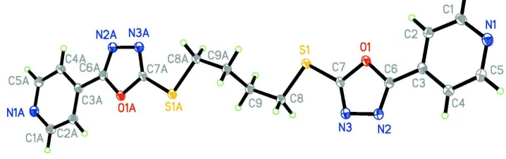
Figure 1
View of the molecule of (I) showing the atom-labelling scheme [symmetry code: (A)-x, -y + 1, -z + 1]. Displacement
ellipsoids are drawn at the 50% probability level.
4-{5-[(4-{[5-(pyridin-4-yl)-1,3,4-oxadiazol-2-yl]sulfanyl}butyl)sulfanyl]- 1,3,4-oxadiazol-2-yl}pyridine
Crystal data
C18H16N6O2S2
Mr = 412.49
Monoclinic, P21/c Hall symbol: -P 2ybc
a = 4.9780 (6) Å
b = 5.7933 (7) Å
c = 31.003 (4) Å
β = 92.588 (5)°
V = 893.20 (18) Å3
Z = 2
F(000) = 428
Dx = 1.534 Mg m−3
Mo Kα radiation, λ = 0.71073 Å Cell parameters from 2798 reflections
θ = 2.6–27.9°
µ = 0.33 mm−1
T = 113 K
Prism, light-yellow 0.20 × 0.18 × 0.10 mm
Data collection
Rigaku Saturn CCD area-detector diffractometer
Radiation source: rotating anode Multilayer monochromator
Detector resolution: 14.63 pixels mm-1
φ and ω scans
Absorption correction: multi-scan (CrystalClear; Rigaku/MSC, 2005)
Tmin = 0.937, Tmax = 0.962
8437 measured reflections 2128 independent reflections 1811 reflections with I > 2σ(I)
Rint = 0.035
θmax = 27.9°, θmin = 2.6°
h = −4→6
k = −7→7
l = −38→40
Refinement
Refinement on F2 Least-squares matrix: full
R[F2 > 2σ(F2)] = 0.032
wR(F2) = 0.089
S = 1.10 2128 reflections 127 parameters 0 restraints
Primary atom site location: structure-invariant direct methods
Secondary atom site location: difference Fourier map
Hydrogen site location: inferred from neighbouring sites
H-atom parameters constrained
w = 1/[σ2(F
o2) + (0.0531P)2 + 0.054P] where P = (Fo2 + 2Fc2)/3
Special details
Geometry. All e.s.d.'s (except the e.s.d. in the dihedral angle between two l.s. planes) are estimated using the full
covariance matrix. The cell e.s.d.'s are taken into account individually in the estimation of e.s.d.'s in distances, angles and torsion angles; correlations between e.s.d.'s in cell parameters are only used when they are defined by crystal symmetry. An approximate (isotropic) treatment of cell e.s.d.'s is used for estimating e.s.d.'s involving l.s. planes.
Refinement. Refinement of F2 against ALL reflections. The weighted R-factor wR and goodness of fit S are based on F2,
conventional R-factors R are based on F, with F set to zero for negative F2. The threshold expression of F2 > σ(F2) is used only for calculating R-factors(gt) etc. and is not relevant to the choice of reflections for refinement. R-factors based on F2 are statistically about twice as large as those based on F, and R- factors based on ALL data will be even larger.
Fractional atomic coordinates and isotropic or equivalent isotropic displacement parameters (Å2)
x y z Uiso*/Ueq
S1 0.25227 (6) 0.35176 (5) 0.422550 (9) 0.01776 (12)
O1 0.61211 (17) 0.17771 (15) 0.37253 (3) 0.0163 (2)
N1 1.3455 (2) −0.0526 (2) 0.27364 (3) 0.0197 (2)
N2 0.7346 (2) −0.15583 (19) 0.40229 (3) 0.0186 (2)
N3 0.5348 (2) −0.05632 (19) 0.42735 (3) 0.0189 (2)
C1 1.1858 (3) 0.1307 (2) 0.27715 (4) 0.0190 (3)
H1 1.2063 0.2551 0.2576 0.023*
C2 0.9910 (3) 0.1507 (2) 0.30774 (4) 0.0179 (3)
H2 0.8797 0.2836 0.3086 0.021*
C3 0.9634 (2) −0.0287 (2) 0.33688 (4) 0.0155 (3)
C4 1.1268 (3) −0.2220 (2) 0.33375 (4) 0.0185 (3)
H4 1.1125 −0.3479 0.3531 0.022*
C5 1.3118 (3) −0.2262 (2) 0.30146 (4) 0.0199 (3)
H5 1.4206 −0.3598 0.2990 0.024*
C6 0.7718 (2) −0.0138 (2) 0.37112 (4) 0.0153 (3)
C7 0.4729 (2) 0.1379 (2) 0.40882 (4) 0.0152 (3)
C8 0.1260 (3) 0.2236 (2) 0.47114 (4) 0.0177 (3)
H8A 0.0331 0.0764 0.4640 0.021*
H8B 0.2770 0.1908 0.4921 0.021*
C9 −0.0695 (3) 0.3927 (2) 0.49063 (4) 0.0178 (3)
H9A −0.2052 0.4403 0.4680 0.021*
H9B −0.1652 0.3137 0.5137 0.021*
Atomic displacement parameters (Å2)
U11 U22 U33 U12 U13 U23
S1 0.0216 (2) 0.01715 (19) 0.01492 (18) 0.00366 (11) 0.00549 (12) 0.00101 (11)
O1 0.0174 (4) 0.0179 (5) 0.0140 (4) 0.0017 (3) 0.0045 (3) 0.0003 (3)
N1 0.0180 (5) 0.0223 (6) 0.0191 (5) −0.0024 (4) 0.0030 (4) −0.0030 (4)
N2 0.0208 (6) 0.0179 (6) 0.0176 (5) 0.0020 (4) 0.0053 (4) −0.0002 (4)
N3 0.0201 (5) 0.0193 (6) 0.0177 (5) 0.0022 (4) 0.0053 (4) −0.0003 (4)
C1 0.0194 (7) 0.0202 (7) 0.0176 (6) −0.0024 (5) 0.0037 (5) 0.0011 (5)
C2 0.0178 (6) 0.0173 (6) 0.0186 (6) 0.0012 (5) 0.0023 (5) −0.0005 (5)
C3 0.0146 (6) 0.0185 (6) 0.0132 (5) −0.0014 (5) −0.0001 (4) −0.0016 (4)
C4 0.0205 (6) 0.0185 (6) 0.0166 (6) 0.0009 (5) 0.0022 (5) 0.0009 (5)
C6 0.0142 (6) 0.0160 (6) 0.0158 (6) 0.0011 (4) 0.0003 (4) −0.0017 (4)
C7 0.0155 (6) 0.0188 (6) 0.0114 (5) −0.0013 (4) 0.0028 (4) −0.0014 (4)
C8 0.0215 (6) 0.0162 (6) 0.0158 (6) −0.0010 (5) 0.0055 (5) −0.0007 (5)
C9 0.0173 (6) 0.0182 (6) 0.0183 (6) −0.0013 (5) 0.0042 (5) −0.0030 (5)
Geometric parameters (Å, º)
S1—C7 1.7218 (13) C2—H2 0.9500
S1—C8 1.8171 (12) C3—C4 1.3902 (17)
O1—C6 1.3669 (14) C3—C6 1.4615 (16)
O1—C7 1.3675 (14) C4—C5 1.3906 (17)
N1—C1 1.3337 (17) C4—H4 0.9500
N1—C5 1.3404 (17) C5—H5 0.9500
N2—C6 1.2888 (16) C8—C9 1.5251 (17)
N2—N3 1.4121 (15) C8—H8A 0.9900
N3—C7 1.2941 (16) C8—H8B 0.9900
C1—C2 1.3914 (18) C9—C9i 1.525 (2)
C1—H1 0.9500 C9—H9A 0.9900
C2—C3 1.3878 (17) C9—H9B 0.9900
C7—S1—C8 99.16 (6) C4—C5—H5 118.1
C6—O1—C7 101.91 (9) N2—C6—O1 113.00 (10)
C1—N1—C5 116.91 (11) N2—C6—C3 128.89 (11)
C6—N2—N3 106.29 (10) O1—C6—C3 118.07 (10)
C7—N3—N2 105.69 (10) N3—C7—O1 113.10 (11)
N1—C1—C2 123.91 (12) N3—C7—S1 131.13 (10)
N1—C1—H1 118.0 O1—C7—S1 115.76 (9)
C2—C1—H1 118.0 C9—C8—S1 108.47 (9)
C3—C2—C1 118.29 (12) C9—C8—H8A 110.0
C3—C2—H2 120.9 S1—C8—H8A 110.0
C1—C2—H2 120.9 C9—C8—H8B 110.0
C2—C3—C4 118.84 (11) S1—C8—H8B 110.0
C2—C3—C6 121.06 (11) H8A—C8—H8B 108.4
C4—C3—C6 120.08 (11) C9i—C9—C8 112.82 (13)
C3—C4—C5 118.21 (12) C9i—C9—H9A 109.0
C3—C4—H4 120.9 C8—C9—H9A 109.0
C5—C4—H4 120.9 C9i—C9—H9B 109.0
N1—C5—C4 123.80 (12) C8—C9—H9B 109.0
N1—C5—H5 118.1 H9A—C9—H9B 107.8
C6—N2—N3—C7 −0.50 (14) C2—C3—C6—N2 −174.70 (13)
C5—N1—C1—C2 0.29 (19) C4—C3—C6—N2 3.7 (2)
N1—C1—C2—C3 1.2 (2) C2—C3—C6—O1 2.71 (17)
C1—C2—C3—C4 −1.37 (19) C4—C3—C6—O1 −178.93 (11)
C1—C2—C3—C6 177.01 (11) N2—N3—C7—O1 0.93 (14)
C2—C3—C4—C5 0.23 (18) N2—N3—C7—S1 −177.95 (10)
C6—C3—C4—C5 −178.17 (11) C6—O1—C7—N3 −0.96 (13)
C3—C4—C5—N1 1.3 (2) C8—S1—C7—N3 −0.56 (14)
N3—N2—C6—O1 −0.09 (14) C8—S1—C7—O1 −179.42 (9)
N3—N2—C6—C3 177.42 (12) C7—S1—C8—C9 177.46 (9)
C7—O1—C6—N2 0.61 (13) S1—C8—C9—C9i −69.35 (15)
C7—O1—C6—C3 −177.20 (11)