Research Center for Cancer, Key Laboratory of Cancer Prevention and Therapy Tianjin, Tianjin 300211, China. *Equal contributors.
Received April 20, 2015; Accepted June 26, 2015; Epub October 1, 2015; Published October 15, 2015
Abstract: Tubeimoside-1 (TBMS1) is considered to have anti-tumor properties. However, the role of TBMS1 on hu-man colorectal cancer (CRC) is still unclear. Therefore, in this study, we investigated the role of TBMS1 on huhu-man CRC and explored the underlying mechanism. The cell proliferation of CRC cells was detected by MTT assay. Cell mi-gration and invasion were assessed by Boyden chamber assay, and the involvement of molecular mechanisms was examined by western blot. In this study, we found that TBMS1 inhibited the proliferation, migration/invasion of CRC
cells, and it reduced β-catenin expression in CRC cells. Furthermore, overexpression of β-catenin rescued TBMS1-induced proliferation and invasion inhibition, and knockdown of β-catenin potentiated TBMS1-TBMS1-induced proliferation
and invasion inhibition. Taken together, our results demonstrate that TBMS1 inhibited CRC cell proliferation and
invasion via suppressing the Wnt/β-catenin signaling pathway. Therefore, TBMS1 may represent a chemopreventive
and/or therapeutic agent in the prevention of CRC.
Keywords: Tubeimoside-1, colorectal cancer, proliferation, invasion, Wnt/β-catenin signaling pathway
Introduction
Colorectal cancer (CRC) is the second most cause of cancer-related death [1]. Despite very aggressive treatment including surgery and combined radio and chemotherapy, most patients are initially diagnosed at the advanced stages, there is no effective therapeutic treat-ment, resulting in short survival time and poor prognosis [2]. Therefore, it is necessary to develop more effective therapeutic agents for CRC.
In recent years, the natural medicine was wide-ly used in clinical and medical research because of its low toxicity and their high biologi-cal activity. Tubeimoside-1 (TBMS1), a natural compound isolated from the tuber of Chine- se medicinal herb Bolbostemma paniculatum (Maxim) Franquet (Cucurbitaceae), has sugar chains that are connected with 3-hydroxy-3-methylglutaric acid to form a unique macro
-cyclic structure [3]. It exerts a broad range of important biological actions including anti-inflammatory, antiviral and immunosuppressive effects [4, 5]. In addition, a growing body of evi-dence indicates that TBMS1 has significant antitumor effects, such as inhibit the cell prolif-eration, arrest the cell cycle, and promote apop-tosis of the human liver [6], squamous esopha -geal carcinoma [7], choriocarcinoma [8] or lung cancer cells [9].
However, the role of TBMS1 on human CRC is still unclear. Therefore, in this study, we investi-gated the role of TBMS1 on human CRC and explored the underlying mechanism.
Materials and methods
Materials
Tubeimoside-1 inhibits the growth and invasion of CRC cells
Products (China), dissolved in phosphate
[image:2.612.94.521.76.224.2]buff-ered saline (PBS) and stored at -20°C. All other chemicals, except otherwise noted, were pur-chased from Sigma. Figure 1. Tubeimoside-1 (TBMS1) inhibits CRC cell proliferation. CRC cells were treated with various concentrations of TBMS1 (0, 10, 20 and 50 µg/ml) for 24 h, 48 h, 72 h and 96 h, and the cell proliferation was determined using MTT assay. TBMS1 inhibited proliferation of SW480 (A) and HCT-8 cells (B) in a time- and concentration-dependent manner, respectively. All experiments were repeated at least three times. *P<0.05 vs control group.
[image:2.612.95.524.296.640.2]Cell culture
Human CRC SW480 and HCT-8 cell lines we- re purchased from American Type Culture Collection (ATCC, Manassas, VA) and main-tained in the RPMI-1640 medium (Gibco, Rockville, MD), with supplements of 10% (v/v) fetal bovine serum (FBS; Gibco, Rockville, MD) and 100 units/ml streptomycin and penicillin (Gibco, Rockville, MD) in a humidified atmo -sphere containing 5% CO2 incubator at 37°C. Cell proliferation assay
Cell proliferation was evaluated by MTT assay. Cells were suspended at 1×10⁴ cells/well and 200 µl of suspension was plated onto each well of a 96-well plate. After 24 h, the medium was replaced by various concentrations of TBMS1 (0, 10, 20 and 50 µg/ml). At the end of treatment, the medium was removed, and 20 µl 5 mg/ml MTT in DMEM medium was added. The cells were further incubated in 5% CO2 at 37°C for 4 h. Formazan was solubilized with 100 µl dimethylsulfoxide (DMSO) for 10 min. The absorbance (OD) was measured with a microplate reader (Bio-Rad, Hercules, CA, USA) at a wavelength of 570 nm.
Cell migration and invasion assay
The migration and invasion assays were per-formed as in a 24-well Boyden chamber with 8 μm pore size polycarbonate membrane (Millipore, Boston, MA, USA). For migration assay, 200 μl of serum-free medium was added to the upper compartment of the chamber, and 750 μl RPMI 1640 with 10% FBS was added into the lower compartment. After incubation at 37°C for 24 h, the tumor cells remaining inside the upper chamber were removed with cotton swabs. The cells on the lower surface of the membrane were fixed in 95% ethanol and stained with 0.1% crystal violet. The number of migrated cell was counted under a light micro-scope. The invasion assay was done by the same procedure, except that the membrane was coated with Matrigel to form a matrix bar-rier. The experiments were performed in triplicate.
Quantitative real-time -PCR (qRT-PCR) assay
[image:3.612.92.524.73.302.2]Tubeimoside-1 inhibits the growth and invasion of CRC cells
using TaqMan reverse transcription reagents (Applied Biosystems, Foster City, CA). The fol -lowing primers were used: β-catenin, 5’-GAGT-GCTGAAGGTGCTATCTGTCTG-3’ (sense), 5’-GTTC- TGAAAAGACGTTGACTTGGA-3’ (antisense); and β-actin 5’-CCGTGAAAAGATGACCCAGATC-3’ (se-nse), 5’-CACAGCCTGGATGGCTACGT-3’ (antisen- se). Reactions were carried out using the Step One Plus real-time PCR machine (Applied Bi- osystems, Carlsbad, CA). And the relative ex- pression was calculated with the 2-ΔΔCT equation
[10]. Western blot
The cells were lysed with lysis buffer containing 0.5% NP-40, 50 mM Tris-Cl (pH 7.5), 1 mM eth-ylenediaminetetraacetic acid (EDTA), and prote-ase inhibitor cocktail (Santa Cruz Biotechnology, Santa Cruz, CA, USA). The samples were incu
[image:4.612.97.310.71.493.2]-bated on ice for 20 min and then centrifuged at 20,000 g for 10 min. The supernatants were collected and the protein concentration was determined using a Bradford protein assay kit (Bio-Rad, Hercules, CA, USA). 25 μg of protein was loaded and separated in 10% sodium dodecyl sulfate polyacrylamide gel electropho-resis (SDS-PAGE) gel and transferred to polyvi-nylidine difluoride membranes (Millipore, Bed-ford, MA). Then, membranes were blocked with 5% fat-free milk, and incubated with primary antibodies (anti-β-catenin or β-actin) (Santa Cruz Biotechnology, Santa Cruz, CA, USA) fol -lowed by horseradish peroxidase-conjugated secondary antibodies (Santa Cruz Biotechno-logy, Santa Cruz, CA, USA). The signals were determined using an enhanced chemilumines-cence (Gibco, Rockville, MD), and the anti-β-actin antibody was used as a loading control.
Vector construction and transfection
Human full-length β-catenin cDNA was ampli -fied by reverse transcription polymerase chain reaction using mRNA extracted from SW480 cells. Then, the open reading frame of β-catenin cDNA was cloned into the pcDNA3.1 vector (Invitrogen, Carlsbad, CA, USA) to generate the recombinant pcDNA3.1-β-catenin expression vector. The small interfering RNA expression vector that expresses β-catenin was purchased from GenePharma Co., Ltd (Shanghai, China). The β-catenin overexpression and siRNA vec -tors were transfected into SW480 cells using the Lipofectamine 2000 reagent (Invitrogen, Carlsbad, CA, USA) according to the manufac-turer’s protocol. Results were checked by west-ern blot at 48 h after transfection.
Statistical analysis
Data are expressed as mean ± SD of triplicate samples. The data significance was evaluated
by using Student’s t-test. P<0.05 was consid-ered a statistically significant difference.
Results
TBMS1 inhibits the proliferation of CRC cells
To determine whether TBMS1 affected CRC cell proliferation, MTT assay was performed and results demonstrated that TBMS1 dramatically inhibited proliferation of SW480 cells in a time- and concentration-dependent manner, com-pared to that of control cells (Figure 1A). Similarly, TBMS1 also suppressed the prolifera-tion of HCT-8 cells (Figure 1B).
TBMS1 inhibits the migration and invasion of CRC cells
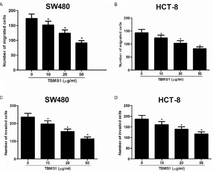
[image:5.612.90.521.75.460.2]To determine whether TBMS1 affected cell motility, the transwell without Matrigel migra-tion assay was carried out. As shown in Figure 2, the mean number of migrated cells per field
Tubeimoside-1 inhibits the growth and invasion of CRC cells
of view was significantly less in SW480 cells treated with TBMS1 than that in control groups (Figure 2A). Likewise, TBMS1 obviously inhibit-ed migration of HCT-8 cells comparinhibit-ed to that of control cells (Figure 2B). Furthermore, the tran -swell Matrigel invasion assay also demonstrat-ed that TBMS1 rdemonstrat-educdemonstrat-ed invasion activities of SW480 (Figure 2C) and HCT-8 cells (Figure 2D). TBMS1 reduced β-catenin expression in CRC cells
The Wnt/β-catenin signaling pathway was re-ported to play important roles in CRC prolifera-tion and invasion [11-13]. To understand the molecular mechanism involved in TBMS1-induced proliferation inhibition, we investigated the effect of TBMS1 on β-catenin involved in Wnt/β-catenin signaling pathway. β-catenin expression in SW480 cells treated with increas-ing concentrations of TBMS1 for 48 h was assessed using western blot. As shown in Figure 3, TBMS1 obviously inhibited the expres-sion of β-catenin in SW480 cells.
Overexpression of β-catenin rescued TBMS1-induced proliferation and invasion inhibition
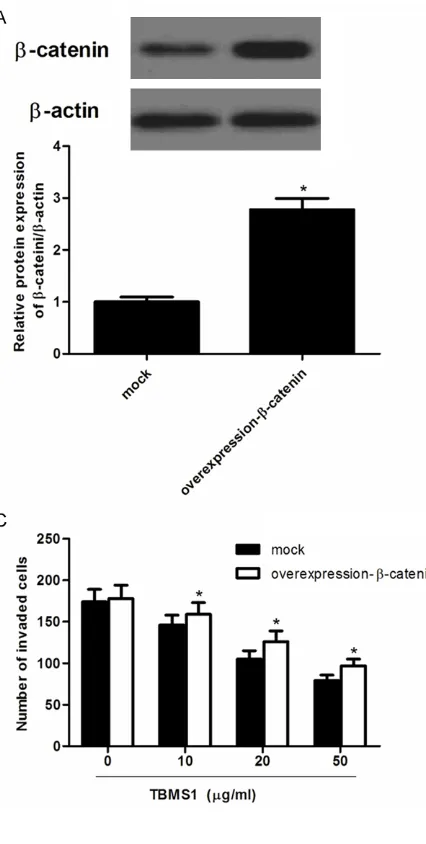
Next, we investigated the effect of β-catenin overexpression on TBMS1-induced prolifera-tion and invasion inhibiprolifera-tion. SW480 cells trans-fected β-catenin overexpression were treated with 15 lm TBMS1 for 48 h. As shown in Figure 4A, β-catenin protein level in SW480 cells transfected with β-catenin overexpression was obviously increased compared with the nega-tive group. In addition, β-catenin overexpres -sion significantly increased the cancer cell pro -liferation and invasion, and β-catenin over-expression rescued TBMS1-induced cell growth (Figure 4B) and invasion inhibition (Figure 4C). Knockdown of β-catenin potentiated TBMS1-induced proliferation and invasion inhibition
To further confirm the role of β-catenin in the proliferation and invasion of CRC cells, we per-formed a gene knockdown experiment in SW480 cells. As shown in Figure 5A, β-catenin protein level in SW480 cells transfected with siRNA-β-catenin was obviously decreased com -pared with the negative group. In addition, knockdown of β-catenin significantly potentiat -ed TBMS1-induc-ed proliferation and invasion inhibition (Figure 5B and 5C).
Discussion
TBMS1 has been reported to possess antican-cer properties. However, the role of TBMS1 on human CRC is still unclear. In this study, we found that TBMS1 inhibited the proliferation, migration/invasion of CRC cells, and it reduced β-catenin expression in CRC cells. Furthermore, overexpression of β-catenin rescued TBMS1-induced proliferation and invasion inhibition, and knockdown of β-catenin potentiated TBM-S1-induced proliferation and invasion inhibi- tion.
TBMS1 significantly inhibited the expression of β-catenin in SW480 cells. In addition, overex -pression of β-catenin rescued TBMS1-induced proliferation and invasion inhibition, and knock-down of β-catenin potentiated TBMS1-induced proliferation and invasion inhibition. All these results suggest that TBMS1 inhibited CRC cell proliferation and invasion via the Wnt/β-catenin signaling pathway.
In summary, this study demonstrated that TBMS1 inhibited CRC cell proliferation and invasion via suppressing the Wnt/β-catenin sig -naling pathway. Therefore, TBMS1 may repre-sent a chemopreventive and/or therapeutic agent in the prevention of CRC.
Acknowledgements
This research was funded by the Health Bure- au and Technology Foundation of Tianjin (No. 2012KZ063).
Disclosure of conflict of interest None.
Address correspondence to: Dr. Baoji Song, De- partment of General Surgery, Tianjin Hospital, Tian- jin 300211, China. Tel: +86-22-28332917; E-mail: songbao_ji@163.com
References
[1] Lee SJ, Moon GS, Jung KH, Kim WJ, Moon SK.
c-Jun N-terminal kinase 1 is required for cordy -cepin-mediated induction of G2/M cell-cycle
arrest via p21WAF1 expression in human co
-lon cancer cells. Food Chem Toxicol 2010; 48:
277-283.
[2] Kriza C, Emmert M, Wahlster P, Niederländer
C, Kolominsky-Rabas P. Cost of illness in co- lorectal cancer: an international review. Phar-macoeconomics 2013; 31: 577-588.
Cai Y, Zou G. NF-κB, JNK and p53 pathways are
involved in tubeimoside-1-induced apoptosis in HepG2 cells with oxidative stress and G 2/M
cell cycle arrest. Food Chem Toxicol 2011; 49:
3046-3054.
[7] Xu Y, Wang G, Chen Q, Lin T, Zeng Z, Luo Q, Liu
J, Sun C. Intrinsic apoptotic pathway and G2/M cell cycle arrest involved in tubeimoside I-in-duced EC109 cell death. Chinese J Cancer Res 2013; 25: 312-321.
[8] Huang P, Yu C, Liu XQ, Ding YB, Wang YX, He JL.
Cytotoxicity of tubeimoside I in human chorio-carcinoma JEG-3 cells by induction of cyto-chrome c release and apoptosis via the mito-chondrial-related signaling pathway. Int J Mol Med 2011; 28: 579-587.
[9] Zhang Y, Xu X, He P. Tubeimoside-1 inhibits
proliferation and induces apoptosis by
increas-ing the Bax to Bcl-2 ratio and decreasincreas-ing COX-2
expression in lung cancer A549 cells. Mol Med Rep 2011; 4: 25-29.
[10] Pfaffl MW. A new mathematical model for rela
-tive quantification in real-time RT-PCR. Nucleic
Acids Res 2001; 29: e45-e49.
[11] Guo Q, Wu M, Lian P, Liao M, Xiao Z, Wang X,
Shen S. Synergistic effect of indomethacin and
NGX6 on proliferation and invasion by human
colorectal cancer cells through modulation of
the Wnt/β-catenin signaling pathway. Mol Cell
Biochem 2009; 330: 71-81.
[12] He B-C, Gao JL, Zhang BQ, Luo Q, Shi Q, Kim
SH, Huang E, Gao Y, Yang K, Wagner ER. Tet
-randrine inhibits Wnt/β-catenin signaling and
suppresses tumor growth of human colorectal cancer. Mol Pharmacol 2011; 79: 211-219. [13] Park CH, Chang JY, Hahm ER, Park S, Kim H-K,
Yang CH. Quercetin, a potent inhibitor against β-catenin/Tcf signaling in SW480 colon cancer
cells. Biochem Bioph Res Co 2005; 328: 227-234.
[14] Zhang Y, Xu XM, Zhang M, Qu D, Niu HY, Bai X,
Tubeimoside-1 inhibits the growth and invasion of CRC cells
[15] Guan YY, Liu HJ, Luan X, Xu JR, Lu Q, Liu YR, Gao YG, Zhao M, Chen HZ, Fang C. Raddeanin
A, a triterpenoid saponin isolated from Ane- mone raddeana, suppresses the angiogene- sis and growth of human colorectal tumor by
inhibiting VEGFR2 signaling. Phytomedicine
2015; 22: 103-110.
[16] Zhang Z, Li Z, Wu X, Zhang CF, Calway T, He TC, Du W, Chen J, Wang CZ, Yuan CS. TRAIL path -way is associated with inhibition of colon can-cer by protopanaxadiol. J Pharmacol Sci 2015; 127: 83-91.
[17] Urakami S, Shiina H, Enokida H, Kawakami T,
Tokizane T, Ogishima T, Tanaka Y, Li LC, Ri
-beiro-Filho LA, Terashima M. Epigenetic inacti -vation of Wnt inhibitory factor-1 plays an im-portant role in bladder cancer through aberrant
canonical Wnt/β-catenin signaling pathway.
Clin Cancer Res 2006; 12: 383-391.
[18] Fodde R, Brabletz T. Wnt/β-catenin signaling in
cancer stemness and malignant behavior. Curr Opin Cell Biol 2007; 19: 150-158.
[19] Saydam O, Shen Y, Würdinger T, Senol O, Boke E, James MF, Tannous BA, Stemmer-Rachami
-mov AO, Yi M, Stephens RM. Downregulated
microRNA-200a in meningiomas promotes tu-mor growth by reducing E-cadherin and
activat-ing the Wnt/β-catenin signalactivat-ing pathway. Mol
Cell Biol 2009; 29: 5923-5940.
[20] Major MB, Camp ND, Berndt JD, Yi X, Golden -berg SJ, Hubbert C, Biechele TL, Gingras AC, Zheng N, MacCoss MJ. Wilms tumor
suppres-sor WTX negatively regulates WNT/ß-catenin
signaling. Science 2007; 316: 1043-1046. [21] Clevers H, Nusse R. Wnt/β-catenin signaling
and disease. Cell 2012; 149: 1192-205. [22] Zhen T, Dai S, Li H, Yang Y, Kang L, Shi H,
Zhang F, Yang D, Cai S, He Y. MACC1 promot-es carcinogenpromot-esis of colorectal cancer via
β-catenin signaling pathway. Oncotarget 2014; 5: 3756-3769.
[23] Gao ZH, Lu C, Wang MX, Han Y, Guo LJ. Differ
-ential β-catenin expression levels are associ -ated with morphological features and progno-sis of colorectal cancer. Oncol Lett 2014; 8: 2069-2076.
[24] Han J, Gao B, Jin X, Xu Z, Li Z, Sun Y, Song B.
Small interfering RNA-mediated downregula-tion of beta-catenin inhibits invasion and mi-gration of colon cancer cells in vitro. Med Sci Monit 2012; 18: BR273-80.