Original Article
Smooth muscle immaturity in the carotid arterial
neointima as a prognostic marker for systemic
atherogenic cardiovascular events in the Asian male
Hirotsugu Hashimoto1,2, Atsushi Kurata1, Tamaki Nashiro2, Shigeru Inoue3, Tomonori Ushijima2, Koji Fujita1,
Toshikazu Kimura4, Kensuke Kawai4, Hajime Horiuchi2, Masahiko Kuroda1
1Department of Molecular Pathology, Tokyo Medical University, Tokyo, Japan; 2Department of Diagnostic
Pathology, NTT Medical Center Tokyo, Tokyo, Japan; 3Department of Preventive Medicine and Public Health, Tokyo Medical University, Tokyo, Japan; 4Department of Neurosurgery, NTT Medical Center Tokyo, Tokyo, Japan
Received September 28, 2015; Accepted October 28, 2015; Epub November 1, 2015; Published November 15, 2015
Abstract: Although immaturity of neointimal smooth muscle cells (SMCs) in coronary arteries has recently been demonstrated to be associated with acute coronary syndrome, the carotid arterial counterpart has not been in-vestigated. We hypothesized that the same investigation of carotid endarterectomy specimens might contribute to living patients. Carotid endarterectomy specimens from 33 Asian males who underwent a 5-year follow-up were examined. Age, atherosclerotic risk factors, and percentage stenosis were investigated. Histologically, the fibrous cap/lipid core ratio was measured. Maturation of SMCs was assessed by the h-caldesmon/smooth muscle actin (SMA) ratio by immunohistochemistry in 3 different regions (luminal, medial, and opposite side of lipid core) in the neointima. Associations of these factors with preoperative symptoms along with postoperative systemic athero-genic cardiovascular events were analyzed. It was revealed that fibrous cap/lipid core ratio was significantly lower in symptomatic than in asymptomatic patients, while the h-caldesmon/SMA ratio was significantly lower in pa -tients with than without postoperative systemic atherogenic cardiovascular events by the Student’s t-test (P<0.05). Logistic regression model demonstrated that younger age and a lower h-caldesmon/SMA ratio were associated with postoperative systemic atherogenic cardiovascular events (P<0.05). This result was not different when 3 different regions were each analyzed instead. Immaturity of neointimal SMCs shown by a lower h-caldesmon/SMA ratio by immunohistochemistry was associated with systemic atherogenic cardiovascular events. Thus, this finding may be predictive of these events after carotid endarterectomy. Uniform results among different neointimal regions suggest that immaturity of neointimal SMCs causes plaque instability and does not occur secondarily to plaque instability.
Keywords: Carotid endarterectomy, immunohistochemistry, smooth muscle, differentiation, prognosis
Introduction
Stroke is the fourth leading cause of mortality and a major source of chronic disability in the United States and Japan, although its incidence has decreased steadily since the 1960s [1, 2]. Extracranial carotid artery stenosis is a factor in 20%-30% of all strokes [1], and surgical endovascular revascularization such as carotid artery stenting or carotid endarterectomy (CEA) is recommended in patients with stenosis
≥60% and with a life expectancy >5 years [1,
3]. Apart from the risk for stroke, the presence of severe asymptomatic carotid artery stenosis is a predictor of systemic cardiovascular events,
reflecting the severity of systemic atherosclero -sis [1-4].
The direct trigger of stroke associated with carotid artery stenosis is mainly plaque rupture due to the vulnerability of atherosclerotic plaque leading to thrombus formation and embolization of the intracranial cerebral vascu-lature [2, 5]. The histological characteristics of rupture-prone vulnerable plaques in the carotid artery were noted to be a large necrotic core,
thin fibrous cap, and abundant macrophages
However, little is known about the association of plaque stability with smoo- th muscle cells (SMCs), the main component of plaque. Although a smaller quantity of SMCs was reported to be associated with vulnerable plaques than with stable plaque in the coronary ar- teries [8], the quality of neointimal SMCs in associ-ation with plaque stability rarely has been described. Indeed, it is believed that neointimal SMCs have a uniformly “synthetic” phe-notype in contrast to a “contractile” phenotype in the media [9]. The synthet-ic phenotype is immunohis-tochemically characterized by lack of staining of some differentiated SMC mark-ers such as desmin and smoothelin [10]. Neointimal SMCs were reported to have an almost exclusively synthetic phenotype not only in atherosclerosis but also in Moyamoya disease [11] and restenosis after coronary stent implanta-tion [10]. We formerly dem-onstrated this phenome-non also in the neointima of Buerger’s disease and thromboembolism [12]. We previously evaluated SMC maturation in uterine muscular neoplasm by im- munostaining for h-caldes-mon [13], and found that this marker was useful for assessment of SMCs be- yond intermediate matura-tion. Also, we recently sho- wed by an autopsy study that neointimal SMCs have divergent phenotypes in differentiation in the coro-nary artery [14]. That is, an h-caldesmon-negative rela-tively immature phenotype
and an h-caldesmon-posi-tive relah-caldesmon-posi-tively mature phe-notype exist, and the for-mer was associated with acute coronary syndrome. Autopsy studies, however,
are not beneficial to pre -mortem patients. In con-trast, CEA specimens pre-sented to pathological de-
partments may benefit liv -ing patients. To the best of our knowledge, evaluation of neointimal SMCs by immunostaining of h-calde-smon in the carotid artery has not been performed. Therefore, the purpose of the present study was to assess whether results of immunohistochemistry for h-caldesmon in CEA mate-rial are associated with pre-operative symptoms and are predictive of systemic cardiovascular events.
Material and methods
Patients
The local Ethics Committee approved the protocol of the present study. A total of 51 consecutive CEAs from 48 patients (44 males and 4 females, aged 52-84 years) performed between November 2003 and De- cember 2008 at NTT Me- dical Center Tokyo were
[image:3.629.98.397.77.648.2]identified from the data -base at that institution. Detailed clinical data had been recorded for each patient, including age and sex, along with past history
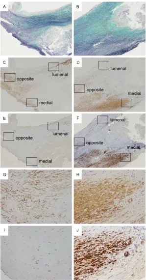
Figure 2. Elastica Masson staining (A, B) as well as Immunohistochemistry for α-SMA (C, D, G, H) and h-caldesmon (E, F, I, J) in case with postoperative cardio -vascular events (A, C, E: identical fields) and in a non-event case (B, D, F: identi -cal fields). (A, B) Lipid core, fibrous cap, and other neointimal components are observed. (C, D) Abundant α-SMA-positive SMCs are identified in the neointima. (E) Most of the neointimal SMCs are not immunostained by h-caldesmon. (F) Most of neointimal SMCs are also positive for h-caldesmon. (G-J) High-power
and/or presence of smoking, hypertension, dia-betes, or dyslipidemia based on current Japanese criteria. Percentage stenosis had been measured by ultrasonic and/or magnetic resonance angiography according to the North American Symptomatic Carotid Endarterectomy Trial (NASCET). Preoperative occurrence of cerebral infarction and transient ischemic attack (TIA) had been recorded. Furthermore, we searched for systemic cardiovascular events that had occurred during a postoperative fol-low-up period of 5 years. Of note, in the present study, cardiovascular events associated with atherosclerosis were adopted, such as re-ste-nosis, cerebral infarction, contralateral carotid stenosis, acute coronary syndrome, arterio-sclerosis obliterans, and aortic aneurysm. Eight
paraffin-embedded specimens in which the
follow-up period was less than 5 years were excluded from the present study. However, specimens from patients in whom the systemic atherogenic cardiovascular events were recog-nized within 5 years were included, even if the follow-up period was less than 5 years. Two specimens from patients with the complication
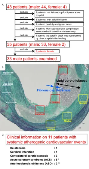
of atrial fibrillation were excluded, since throm -boembolism regardless of atherosclerosis may occur. One specimen from a postoperative death from another cause, 1 from a patient with a postoperative local complication (early rupture), and 1 specimen that had been bor-rowed and not returned were excluded. Three specimens from a CEA of contralateral carotid stenosis were excluded, but preceding CEA specimens were used and regarded as from cases with postoperative systemic atherogenic cardiovascular events. Thus, 35 specimens from 35 patients (33 males and 2 females) were adopted. Due to the remarkable sex dif-ference, only the 33 specimens from the males were included in the present study (Figure 1A). In all 33 cases, antiplatelet drugs had been prescribed post-operatively.
Grouping
Grouping of the cases was performed by 2 indi -vidual methods. First, cases were divided into a symptomatic or asymptomatic group based on the presence or absence, respectively, of pre-operative symptoms including cerebral infarc-tion and TIA. Second, cases were divided into an event or non-event group by the presence or absence, respectively, of postoperative
occur-rence of one or more of the systemic athero-genic cardiovascular events thus far men- tioned.
Pathological study and immunohistochemistry
The surgically removed carotid plaques were
fixed in 10% formalin solution for 24 h, decalci
-fied by formic acid, and embedded in paraffin.
The specimen with the most narrowed artery was selected for each case. Prior to immunos-taining, selected tissue sections were re-stained with hematoxylin-eosin and elas- tica Masson staining. Immunohistochemistry
was performed using paraffin-embedded se-ctions, 4 μm thick, using the
avidin-biotin-peroxidase complex according to st- andard methods. The monoclonal antibody to
alpha-smooth muscle actin (α-SMA, clone 1A4, working dillution 1:100, Dako, Glostrup,
Denmark) was used to identify all SMCs, and h-caldesmon (clone h-CD, working dillution 1:100, Dako) was used to identify SMCs beyond
intermediate differentiation. Deparaffinized
and dehydrated sections were treated with 0.3% hydrogen peroxide in methanol for 30 min to block endogenous peroxidase activity. For immunostaining of h-caldesmon, sections were autoclaved in 10 mmol/L sodium citrate buffer (pH 6.0) at 121°C for 10 min to expose anti-gens and were cooled for 30 min. After rinsing in 0.1 mol/L phosphate buffered saline (PBS, pH 7.4), the sections were incubated with affin
-ity purified primary antibodies overnight
at 4°C. Thereafter, they were incubated with Envision (+) rabbit peroxidase (Dako, Carpinteria, CA, USA) for 30 min. The peroxi-dase reaction was performed using 0.02% 3,3’-diaminobenzidine tetrahydrochloride and 0.01% hydrogen peroxide in 0.1 mol/L PBS (pH 7.4). Finally, nuclear counterstaining was per-formed with Mayer’s hematoxylin.
Assessment
For each patient, the number of risk factors present was counted from among past history and/or presence of smoking, hypertension, dia-betes, and dyslipidemia.
Light microscopy performed with a Carl Zeiss HAL 100 instrument and a W-PI 10×/23 ocular lens was used to analyze and quantitate the hisitological data. The narrowest thickness of
core of each vessel were measured using elas-tica Masson staining in the region where the lipid core was widest (Figure 1B). In specimens with obviously ruptured plaque, the thickness
of the fibrous cap was counted as 0. Then, the ratio of fibrous cap thickness to lipid core thick
-ness was calculated (fibrous cap/lipid core
ratio).
In assessing immunohistochemical results, the luminal, medial, and opposite (neointima but devoid of lipid core) sides of the lipid core were
identified from the neointima. Then 3 different fields from each of these 3 regions were select -ed in each specimen (Figure 1B). Identical
fields were selected from α-SMA and h-caldes -mon immunostaining. Digital images of
immu-nohistochemistry of these 9 fields (×200) in 2
immunostainings were captured by the NY-D5000 super system (Microscope Network, Nikon, Tokyo, Japan) and were printed by a true-color printer (IPSiO SP C420, Ricoh, Tokyo, Japan). In every image, a separated positive area was regarded as individual positive cells, but a positive area less than the size of a mus-cular nucleus was discarded. Total number of
positive cells was counted in each α-SMA and
h-caldesmon immunostained image, respec-tively. Then, the average ratio of h-caldesmon+
cells to α-SMA+ cells (h-caldesmon/SMA ratio)
was counted in each of 3 regions. Further, the total average h-caldesmon/SMA ratio was cal-culated in each specimen.
Statistical analyses
As statistical analyses, comparison of average values for data including clinicopathological
and immunohistochemical results was first per -formed by the Student’s t-test between symp-tomatic and asympsymp-tomatic groups as well as between event and non-event groups. Then,
logistic regression analyses were performed using the SPSS software package (ver17.0, Tokyo, Japan) since outcome variables were dichotomous, and multiple explanatory vari-ables were to be analyzed simultanously. Outcome variables were established as symp-tomatic vs. asympsymp-tomatic groups (sympsymp-tomatic =1, asymptomatic =0) as well as event vs. non-event groups (non-event =1, non-non-event =0). As explanatory variables, 5 variables were estab-lished and used in each analysis: 1, age; 2, number of atherosclerotic risk factors; 3,
per-centage stenosis by NASCET; 4, fibrous cap/
lipid core ratio; and 5, total average h-caldes-mon/SMA ratio. These analyses were also per-fomed by changing total average h-caldesmon/ SMA ratio to the average ratio in each of 3
dif-ferent regions. The level of significance was set
at P<0.05.
Results
Patients’ baseline characteristics and average values of assessed data
Specimens for the present study were obtained from 33 males (age 52-84, mean 68.1±6.8 y). Past history and/or presence of smoking, hypertension, diabetes, and dyslipidemia were
identified in 30, 29, 15, and 17, respectively, of
the 33 patients. The number of these risk fac-tors ranged from 1 to 4 (mean 2.76±0.83) among the 33 patients. Percentage stenosis of the carotid artery by NASCET ranged from 50 to 99% (mean 80.2±12.1%). Fibrous cap thick-ness ranged from 0 to 1200 (mean 267±274)
μm, while lipid core thickness ranged from 200 to 7500 (mean 2249±1916) μm. Fibrous cap/
lipid core ratio ranged from 0 to 400% (mean 31±71%). Immunohistochemical results of SMA+ cells ranged from 2068-7861 (mean
[image:6.629.94.533.95.186.2]4828±1563), while h-caldesmon+ cells ranged
Table 1. Odds ratios (ORs) according to grouping by preoperative symptoms or postoperative events Symptomatic vs. Asymptomatic Event vs. Non-event
OR (95% CI) P OR (95% CI) P
Age 1.01 (0.86-1.20) 0.87 0.86 (0.74-1.00) 0.047
from 269-4083 (mean 1466±1038) for the
sum of 9 analyzed fields. Then, the h-caldes -mon/SMA ratio ranged from 8.7 to 78.9% (mean 29.5±17.5%) according to the total aver-age of 3 different regions. Results of
sub-clas-sifications of these values were 3.9-78.3%
(mean 23.1±17.8%) for the luminal side, 4.2-68.8% (mean 27.4±15.3%) in the medial side, and 8.9-85.3% (mean 34.4±21.2%) in the opposite side of lipid core. Although the h-caldesmon/SMA ratio varied among different specimens, this ratio tended to be similar among different regions in the same specimen.
Figure 2 shows specimens with lower and
high-er h-caldesmon/SMA ratios by low powhigh-er fields and representative high power fields (medial
side).
Preoperatively, cerebral infarction was detect-ed in 19 cases, and TIA only was detectdetect-ed in 7
cases; no symptoms were identified in 7 cases.
Thus, the symptomatic group and asymptom-atic group were comprised of 26 and 7 cases, respectively. Postoperative events were
identi-fied in 11 patients (Figure 1C), so that the event and non-event groups were comprised of 11 and 22 patients, respectively.
Differences in assessed values according to preoperative symptoms
Comparisons of representative data according
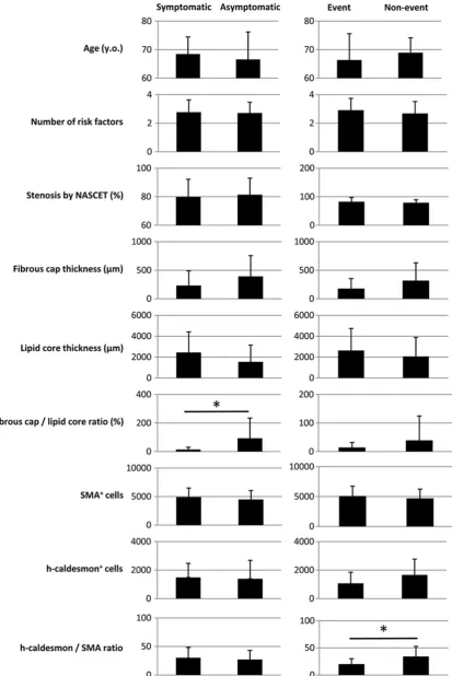
to classifications are charted in Figure 3. Average age ± S.D. of the symptomatic vs. asymptomatic group was 68.5±6.0 vs. 66.6±9.6 y. Differences in average values ± S. Ds. between the symptomatic vs. asymptomat-ic groups were 2.77±0.86 vs. 2.71±0.76 for ath-erosclerotic risk factors, 79.9±12.4% vs. 81.4±11.6% for stenosis by NASCET, 233±257
vs. 393±364 μm for fibrous cap thickness, 2438±1976 vs. 1543±1606 μm for lipid core thickness, 14±16% vs. 93±142% for the
fibr-ous cap/lipid core ratio, 4918±1578 vs.
4490±1575 for SMA+ cells, 1486±987 vs.
1390±1297 for h-caldesmon+ cells, and
30.2±18.1% vs. 27.0±16.1% for the h-caldes-mon/SMA ratio. Among these comparisons,
only the fibrous cap/lipid core ratio was statisti
-cally significant by the Student’s t-test (P< 0.05).
Differences in assessed values according to postoperative events
Average age ± S.D. in the event group vs. non-event group was 66.4±9.2 vs. 68.9±5.2 y. Differences in average values ± S.Ds. between the event vs. the non-event group were 2.91±0.83 vs. 2.68±0.84 for atherosclerotic risk factors, 82.5±15.4% vs. 79.1±10.3% for
stenosis by NASCET, 173±182 vs. 314±317 μm for fibrous cap thickness, 2632±2115 vs. 2057±1831 μm for lipid core thickness, 14±18% vs. 39±86% for the fibrous cap/lipid
core ratio, 5086±1644 vs. 4698±1544 for SMA+ cells, 1072±777 vs. 1663±1110 for
h-caldesmon+ cells, and 20.1±10.0% vs.
34.3±18.6% for the h-caldesmon/SMA ratio. Of these comparisons, only the h-caldesmon/
SMA ratio was statistically significant by the
Student’s t-test (P<0.05).
Results of logistic regression analyses
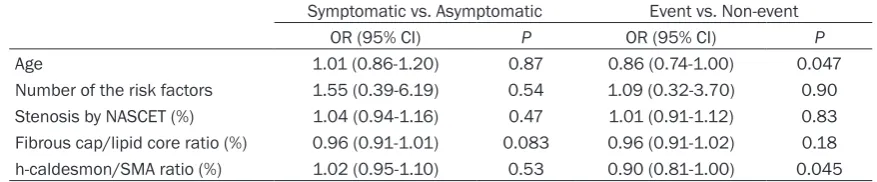
The results of logistic regression analyses using the total average h-caldesmon/SMA ratio are summarized in Table 1. There was no
sig-nificantly related variable in the analysis of the
symptomatic versus the asymptomatic group. However, the analysis of the event vs. the non-event group showed that age and the
h-caldes-mon/SMA ratio were significantly related to
[image:7.629.96.532.94.173.2]each other (OR 0.86, 95% CI 0.74 to 1.00, P=0.047; and OR 0.90, 95% CI 0.81 to 1.00, P=0.045, respectively), meaning that younger age and a lower h-caldesmon/SMA ratio were
Table 2. Odds ratios (ORs) of h-caldesmon/SMA ratio according to different evaluated regions Symptomatic vs. Asymptomatic Event vs. Non-event
OR (95% CI) P OR (95% CI) P
Average of 3 regions 1.02 (0.95-1.10) 0.53 0.90 (0.81-1.00) 0.045 Luminal side of lipid core 1.05 (0.96-1.16) 0.31 0.90 (0.81-1.00) 0.056 Medial side of lipid core 1.02 (0.94-1.11) 0.57 0.94 (0.87-1.01) 0.11 Opposite side of lipid core 1.01 (0.96-1.07) 0.67 0.93 (0.86-1.00) 0.057
significantly related to postoperative systemic
atherogenic cardiovascular events.
The above-mentioned 2 logistic logistic regres-sion analyses were further performed by chang-ing the total average h-caldesmon/SMA ratio to the ratio in each of 3 different regions. As shown in Table 2, no significant results were
obtained concerning analyses of symptomatic versus asymptomatic groups. Although no
sig-nificant results were obtained concerning anal -yses of the event vs. the non-event group either, similar results were obtained for analyses based on the luminal side (OR 0.90, 95% CI 0.81 to 1.00, P=0.056), medial side (OR 0.94, 95% CI 0.87 to 1.01, P=0.11), and opposite side (OR 0.93, 95% CI 0.86 to 1.00, P=0.057).
Discussion
In the present study, we showed heterogeneity in the degree of neointimal SMC maturation assessed by the immunohistochemical h-calde-smon/SMA ratio in the carotid artery for the
first time, even if the extent of stenosis in the
arteries was to the same degree. An especially
important finding was that this ratio was signifi -cantly associated with postoperative systemic atherogenic cardiovascular events. This means that when we regard atherosclerosis as a sys-temic disease [2-4], we can predict the cardio-vascular prognosis by examining CEA material. Therefore, immaturity of neointimal SMCs may not only be associated with acute coronary syn-drome when assessed in the coronary arteries as we have recently reported [14], but also may be an indicator of systemic complications when assessed in the carotid arteries.
The Student’s t-test demonstrated that only the
fibrous cap/lipid core ratio was significantly dif -ferent between symptomatic and asymptomat-ic patients, with this ratio being lower in the symptomatic patients. This result is in accor-dance with a histological study of a large cohort by other authors in which unstable plaques
indicated by a thin fibrous cap and large lipid core were significantly more prevalent in symp -tomatic cases than in asymp-tomatic cases [5]. Another study also showed that a large lipid
core and macrophage infiltration were associ -ated with plaque instability [6].
Although our previous study demonstrated that immaturity of neointimal SMCs assessed by
the h-caldesmon/SMA ratio was associated with acute coronary syndrome in coronary
arteries [14], this ratio did not differ significant -ly between symptomatic and asymptomatic patients. This may be due to a difference in pathogenesis between cerebral and myocardial infarction. That is, the major pathogenesis of cerebral infarction associated with carotid ste-nosis is not local acute thrombotic occlusion as in myocardial infarction, but artery-to-artery embolism [2]. In addition, hemodynamics, including the development of collaterals, is also concerned with the occurrence of cerebral infarction [2]. Furthermore, sample bias due to investigation of only CEA material may be of concern since extracranial carotid artery steno-sis is a factor in only 20%-30% of all strokes [1]. When data based on the grouping according to postoperative events were compared, the
fibrous cap/lipid core ratio was not significantly
different between event and non-event patients
as verified by a higher standard deviation. This
may be due to the relatively smaller number of enrolled subjects, but, alternatively, this
param-eter may not influence systemic complications
although it affects local manifestations. Instead, the Student’s t-test demonstrated that
only the h-caldesmon/SMA ratio was signifi -cantly different between the event and non-event groups. Furthermore, logistic regression analysis revealed that younger age and a lower
h-caldesmon/SMA ratio were significantly relat -ed to postoperative systemic atherogenic car-diovascular events. Although the average age was not greatly different between the event and non-event groups, the lower standard
devi-ation may have resulted in a significant value
for this variable in the logistic regression analy-sis. Indeed, other authors have also reported results of CEA studies showing that younger patients were more likely to develop recurrent carotid stenosis and cardiovascular events than their older counterparts [15, 16]. On the other hand, independently of age, the
h-caldes-mon/SMA ratio was significantly related to
postoperative atherogenic events. Therefore, the degree of maturation of neointimal SMCs in the carotid artery may be paralleled by that in the systemic vasculature, where immature neo-intimal SMCs are likely to be associated with atherogenic events.
that at least 2 possibilities exist. First, intra-plaque leukocytes, including macrophages, secrete enzymes such as matrix metallopro-teinase, which inhibit maturation of SMCs in the same manner as the development of plaque
instability through fragilization of the fibrous
cap [2, 5]. Second, a genetic or acquired unknown factor regulates maturity or at least the maturation speed of SMCs. Then, immature SMCs cannot inhibit the secondary enlarge-ment of the lipid core in contrast to mature SMCs that suppress growth of the lipid core. We postulate that the second hypothesis is likely, since additional logistic regression analy-ses in which 3 different neointimal regions were individually analyzed demonstrated simi-lar results among analyses based on luminal, medial, and opposite side of the lipid core. If
the first hypothesis were true, the luminal or
medial side of the lipid core, where macro-phages were near SMCs, should have been
more significantly related to the outcome than the opposite side of the lipid core. Genetic pre -disposition has been suggested to account for 40% to 60% of human susceptibility to coro-nary artery disease and thus to atherosclero-sis, but genome-wide association studies dem-onstrated that most genetic risk variants are mediated through unknown mechanisms [17]. Whether or not maturation of SMCs is associ-ated with these unknown mechanisms will be elucidated in the future.
The limitation of the present study is that analy-ses of the individual atherosclerotic risk factors of smoking history, hypertension, diabetes, and dyslipidemia were not performed since the number of enrolled patients was relatively small, aggregation bias was relatively higher than in usual studies in which the positivity ratio of each factor is around 50% (e.g. smoking was found 30 out of 33 subjects), and patho-logical risk factors other than atherosclerotic risk factors were mainly analyzed. However, one study reported that none of these factors
was significantly different between a symptom -atic and asymptom-atic group [5]. In addition, female subjects were excluded from the pres-ent study because of the extremely small
num-ber of females identified in our examination of
the database. This small number was probably because of estrogen-related anti-atherosclerot-ic mechanisms. Future study is necessary to investigate SMC maturation and sex differences.
In conclusion, we have conducted a clinico-pathological retrospective cohort study using CEA material from Asian males. Plaque
instabil-ity indicated by a lower fibrous cap/lipid core
ratio was shown more frequently in symptom-atic than in asymptomsymptom-atic patients, whereas immature neointimal SMCs indicated by a lower h-caldesmon/SMA ratio was associated with postoperative systemic atherogenic cardiovas-cular events. Thus, immunostaining of h-calde-smon and SMAs in CEA material may predict systemic atherogenic events. Uniform results among different neointimal regions suggested that immaturity of neointimal SMCs is not a result of but a cause of plaque instability.
Acknowledgements
We thank Goichiro Yanagi for his skillful techni -cal assistance. Further, we thank Dr. Yumiko Yamaoka for her academic advice.
Disclosure of conflict of interest
None.
Address correspondence to: Dr. Atsushi Kurata, Department of Molecular Pathology, Tokyo Medical University, 6-1-1 Shinjuku, Shinjuku-ku, Tokyo 160-8402, Japan. Tel: + 81 3 3351 6141 (393); Fax: + 81 3 3352 6335; E-mail: [email protected]
References
[1] O’Brien M, Chandra A. Carotid revasculariza-tion: risks and benefits. Vasc Health Risk Manag 2014; 10: 403-16.
[2] Yamazaki M, Uchiyama S. Pathophysiology of carotid stenosis. Brain Nerve 2010; 62: 1269-75.
[3] Ammirati E, Moroni F, Norata GD, Magnoni M, Camici PG. Markers of inflammation associat -ed with plaque progression and instability in patients with carotid atherosclerosis. Mediators Inflamm 2015; 2015: 718329. [4] Lamina C, Meisinger C, Heid IM, Löwel H,
Rantner B, Koenig W, Kronenberg F; Kora Study Group. Association of ankle-brachial in -dex and plaques in the carotid and femoral ar-teries with cardiovascular events and total mortality in a population-based study with 13 years of follow-up. Eur Heart J 2006; 27: 2580-7.
[6] Redgrave JN, Lovett JK, Gallagher PJ, Rothwell PM. Histological assessment of 526 symptom-atic carotid plaques in relation to the nature and timing of ischemic symptoms: the Oxford plaque study. Circulation 2006; 113: 2320-8. [7] Virmani R, Burke AP, Farb A, Kolodgie FD.
Pathology of the vulnerable plaque. J Am Coll Cardiol 2006; 47 Suppl: C13-8.
[8] Falk E, Nakano M, Bentzon JF, Finn AV, Virmani R. Update on acute coronary syndromes: the pathologists’ view. Eur Heart J 2013; 34: 719-28.
[9] Rudijanto A. The role of vascular smooth mus-cle cells on the pathogenesis of atherosmus-clero- atherosclero-sis. Acta Med Indones 2007; 39: 86-93. [10] Chaabane C, Otsuka F, Virmani R,
Bochaton-Piallat ML. Biological responses in stented ar-teries. Cardiovasc Res 2013; 99: 353-63. [11] Masuda J, Ogata J, Yutani C. Smooth muscle
cell proliferation and localization of macro-phages and T cells in the occlusive intracranial major arteries in moyamoya disease. Stroke 1993; 24: 1960-7.
[12] Kurata A, Machinami R, Schulz A, Fukayama M, Franke FE. Different immunophenotypes in Buerger’s disease. Pathol Int 2003; 53: 608-15.
[13] Horita A, Kurata A, Maeda D, Fukayama M, Sakamoto A. Immunohistochemical character-istics of atypical polypoid adenomyoma with special reference to h-caldesmon. Int J Gynecol Pathol 2011; 30: 64-70.
[14] Horita A, Kurata A, Ohno S, Shimoyamada H, Saito I, Kamma H, Kuroda M. Immaturity of smooth muscle cells in the neointima is asso-ciated with acute coronary syndrome. Cardiovasc Pathol 2015; 24: 26-32.
[15] Valentine RJ, Myers SI, Hagino RT, Clagett GP. Late outcome of patients with premature ca-rotid atherosclerosis after caca-rotid endarterec-tomy. Stroke 1996; 27: 1502-6.