Int J Clin Exp Pathol 2015;8(12):15801-15807 www.ijcep.com /ISSN:1936-2625/IJCEP0017101
Original Article
Expression of TAK1/TAB1 expression in non-small
cell lung carcinoma and adjacent normal
tissues and their clinical significance
Jiang Zhu, Qiang Li, Jin-Tao He, Guang-Yuan Liu
Department of Pulmonary Oncology, Sichuan Cancer Hospital, Chengdu 610041, P. R. China
Received September 29, 2015; Accepted November 24, 2015; Epub December 1, 2015; Published December 15, 2015
Abstract:The purpose of this study was to investigate the expression of transforming growth factor beta-activated kinase 1 (TAK1) and its activation ligand, TAK1-binding protein 1 (TAB1), in non-small cell lung carcinoma (NSCLC) and adjacent normal tissues and to analyze the relevance between TAK1 and TAB1 protein expression and the pathological features of NSCLC patients. Surgical resection NSCLC specimens were collected from 74 patients un-dergoing surgery in our hospital from September 2003 to July 2008; tumor-adjacent normal tissue specimens were
collected as controls. All cases were pathologically confirmed after surgery, and pathological data were complete
for all patients. The expression of TAK1/TAB1 proteins in NSCLC and adjacent cancer tissues was detected by im-munohistochemical analysis. The correlation between TAK1/TAB1 protein expression and the clinicopathological features and outcome of NSCLC was assessed. The positive expression ratio of TAK1 in NSCLC tissue was 63.5%,
which was significantly higher than that in tumor-adjacent normal tissue (31.1%). The positive expression ratio of TAB1 in NSCLC tissue was 51.4%, which was significantly higher than that in tumor-adjacent normal tissue (24.3%).
Further analysis showed that positive protein expression of TAK1 and TAB1 was unrelated to patient gender, age, tumor size, degree of differentiation, and history of smoking (P>0.05) but was significantly related to clinical stage
and lymph node metastasis (P<0.05). Additionally, the expression of TAK1 as well as TAB1 was negatively related
to NSCLC patient prognosis, and patients with positive protein expression had a significantly lower 5-year survival
rate than those with negative protein expression (P<0.05). TAK1/TAB1 expression in NSCLC tissue is significantly
increased and closely associated with patient clinical prognosis. These two proteins are likely to become new thera-peutic targets for the treatment of NSCLC.
Keywords: Non-small cell lung carcinoma, TAK1, TAB1
Introduction
Non-small cell lung carcinoma (NSCLC) ac- counts for more than 85% of all lung cancers and is a malignant tumor that seriously threat-ens human life and health. Although new meth-ods for NSCLC diagnosis and treatment have continuously emerged, and the overall thera-peutic effect on NSCLC has improved, the 5- year survival rate of NSCLC is still approximate-ly 15% [1]. Therefore, improving patient prog- nosis through an in-depth understanding of the exact molecular mechanisms of NSCLC development and progression and identifying specific targets for medication treatment is critical. Transforming growth factor beta-acti-vated kinase 1 (TAK1) shows high expression in a variety of tumor tissues, and this protein is
closely related to the development, progres-sion, and invasion of various tumors [2]. The present study investigated the expression of TAK1 and its activation ligand, TAK1-binding protein 1 (TAB1), in NSCLC tissue. We further analyzed the relevance of TAK1/TAB1 expres-sion and the clinicopathological features and prognosis of NSCLC patients to determine new areas of interest for NSCLC treatment.
Materials and methods
Specimens
tis-sues were collected for testing. Prior to surgery, none of the patients underwent comprehensive therapy, such as radiotherapy, chemotherapy, and biological therapy. All cases were con-firmed by pathological diagnosis after surgery. Clinical data were complete for all patients, and all subjects signed an informed consent form.
Immunohistochemical and hematoxylin-eosin staining
All antibodies used for immunohistochemical staining were purchased from Santa Cruz Bio- technology (Santa Cruz, CA, USA). Tissue speci-mens from surgical resection were immediately immersed in neutral formalin and kept at 4°C for 1 d. The fixed specimens were embedded in paraffin and then sliced into 5 µm tissue sec -tions using a microtome. Section specimens were deparaffinized with xylene and hydrated with graded ethanol, followed by antigen re- trieval using antigen retrieval solution. There-
immune sera and incubated with TAK1 and TAB1 antibodies in a 37°C incubator for 2 h. After thoroughly washing with phosphate-buff-ered saline (PBS), the specimens were incubat-ed with the corresponding secondary antibod-ies for 1 h, thoroughly washed with PBS on a shaker, and incubated with horseradish peroxi-dase-labeled streptavidin. After 15 min of reac-tion, the specimens were washed with PBS fol-lowed by coloration with diaminobenzidine, counter-staining with hematoxylin, and micro-scopic examination. The negative control was prepared with PBS replacing primary antibo- dies.
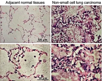
[image:2.629.99.532.81.425.2]Therapeutic targets for NSCLC-TAK1/TAB1 expression
in 15 fields of view; the ratio of positive ce-lls was calculated from the mean number of TAK1/TAB1-positive cells in 100 cells; the mean value from 15 fields of view was taken as the final result of each pathological section; four section specimens were selected from each patient, and the mean value of the four sections was taken as the final result of the patien (Figure 1). Further, immunohistochemi-cal data were interpreted according to previous reports, mainly based on the following two points: 1) the percentage of positive cells with <1% for 0 points; 1-10% for 1 point, 10-30% for 2 points, 30-60% for 3 points, and >60% for 4 points; and 2) the staining intensity of positive cells with negative for 0 points, light yellow for 1 point, yellow for 2 points, and brownish yellow for 3 points. The final result was interpreted as 0-2 points for negative and 3-7 points for posi-tive [4].
Statistical analyses
Statistical analyses were performed using SP- SS 13.0 (SPSS Inc., Chicago, IL, USA). The count data were analyzed using χ2 test. Survival anal-ysis was performed using the Kaplan-Meier method. P<0.05 was considered significant. Results
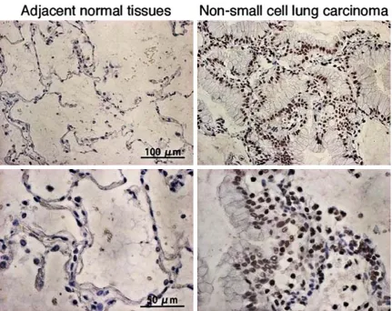
TAK1 and TAB1 protein expression in NSCLC and adjacent normal tissues
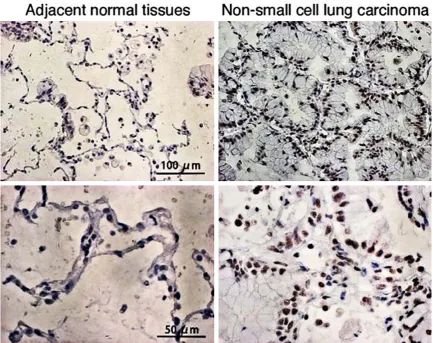
[image:3.629.100.532.80.422.2]adjacent normal tissue, showing a significant difference between groups (Table 1, P<0.05). TAB1 had a positive expression ratio of 51.4% in NSCLC tissue, which was significantly higher than that in tumor-adjacent normal tissue, 24.3% (Table 1, P<0.05).
Correlation of TAK1/TAB1 expression and clinicopathological features of NSCLC
TAK1 expression was unrelated to patient gen-der, age, tumor size, degree of differentiation,
[image:4.629.98.532.81.424.2]and smoking history (P>0.05) but was positi- vely related to clinical stage and lymph node metastasis (P<0.05, Table 2). Similarly, positive TAB1 expression was unrelated to patient gen-der, age, tumor size, degree of differentiation, and history of smoking (P>0.05) but was close-ly related to clinical stage and close-lymph node metastasis (P<0.05, Table 2). The correlation of TAK1 and TAB1 expression to the clinicopa- thological features of NSCLC patients was consistent.
Figure 3. Representative microphotographs of immunohistochemical staining of TAB1 in non-small cell lung carci-noma and adjacent normal tissues.
Table 1. Data analysis of TAK1 and TAB1 expression in non-small cell lung carcinoma (NSCLC) and adjacent normal tissues
Group casesTotal
TAK1
P
TAB1
P
Positive
cases (%) cases (%)Negative cases (%)Positive cases (%)Negative
NSCLC 74 47 (63.5) 27 (36.5) <0.001 38 (51.4) 47 (63.5) 0.0007 Adjacent normal tissue 74 23 (31.1) 51 (68.9) 18 (24.3) 23 (31.1)
[image:4.629.96.533.502.566.2]Therapeutic targets for NSCLC-TAK1/TAB1 expression
Correlation between TAK1/TAB1 expression and clinical outcome of NSCLC
The results showed that patients with positi- ve TAK1 expression had a significantly lower 5-year survival rate than those with negative TAK1 expression (Figure 4, P<0.05). Similarly, patients associated with positive TAB1 expres-sion had a significantly lower 5-year survival
rate than those with negative TAB1 expression (Figure 5, P<0.05).
Discussion
[image:5.629.101.530.105.422.2]TAK1 is a serine/threonine protein kinase be- longing to the mitogen-activated protein kinase (MAPK) kinase (MAPKKK) protein family. Early Table 2. Correlation between TAK1/TAB1 expression and clinicopathological grading of non-small cell lung carcinoma
Group casesTotal TAK1 P TAB1 P
Positive cases Negative cases Positive cases Negative cases
Gender 0.21 0.88
Male 54 32 22 28 26
Female 20 15 5 10 10
Age (year) 0.37 0.24
<65 38 26 12 17 21
≥65 36 21 15 21 15
Degree of differentiation 0.61 0.79
Low 18 13 5 10 8
Moderate 29 20 9 16 13
High 17 14 3 11 6
Clinical stage 0.0002 <0.0001
I, II 39 17 22 11 28
III, IV 35 30 5 27 8
Lymph node metastasis 0.005 0.001
Yes 43 33 10 29 14
No 31 14 17 9 22
Smoking 0.32 0.97
Yes 41 24 17 21 20
No 33 23 10 17 16
Tumor size (cm) 0.58 0.24
<3 38 23 15 17 21
≥3 36 24 12 21 15
TAK1: transforming growth factor beta-activated kinase 1; TAB1: TAK1-binding protein 1.
Figure 4. Correlation between TAK1 expression and
[image:5.629.100.295.454.580.2] [image:5.629.333.530.458.590.2]studies have shown that TAK1 plays an impor-tant role in signal transduction of TGF-β and bone morphogenetic proteins. Recently, numer-ous studies have shown that the TAK1-mediated signal transduction pathway plays a critical role in tumor development and progression [5]. In the TLR, TCR, BCR, and ILR signal transduction cascade, TAK1 is located at a key node that regulates the activities of nuclear factor-kappa beta (NF-κB) and MAPK. After the binding of IL-1β to the IL-1 receptor (IL-1R), the activated IL-1R activates IL-1R-associated kinases th- rough an adaptor protein, myeloid differentia-tion factor 88, while the latter further activates TNF-α receptor-associated factor 6. The acti -vated TNF-α receptor-associated factor 6 re-cruits and activates TAK1 and TAB1, and the activated TAK1 further activates NF-κB-indu-cing kinase and IκB kinase, as well as p38 MAPK, c-Jun N-terminal kinase (JNK), and ex- tracellular-signal-regulated kinase (ERK) [6]. The present study showed that the expression of TAK1 and its desired activation ligand, TAB1, was significantly increased in NSCLC tissue. The expression of these two proteins was clo- sely related to tumor stage and metastasis, and their high expression status severely af- fected NSCLC patient prognosis. These results suggest that TAK1 is an important participant in NSCLC development and progression. Addi- tionally, this study found that the positive ex- pression rate of TAK1 was higher than that of TAB1 in NSCLC tissue. A possible reason is that TAB1 is not the only activation ligand of TAK1; the other activation ligands include TAB2 and TAB3.
Previous studies have found that members of the MAPK family are important components involved in NSCLC development, progression, and invasion. It has been confirmed that an increase in ERK activity upregulates the expres-sion of matrix metalloproteinases 2 and 9, thereby promoting NSCLC cell invasion and pro-liferation [7]. Additionally, ERK can synergize with vascular endothelial growth factor, and inhibition of ERK activity downregulates the expression of vascular endothelial growth fac-tor, thereby inhibiting tumor angiogenesis [7, 8]. Recent studies have also found that inhibition of ERK activity enhances the chemosensitivity of NSCLC [9], whereas inhibition of JNK activity inhibits cap-dependent translation in tumor cells [10]. Moreover, p38 expression is signifi -cantly higher in lung cancer tissue, and
inhibi-tion of p38 activity significantly inhibits the pro -liferation of tumor cells [11].
The transcription factor NF-κB also plays an important role in NSCLC development and pro-gression, and inhibition of NF-κB activity sup -presses the development and growth of tumor tissue. TAK1 simultaneously regulates the two signaling pathways in a variety of tumor cells. High TAK1 expression has been demonstrated in various tumor tissues and is closely associ-ated with tumor cell proliferation and invasion. Inhibition of TAK1 activity significantly increas -es apoptosis and suppr-ess-es proliferation of tumor cells. Given the pivotal role of TAK1 in the MAPK and NF-κB signaling pathways, and tak -ing into account the results from the present study, we predict that TAK1 is likely to become a new therapeutic target for the treatment of NSCLC. This study provides a new area of inter-est for NSCLC treatment, which needs to be verified in future studies through in vitro cell culture and animal experiments to lay a solid foundation for guiding clinical therapy.
In conclusion, TAK1 and TAB1 protein expres-sion is significantly increased in NSCLC tissue. TAK1/TAB1-targeted therapy may have great implications for the treatment of lung cancer.
Disclosure of conflict of interest
None.
Address correspondence to: Dr. Jiang Zhu, De- partment of Pulmonary Oncology, Sichuan Cancer Hospital, 55 4th Section of South Renmin Road, Chengdu 610041 P. R. China. Tel: +86-1806250- 8663; E-mail: zhujiang1219@163.com
References
[1] D’Arcangelo M and Hirsch FR. Clinical and comparative utility of afatinib in non-small cell lung cancer. Biologics 2014; 8: 183-192. [2] Sakurai H. Targeting of TAK1 in inflammatory
disorders and cancer. Trends Pharmacol Sci 2012; 33: 522-530.
[3] Marsit CJ, Zheng S, Aldape K, Hinds PW, Nel-son HH, Wiencke JK, Kelsey KT. PTEN expres-sion in non-small-cell lung cancer: evaluating its relation to tumor characteristics, allelic loss, and epigenetic alteration. Hum Pathol 2005; 36: 768-776.
Therapeutic targets for NSCLC-TAK1/TAB1 expression
poor prognosis. Lung Cancer 2006; 51: 181-191.
[5] Sakurai H. Targeting of TAK1 in inflammatory
disorders and cancer. Trends Pharmacol Sci 2012; 33: 522-530.
[6] Ajibade AA, Wang HY, Wang RF. Cell
type-spe-cific function of TAK1 in innate immune signal -ing. Trends Immunol 2013; 34: 307-316. [7] Buonato JM, Lazzara MJ. ERK1/2 blockade
prevents epithelial-mesenchymal transition in lung cancer cells and promotes their sensitivity to EGFR inhibition. Cancer Res 2014; 74: 309-319.
[8] Vicent S, Lopez-Picazo JM, Toledo G, Lozano MD, Torre W, Garcia-Corchón C, Quero C, Soria JC, Martín-Algarra S, Manzano RG, Montuenga LM. ERK1/2 is activated in non-small-cell lung cancer and associated with advanced tu-mours. Br J Cancer 2004; 90: 1047-1052.
[9] Li H, Schmid-Bindert G, Wang D, Zhao Y, Yang X, Su B, Zhou C. Blocking the PI3K/AKT and MEK/ERK signaling pathways can overcome
gefitinib-resistance in non-small cell lung can -cer cell lines. Adv Med Sci 2011; 56: 275-284. [10] Song W, Ma Y, Wang J, Brantley-Sieders D,
Chen J. JNK signaling mediates EPHA2-depen-dent tumor cell proliferation, motility, and can-cer stem cell-like properties in non-small cell lung cancer. Cancer Res 2014; 74: 2444-54. [11] Greenberg AK, Basu S, Hu J, Yie TA,