Introduction
Type 2 diabetes is tightly linked to obesity char-acterized by hyperlipidemia and elevated circu-lating free fatty acids (FFAs) [1;2]. Excessive accumulation of unoxidized long-chain fatty ac-ids can lead to the overflow of lipid to non-adipose tissues such as pancreatic islets [3-5]. Intracellular accumulation of lipid in the pancre-atic islets is associated with β cellular dysfunc-tion and death and ultimately contributes to the pathogenesis of type 2 diabetes [6-8]. There-fore, a progressive deterioration of β-mass and reduced β-cell function were observed in pa-tients with type 2 diabetes [9]. Studies have shown that islet function was about 50% of nor-mal upon the onset of diabetes, while β- mass remained only about 40% of normal [10]. There is compelling evidence that the reduction of β -mass is attributable to the accelerated apop-tosis [11-13]. Factors responsible for the
pro-gressive loss of β-cell function and β-mass in-clude glucotoxicity [14], lipotoxicity [15-17], pro-inflammatory cytokines [18;19], leptin [20;21], and islet amyloidosis [22]. Conventionally, type 2 diabetes has been defined in a glucocentric perspective (insulin resistance), while elevated systemic levels of fatty acids has now been con-sidered a significant contributor towards the pathophysiological aspects, as dysregulated lipid homeostasis not only contributes to the development of insulin resistance but also plays a primary role in the progressive loss of pancre-atic β cells [16;23]. As a result, chronic expo-sure to long-chain FFAs is associated with re-duced insulin content, defective insulin secre-tionand β-cell apoptosis (lipoapoptosis). Despite recent extensive studies, the underlying molecu-lar mechanisms leading to lipoapoptosis, how-ever, remained poorly understood.
MicroRNAs (miRNAs) are short, noncoding RNAs
Original Article
miR-375 enhances palmitate-induced lipoapoptosis in
insulin-secreting NIT-1 cells by repressing myotrophin
(V1) protein expression
Yan Li1, Xuejuan Xu2, Ying Liang1, Shanying Liu1, Huisheng Xiao1, Feng Li1, Hua Cheng1, Zuzhi Fu1
1Department of Endocrinology, The Second Affiliated Hospital, Sun Yat-Sen University, Guangzhou, China;
2Department of Endocrinology, The First Affiliated Hospital in Foshan, Sun Yat-Sen University, Guangzhou, China.
Received December 29, 2009, accepted January 20, 2010, available online: January 25, 2010
Abstract: Lipoapoptosis of pancreatic β cells caused by elevated circulating free fatty acids (FFAs) has now been rec-ognized to be a pivotal factor contributing to β cellular dysfunction and β-mass lose in type 2 diabetes. Although re-cent studies suggested an important role for the ceramide pathway in the late destructive phase of lipid overload in the pancreatic β cells, the overall underlying mechanisms leading to lipoapoptosis, however, remained poorly under-stood. mir-375 was recently characterized to be a pancreatic islet-specific miRNA implicated in the regulation of insu-lin secretion and β-mass turnover. In the present study we further examined its effect on palmitate-induced lipoapop-tosis in NIT-1 cells, a NOD-derived β-cell line. It was found that NIT-1 cells with ectopic mir-375 expression were much more susceptible to palmitate-induced lipoapoptosis. In contrast, knockdown of endogenous pri-mir-375 expression by a modified antisense oligo, 2′-O-me-375, almost completely protected NIT-1 cells from palmitate-induced lipoapoptosis. We further demonstrated that mir-375 could target V1 mRNA and repress its translation. Consistent with this as-sumption, NIT-1 cells transfected with 2′-O-me-375 showed significant higher levels of V1 protein after palmitate induction. Together, our data suggest that mir-375 could be a potential therapeutic target for prevention and intervention of β -cell dysfunction and β-mass lose in type 2 diabetes.
that have recently been found to be pivotal for the regulation of gene expressions [24;25]. miRNA genes are transcribed primarily by RNA polymeraseII into long precursor molecules which are then processed via RNase III enzymes Drosha and Dicer into the matured 21 - 23 nu-cleotidemiRNA.Matured miRNAs subsequently bind to the specific sequences in the 3'UTR of mRNAs to regulate protein translation or mRNA stability. miRNAs are now known to be virtually ubiquitousamong vertebrates, and are involved in a remarkable array of key cellular activities includingdifferentiation, proliferation and apop-tosis. Studies in animal model suggested that mir-375 is a pancreatic specific miRNA impli-cated in the maintenance of normal pancreatic
α- and β-mass [26]. More interestingly, recent studies further demonstrated that mir-375 is also important in the regulation of β-cell apop-tosis and insulin secretion [27-30]. Based on these observations, we sought to test the hy-pothesis that mir-375 is also important in the regulation of β-cell lipoapoptosis. Previously, we used NIT-1 cells, a non-obese diabetic (NOD) mouse derived β-cell line, to demonstrate the impact of G protein-coupled receptor 40 (GPR40) on lipoapotosis in β cells [31]. We now also used NIT-1 cells to address the above hy-pothesis. By transfection of NIT-1 cells with 375 duplexes or an antisense oligo specific mir-375 we have clearly demonstrated that mir-mir-375 is a pivotal regulator for β-cell lipoapoptosis and, as a result, it could be a potential thera-peutic target for prevention of β-cell lipoapop-tosis during type 2 diabetes.
Materials and methods
Cell culture and treatment
NIT-1 cells originated from NOD mice were grown in Dulbecco’s modified Eagle’s medium (DMEM) containing 10% (v/v) fetal bovine se-rum (FBS, Hyclone, USA) and 1% penicillin-streptomycin. The cells were incubated at 37oC in a humidified atmosphere of 5% CO2. Culture medium was refreshed every three days and NIT -1 cells with passages 20 - 40 in actively grow-ing condition were used for the experiment. Long chain saturated palmitic fatty acid (palmitate, Sigma, USA) was employed to induce lipoapoptosis in NIT-1 cells. After 2 h of preincu-bation in serum-free condition, NIT-1 cells were cultured in the presence of 500 μM BSA-bound palmitate or control medium for 48 h as previ-ously reported [31].
mir-375 preparation and transfection
mir-375 duplexes (5'- UUU GUU CGU UCG GCU CGC GUG A-3' and 5'-ACG CGA GCC GAA CGA ACA A AU U-3') were commercially synthesized from GenePharma (Shanghai, China). An unre-lated known miRNA (mir-375-NC, 5′-UUC UCC GAA CGU GUC ACG UTT-3′ and 5′-ACG UGA CAC GUU CGG AGA ATT-3′, Invitrogen, China) was used as a negative control. Knockdown of mir-375 was carried out by using 2′-O-me-375 (5′-CAG UAC UUU UGU GUA GUA CAA-3′), and a control oligo named inhibi-tor-NC (5′-CAG UAC UUU UGU GUA GUA CAA-3′) was used as a negative control. NIT-1 cells were pre-pared at 30 - 50% confluence at the time of transfection. Lipofectamine 2000 transfection reagent (Invitrogen, China) was used to trans-fect NIT-1 cells. A total of 80 pmol mir-375 du-plex, 2′-O-me-375, mir-375-NC, and inhibitor-NC were used along with 2 μl Lipofectamine 2000 per well with each containing 5 × 10⁵ cells (six-well plate), respectively. Cells with Lipofec-tamine 2000 only (lipo2000) were used as a control.
MiroRNA qRT-PCR
Relative mir-375 expression levels were deter-mined using a TaqMan MicroRNA Assay kit (ABI, USA) according to the manufacturer’s instruc-tion. Total RNAs from transfected NIT-1 cells were extracted with an RNeasy mini kit (Qiagen, USA), and 10 ng of total RNAs were then used for reverse transcription (RT) using the TaqMan MicroRNA Reverse Transcription kit (ABI, USA) in the presence of TaqMan MicroRNA Assay RT primer (1 μl). The reactions were carried out at 16°C for 30 min followed by 42°C for 30 min and 85°C for 5 min. The resulting cDNA prod-ucts were diluted at 20×, and 1.33 μl of the diluted cDNA were used for PCR reaction (total volume 20 μl) with 1 μl of TaqMan MicroRNA Assay mix and 10 μl of TaqMan 2× Universal PCR Master Mix. The PCR reaction was con-ducted at 95°C 10 min followed by 40 cycles of 95°C 15s and 60°C 60s in an ABI 7900HT fast real-time PCR system. The real-time PCR results were analyzed as relative mir-375 expression of CT (threshold cycle) value to U6 internal control, which were then converted to fold changes.
Apoptosis assay
end-labeling (TUNEL) and flow cytometry. For TUNEL assay, the cells were harvested after 48 h of palmitate treatment. After washing with PBS, the cells were fixed and permeabilized, followed by TUNEL labeling using a One Step TUNEL Apoptosis Assay Kit (Beyotime, China) as instructed. The percentage of apoptotic cells was estimated by the percentage of cells with positive TUNEL staining of five randomly se-lected fields in each slide under a fluorescent microscope. At least 100 cells were assessed in each selected field. Flow cytometry analysis of apoptotic NIT-1 cells was carried out using an
Annexin V-FITC/PI staining kit (BD Biosciences, USA). After washes with cold PBS, the cells were resuspended in 1× binding buffer (0.1 M HEPES/ NaOH, pH 7.4, 1.4 M NaCl, and 25 mM CaCl2) followed by staining with AnnexinV-FITC/ PI at RT in darkness for 15 min. Apoptotic cells were then evaluated by gating both PI and An-nexin V positive cells on a FACSCalibur (BD Bio-science, USA). All experiments were performed in triplicates
Western blotting
Total proteins were prepared from NIT-1 cells using RIPA lysis buffer supplemented with prote-ase inhibitors. Protein concentrations for all preparations were determined using the Brad-ford method (Bio-Rad, USA). Equal amount (25 – 50 μg) of cellular lysates was loaded onto a 12% SDS-polyacrylamide gel and run for 2 h at constant voltage (200V). Resolved proteins were then electrophoretically transferred to polyvinylidene fluoride (PVDF) membranes, which were blocked for 1 h in blocking buffer containing 5% Non-Fat Dry Milk in TBST (10 mM Tris pH 7.6, 150 mM NaCl, 0.05% Tween-20) at room temperature. The membranes were first incubated with a primary antibody (sc-28416, Santa Cruz, USA, 1:500) at 4oC over-night. After washes with TBST buffer, the mem-branes were then incubated with a secondary antibody conjugated to peroxidase (BA1050, Boster, China, 1:10000) for 1 h. After extensive washes, the immunoreactive bands were visual-ized using a chemiluminescent substrate (M502, Jingmei Biotech, China). β-actin was used for normalization. The relative intensity for the target bands was analyzed by the Kodak Digital Science 1D analysis software (version 2.0). The results are present as a ratio with β -actin.
Statistical analysis
All of our data are present as mean ± SD unless otherwise indicated. Comparisons between groups for NIT-1 cell apoptosis were accom-plished by one-way ANOVA using SPS 11.5 for windows. Mean values for microRNA expression and target protein expression were compared by unpaired Student’s t-test. P < 0.05 was consid-ered statistically significant.
Results
Palmitate is a strong inducer for lipoapoptosis in NIT-1 cells
Although previous studies suggested the role of palmitate in the induction of β-cell lipoapop-tosis, its effect on NIT-1 cells in our current sys-tem is unknown. Therefore, we first sought to examine the effect of palmitate on NIT-1 cell lipoapoptosis in our culture system. For this purpose, NIT-1 cells were cultured in the pres-ence of palmitate (500µM) or control medium for 48 h and then harvested for analysis of li-poapoptosis by TUNEL and flow cytometry as-says. Consistent with previous report, palmitate is also a strong lipoapoptosis inducer for NIT-1 cells. Both in situ TUNEL staining and Annexin V/PI staining indicated significant higher levels of NIT-1 cells undergoing apoptosis after palmi-tate treatment. As shown in Figure 1, in average around 15.3% of palmitate treated NIT-1 cells became apoptotic. In sharp contrast, only 3.5% of cells cultured with control medium showing apoptosis (p < 0.0001).
2′-O-me-375 is potent for knockdown of the
endogenous pri-mir-375 expression in NIT-1 cells
cells. As expected, the unrelated control oligo did not show perceptible effect on pri-mir-375 expressions. Of important note, one particular antisense oligo named 2′-O-me-375 showed high potency for knockdown of pri-mir-375 ex-pressions. We then performed similar assays with different doses of 2′-O-me-375 and found that the highest reduction for pri-miRNA was achieved when cells transfected with 60 – 80 pmol of 2′-O-me-375 (data not shown). As can be seen in Figure 2, pri-mir-375 expressions in NIT-1 cells had been reduced by 70% when cells transfected with 80 pmol of 2′-O-me-375. In contrast, the other two 2′-OMe modified oligos, oligo-2 and olig-3, only showed minor effect for knockdown of endogenous pri-mir-375 expres-sions (Figure 2). Together, our data indicate that 2′-O-me-375 possesses high potency for knock-down of pri-mir-375 expressions in NIT- 1 cells.
mir-375 enhances the susceptibility of NIT-1 cells to palmitate-induced lipoapoptosis
To demonstrate the effect of mir-375 on palmi-tate-induced lipoapoptosis in NIT-1 cells, we next transfected NIT-1 cells with 80 pmol of mir-375 duplex (mir-mir-375) and 2′-O-me-375, respec-tively. Transfection of NIT-1 cells with an unre-lated known miRNA (mir-375-NC) was severed
[image:4.612.105.507.86.304.2]as a control. NIT-1 cells cultured with lipofec-tamine 2000 only (lipo2000) or normal medium (normal) were used as negative controls. Follow-ing 72 h of transfection, the cells were then replaced with medium containing 500 µM of palmitate. After culturing the cells with another Figure 1. Palmitate is potent to induce NIT-1 cell lipoapoptosis. A. A representative results for TUNEL staining of apop-totic NIT-1 cells. B. A representative results for flow cytometry analysis of apopapop-totic NIT-1 cells. C. A bar graph showing the average apoptosis of NIT-1 cells cultured in the presence of palmitate and control medium. The data are present as mean ± SD of three independent experiments performed.
[image:4.612.325.530.392.511.2]48 h, the cells were harvested and subjected to analysis of apoptosis by in situ TUNEL staining and flow cytometry analysis as above. It was interestingly found that transfection of NIT-1 cells with mir-375 duplexes significantly en-hanced palmitate-induced lipoapoptosis as compared with that of control cells and cells transfected with control oligos (Figure 3, 31.2 ± 5.3% vs. 15.4 ± 6.5% for Normal; 14.7 ± 4.7% for mir-375-NC; and 16.5 ± 4.1% for lipo2000,
p < 0.001).
In line with above observations, knockdown of endogenous mir-375 expressions by transfec-tion of 2′-O-me-375 almost completely pro-tected NIT-1 cells from palmitate-induced li-poapoptosis. As shown in Figure 3, only about 4.8% of 2′-O-me-375 transfected cells were un-dergoing apoptosis, which is comparable to those cells without palmitate treatment (3.5%, Figure 1). Combining all of these data together,
our results suggest that mir-375 plays a pivotal role in palmitate-induced lipoapoptosis in NIT-1 cells, and as a result, it could be potential thera-peutic target for prevention of β-cell dysfunction and β-mass loss during type 2 diabetes.
mir-375 negatively regulates myotrophin (V1) expression in NIT-1 cells after palmitate treatment
[image:5.612.118.496.84.378.2]Figure 4, the protein levels for V1 in NIT-1 cells were decreased by 2-fold as compared with those NIT-1 cells cultured with control medium. A recent study suggested that V1 could be a specific target for mir-375 [27], the above
re-sults prompted us to check the effect of mir-375 on V1 expression in NIT-1 cells. For this purpose, we performed similar studies as above by transfection of NIT-1 cells with mir-375 du-plexes and 2'-O-Me-375. Transfection of NIT-1 Figure 4. Palmitate treatment is associated with a significant reduction of V1 protein in NIT-1 cells. Left: A repre-sentative of Western blot results for V1 protein levels in NIT-1 cells treated with palmitate or control medium. Right: A bar graph showing the relative V1 expression levels in NIT-1 cells treated with palmitate. The relative inten-sity of each band was determined as a ratio with its corresponding β-actin band. V1 relative expression levels are present as fold changes compared with control cells. The dada are present as mean ± SD of three independent experiments performed.
[image:6.612.140.474.370.632.2]cells with mir-375-NC was used as a negative control. The cells were harvested after 72 h of transfection and then subjected to semi-quantitative Western blot analysis of V1 protein levels. As expected, transfection of mir-375-NC did not show discernible effect on V1 expres-sion. However, Ectopic mir-375 duplexes re-duced V1 protein levels in NIT-1 cells by 3-fold, while inhibition of the endogenous mir-375 ex-pression by transfection of 2'-O-Me-375 in-creased V1 protein levels by 1-fold (Figure 5).
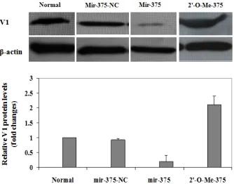
To further confirm the above observations, we next checked the effect of mir-375 on V1 pro-tein expression after palmitate induction. To this end, the cells were treated with 500 µM of palmitate for 48 h after 72 h of transfection. The corresponding cell lysates were then pre-pared for Western blot analysis as above. Con-sistently, only very low levels of V1 protein can
be detected in normal control cells and cells transfected with mir-375-NC. Of important note, V1 protein was almost undetectable in cells with ectopic mir-375 duplexes, while more than 3-fold higher V1 protein was observed in cells transfected with 2'-O-Me-375 (Figure 6). All to-gether, our data strongly suggest that mir-375 could target V1 mRNA and affect its translation, which then promotes pamitate-induced lipo-apoptosis in NIT-1 cells.
Discussion
pivotal predisposing factors responsible for β -cell apoptosis [16;17]. Recent studies high-lighted the importance for one particular islet specific miRNA, mir-375, in the regulation of β -cell function and the maintenance of normal pancreatic α- and β-mass [26;27]. In the pre-sent study, we further examined the role of mir-375 in the regulation of palmitate-induced β-cell lipoapoptosis. We found that NIT-1 cells, a NOD-derived β-cell line, with ectopic mir-375 du-plexes are much more susceptible to palmitate-induced lipoapoptosis. In contrast, NIT-1 cells are remarkably resistant to palmitate-induced lipoapoptosis once the endogenous mir-375 was suppressed by a specific antisense oligo, 2'-O-Me-375. Our study also demonstrates that the expression levels of V1 protein are inversely associated with β-cell lipoapoptosis. Since V1 could be a target for mir-375 [27], our data sug-gest that mir-375 probably enhances β-cell li-poapoptosis by downregulation of V1 expres-sions in β cells.
The cellular population for a particular organ is determined by the balance between the rates of cell division and programmed cell death named apoptosis. Once the apoptosis rate for a particu-lar cell type becomes higher than that of cell replication, a reduction for cell population would occur, which subsequently leads to organ de-compensation. Loss of β-mass late in the course of obesity is a typical example that β-cell apop-tosis exceeds cell division, which then predis-poses to the development of type 2 diabetes. Lipoapoptosis, a type of apoptosis caused by dysregulated lipid metabolism, has recently been recognized to occur in obesity and aging. Excessive accumulation of long-chain fatty acids is associated with altered leptin liporegulation and, as a result, when nonadipose tissues such as pancreatic islets are exposed to an excess of long-chain fatty acids, the cell would undergo lipoapoptosis. The effect of thermal, hypoxic or cytotoxic factors on apoptosis has been well demonstrated [32], while metabolic causes of programmed cell death, particularly lipoapop-tosis, have received less attention. Although recent studies in ZDF (fa/fa) rats suggested an important role for the ceramide pathway in the late destructive phase of lipid overload in the pancreatic β cells [16], the overall underlying mechanisms leading to lipoapoptosis remained poorly understood. Our studies in the current report provided strong evidence indicating a pivotal role for miRNA in the regulation of β-cell
lipoapoptosis. Although we are only beginning to appreciate the immense potential of miRNAs as controllers of gene networks, there is already substantial evidence that these small noncod-ing RNA molecules play a central role in a vari-ety of physiological processes, including tissue differentiation, cell proliferation, and apoptosis. miRNAs are a growing class of non-coding RNAs involved in the regulation of gene expression by translational repression. Islet-specific mir-375 is originally characterized by Poy and colleagues from analysis of miRNAs in endocrine cell types of the pancreas [27]. Recent functional studies indicated that mir-375 plays an indispensable role in normal glucose homeostasis, β-mass turnover in newborns and adaptive β-cell expan-sion in response to increasing insulin demand in insulin resistance [26]. Our data now provide evidence indicating that mir-375 could also act as a proapoptotic factor implicated in the patho-genesis of β-cell lipoapoptosis.
Studies have shown that some evolutionarily conserved proteins characterized in neuronal cells are also present in the pancreatic β cells. V1 is such a protein that is highly expressed in both neuronal cells and pancreatic β cells. V1 (also known as myotrophin) is a 12-kD protein, which was originally identified from the hyper-trophied ventricles of spontaneously hyperten-sive rats and rat cerebellum [33;34]. By 3-dimensional alignment, Knuefermann and col-leagues demonstrated that mammalian V1 re-sembles a truncated IκBα without the signal response domain and PEST sequence [35]. Therefore, V1 was later validated as an an-tiapoptotic factor in cardiac myocytes, neuronal cells and fibroblasts [36-38]. In the current study, we further noticed that the expression levels for V1 in NIT-1 cells are inversely associ-ated with palmitate-induced lipoapoptosis, indi-cating that V1 could also act as an antiapoptotic factor in maintaining β-cell viability.
multiple signaling pathways could be regulated by mir-375, by which mir-375 enhances β-cell lipoapoptosis.
The modified synthetic anti-miRNA oligonucleo-tides (AMOs) were found to be useful tools for specific repression of targeted miRNA expres-sions, thereby helping to unravel the function of miRNAs and their targets. The 2′-O-methyl oli-gonucleotides have been shown to irreversibly inhibit small RNA function in vitro and in intact cells in a sequence-specific fashion, presumably by stoichiometric binding to RISCs containing the cognate miRNA, and thus preventing inter-action with its mRNA targets [41]. Similar to antisense-based oligonucleotides (ASOs), AMOs may contribute to the prioritization of pharma-ceutical targets and have the potential to even-tually progress into a new class of therapeutic agents. Here we have designed a 2′-OMe modi-fied anti-mir-375 oligonucleotide, 2′-O-me-375, and demonstrated its high potency for repres-sion of endogenous mir-375 expresrepres-sions in NIT-1 cells (Figure 2). As 2′-O-me-375 transfected NIT-1 cells were almost completely protected from palmitate-induced lipoapoptosis (Figure 3), 2′-O-me-375 could be a useful “off-target” re-agent for mir-375 with significant implications in both experimental and clinical studies.
Impaired β-cell function and possibly β-mass deficit appear to be reversible, particularly at the early stage of disease when the limiting threshold for reversibility of the decreased β -mass has probably not been passed. Our data in the current report indicate that mir-375, a pancreatic islet-specific miRNA, could be a piv-otal regulator of lipoapoptosis in the pancreatic
β cells. Taking into accountthat mir-375 also regulates insulin secretion, mir-375 could emerge as a potential therapeutic target for prevention and intervention of β-cell dysfunction and β-mass loss in type 2 diabetes.
Acknowledgments
This work was partly supported by the Natural Science Foundation of China (30671974) and by the Guangdong Provincial Natural Science Foundation of China (6021329). All authors declare that they have no competing financial interest.
Please address correspondence to: Dr. Yan Li, De-partment of Endocrinology, The Second Affiliated Hospital, Sun Yat-Sen University, 107th Yanjiang
Western Road, Guangzhou 510120, China. Tel: +86 20 81332286; Fax: +86 20 81332404, E-mail: li-yan19642002@yahoo.cn.
References
[1] Charles MA, Eschwege E, Thibult N, Claude JR, Warnet JM, Rosselin GE, Girard J, Balkau B. The role of non-esterified fatty acids in the deteriora-tion of glucose tolerance in Caucasian subjects: results of the Paris Prospective Study. Diabetolo-gia 1997 Sep;40(9):1101-6.
[2] Schulz LO, Bennett PH, Ravussin E, Kidd JR, Kidd KK, Esparza J, Valencia ME. Effects of tradi-tional and western environments on prevalence of type 2 diabetes in Pima Indians in Mexico and the U.S. Diabetes Care 2006 Aug;29(8):1866-71.
[3] Bluher M. Adipose tissue dysfunction in obesity. Exp Clin Endocrinol Diabetes 2009 Jun;117 (6):241-50.
[4] Surampudi PN, John-Kalarickal J, Fonseca VA. Emerging concepts in the pathophysiology of type 2 diabetes mellitus. Mt Sinai J Med 2009 Jun;76(3):216-26.
[5] Davis N, Forges B, Wylie-Rosett J. Role of obesity and lifestyle interventions in the prevention and management of type 2 diabetes. Minerva Med 2009 Jun;100(3):221-8.
[6] Johnson JD. Proteomic identification of car-boxypeptidase E connects lipid-induced beta-cell apoptosis and dysfunction in type 2 diabetes. Cell Cycle 2009 Jan 1;8(1):38-42.
[7] Gwiazda KS, Yang TL, Lin Y, Johnson JD. Effects of palmitate on ER and cytosolic Ca2+ homeo-stasis in beta-cells. Am J Physiol Endocrinol Me-tab 2009 Apr;296(4):E690-E701.
[8] El-Assaad W, Buteau J, Peyot ML, Nolan C, Ro-duit R, Hardy S, Joly E, Dbaibo G, Rosenberg L, Prentki M. Saturated fatty acids synergize with elevated glucose to cause pancreatic beta-cell death. Endocrinology 2003 Sep;144(9):4154-63.
[9] Funakoshi S, Fujimoto S, Hamasaki A, Fujiwara H, Fujita Y, Ikeda K, Hamamoto Y, Hosokawa M, Seino Y, Inagaki N. Analysis of factors influenc-ing pancreatic beta-cell function in Japanese patients with type 2 diabetes: association with body mass index and duration of diabetic expo-sure. Diabetes Res Clin Pract 2008 Dec;82 (3):353-8.
[10] Wajchenberg BL. beta-cell failure in diabetes and preservation by clinical treatment. Endocr Rev 2007 Apr;28(2):187-218.
[11] Campbell RK. Fate of the beta-cell in the patho-physiology of type 2 diabetes. J Am Pharm Assoc (2003 ) 2009 Sep;49 Suppl 1:S10-S15.
[12] Thomas HE, McKenzie MD, Angstetra E, Camp-bell PD, Kay TW. Beta cell apoptosis in diabetes. Apoptosis 2009 Mar 26.
Beta-cell failure as a complication of diabetes. Rev Endocr Metab Disord 2008 Dec;9(4):329-43.
[14] Poitout V, Amyot J, Semache M, Zarrouki B, Hag-man D, Fontes G. Glucolipotoxicity of the pancre-atic beta cell. Biochim Biophys Acta 2009 Aug 26.
[15] Rutti S, Ehses JA, Sibler RA, Prazak R, Rohrer L, Georgopoulos S, Meier DT, Niclauss N, Berney T, Donath MY, von EA. Low- and high-density lipo-proteins modulate function, apoptosis, and pro-liferation of primary human and murine pancre-atic beta-cells. Endocrinology 2009 Oct;150 (10):4521-30.
[16] Unger RH, Zhou YT. Lipotoxicity of beta-cells in obesity and in other causes of fatty acid spill-over. Diabetes 2001 Feb;50 Suppl 1:S118-S121.
[17] Shimabukuro M, Higa M, Zhou YT, Wang MY, Newgard CB, Unger RH. Lipoapoptosis in beta-cells of obese prediabetic fa/fa rats. Role of serine palmitoyltransferase overexpression. J Biol Chem 1998 Dec 4;273(49):32487-90. [18] Grunnet LG, Aikin R, Tonnesen MF, Paraskevas
S, Blaabjerg L, Storling J, Rosenberg L, Billestrup N, Maysinger D, Mandrup-Poulsen T. Proinflam-matory cytokines activate the intrinsic apoptotic pathway in beta-cells. Diabetes 2009 Aug;58 (8):1807-15.
[19] Sauter NS, Schulthess FT, Galasso R, Castellani LW, Maedler K. The antiinflammatory cytokine interleukin-1 receptor antagonist protects from high-fat diet-induced hyperglycemia. Endocrinol-ogy 2008 May;149(5):2208-18.
[20] Okuya S, Tanabe K, Tanizawa Y, Oka Y. Leptin increases the viability of isolated rat pancreatic islets by suppressing apoptosis. Endocrinology 2001 Nov;142(11):4827-30.
[21] Brown JE, Dunmore SJ. Leptin decreases apop-tosis and alters BCL-2 : Bax ratio in clonal rodent pancreatic beta-cells. Diabetes Metab Res Rev 2007 Sep;23(6):497-502.
[22] Guardado-Mendoza R, Davalli AM, Chavez AO, Hubbard GB, Dick EJ, Majluf-Cruz A, Tene-Perez CE, Goldschmidt L, Hart J, Perego C, Comuzzie AG, Tejero ME, et al. Pancreatic islet amyloido-sis, beta-cell apoptoamyloido-sis, and alpha-cell prolifera-tion are determinants of islet remodeling in type-2 diabetic baboons. Proc Natl Acad Sci U S A 2009 Aug 18;106(33):13992-7.
[23] Kusminski CM, Shetty S, Orci L, Unger RH, Scherer PE. Diabetes and apoptosis: lipotoxicity. Apoptosis 2009 May 8.
[24] Winter J, Jung S, Keller S, Gregory RI, Diederichs S. Many roads to maturity: microRNA biogenesis pathways and their regulation. Nat Cell Biol 2009 Mar;11(3):228-34.
[25] Rogaev EI, Borinskaia SA, Islamgulov DV, Grigorenko AP. [Human microRNA in norm and pathology]. Mol Biol (Mosk) 2008 Sep;42(5):751 -64.
[26] Poy MN, Hausser J, Trajkovski M, Braun M,
Collins S, Rorsman P, Zavolan M, Stoffel M. miR-375 maintains normal pancreatic alpha- and beta-cell mass. Proc Natl Acad Sci U S A 2009 Apr 7;106(14):5813-8.
[27] Poy MN, Eliasson L, Krutzfeldt J, Kuwajima S, Ma X, Macdonald PE, Pfeffer S, Tuschl T, Rajewsky N, Rorsman P, Stoffel M. A pancreatic islet-specific microRNA regulates insulin secretion. Nature 2004 Nov 11;432(7014):226-30. [28] Lynn FC. Meta-regulation: microRNA regulation
of glucose and lipid metabolism. Trends Endocri-nol Metab 2009 Nov;20(9):452-9.
[29] El OA, Baroukh N, Martens GA, Lebrun P, Pipeleers D, van OE. miR-375 targets 3'-phosphoinositide-dependent protein kinase-1 and regulates glucose-induced biological re-sponses in pancreatic beta-cells. Diabetes 2008 Oct;57(10):2708-17.
[30] Walker MD. Role of MicroRNA in pancreatic beta-cells: where more is less. Diabetes 2008 Oct;57 (10):2567-8.
[31] Zhang Y, Xu M, Zhang S, Yan L, Yang C, Lu W, Li Y, Cheng H. The role of G protein-coupled recep-tor 40 in lipoapoptosis in mouse beta-cell line NIT-1. J Mol Endocrinol 2007 Jun;38(6):651-61. [32] Hengartner MO. The biochemistry of apoptosis.
Nature 2000 Oct 12;407(6805):770-6.
[33] Sen S, Kundu G, Mekhail N, Castel J, Misono K, Healy B. Myotrophin: purification of a novel pep-tide from spontaneously hypertensive rat heart that influences myocardial growth. J Biol Chem 1990 Sep 25;265(27):16635-43.
[34] Taoka M, Yamakuni T, Song SY, Yamakawa Y, Seta K, Okuyama T, Isobe T. A rat cerebellar protein containing the cdc10/SWI6 motif. Eur J Biochem 1992 Jul 15;207(2):615-20.
[35] Knuefermann P, Chen P, Misra A, Shi SP, Abdel-latif M, Sivasubramanian N. Myotrophin/V-1, a protein up-regulated in the failing human heart and in postnatal cerebellum, converts NFkappa B p50-p65 heterodimers to p50-p50 and p65-p65 homodimers. J Biol Chem 2002 Jun 28;277 (26):23888-97.
[36] Young D, Popovic ZB, Jones WK, Gupta S. Block-ade of NF-kappaB using IkappaB alpha domi-nant-negative mice ameliorates cardiac hypertro-phy in myotrophin-overexpressed transgenic mice. J Mol Biol 2008 Sep 5;381(3):559-68. [37] Li X, Wang H, Qiu P, Luo H. Proteomic profiling of
proteins associated with methamphetamine-induced neurotoxicity in different regions of rat brain. Neurochem Int 2008 Jan;52(1-2):256-64. [38] Gupta S, Purcell NH, Lin A, Sen S. Activation of
nuclear factor-kappaB is necessary for myotro-phin-induced cardiac hypertrophy. J Cell Biol 2002 Dec 23;159(6):1019-28.
[40] Choi JH, Yang YR, Lee SK, Kim SH, Kim YH, Cha JY, Oh SW, Ha JR, Ryu SH, Suh PG. Potential inhibition of PDK1/Akt signaling by phenothiazi-nes suppresses cancer cell proliferation and survival. Ann N Y Acad Sci 2008 Sep;1138:393-403.