Case Report
Soft tissue perineurioma and other unusual tumors in a
patient with neurofibromatosis type 1
Inga-Marie Schaefer1,2, Philipp Ströbel1, Aung Thiha3, Jan Martin Sohns4, Christian Mühlfeld5, Stefan Küffer1, Gunther Felmerer3, Adam Stepniewski3, Silke Pauli6, Abbas Agaimy7
1Institute of Pathology, 3Division of Plastic Surgery, Clinic of Trauma Surgery, Plastic, and Reconstructive Surgery, 4Institute of Diagnostic and Interventional Radiology, 6Institute of Human Genetics, University Medical Center
Göt-tingen, Germany; 2Department of Pathology, Brigham and Women’s Hospital, Harvard Medical School, Boston MA,
USA; 5Institute of Functional and Applied Anatomy, Hannover Medical School, Biomedical Research in Endstage
and Obstructive Lung Disease Hannover (BREATH), Member of The German Center for Lung Research (DZL), Germany; 7Institute of Pathology, University Hospital Erlangen, Germany
Received September 18, 2013; Accepted October 21, 2013; Epub November 15, 2013; Published December 1, 2013
Abstract: Perineurioma is a rare benign peripheral nerve sheath tumor featuring perineurial differentiation.
Perineurioma occurs sporadically with only one reported case in the setting of neurofibromatosis type 1 (NF-1).
We present a 6.7-cm soft tissue perineurioma of the lower leg in a 51-year-old man with proven NF-1. The tumor
displayed whorled and fascicular pattern with infiltrative margins and expressed EMA, GLUT-1, claudin-1, and CD34. Electron microscopy confirmed diagnosis. Furthermore, lipomatosis, cutaneous angiomatous nodules, vasculopa
-thy, and iliac spine lesion consistent with non-ossifying fibroma were observed. Tumor DNA revealed no NF2 mu-tations or chromosomal aberrations but a germline NF1-deletion (c.449_502delTGTT) was detected in his blood
sample. His brother displayed neurofibromas, duodenal ganglioneuroma and colonic juvenile polyp, and his mother a neurofibroma, cutaneous squamous cell carcinoma, and jejunal gastrointestinal stromal tumor (GIST); both were
affected by NF-1.In conclusion, perineurioma may rarely be NF-1 related and should be included in the spectrum of neoplasms occurring in this disorder.
Keywords: Perineurioma, soft tissue, neurofibromatosis, vasculopathy, NF1
Introduction
Perineurioma is a rare benign peripheral nerve sheath tumor displaying perineurial cell differ-entiation throughout. Since their first descrip-tion by Lazarus and Trombetta in 1978 [1] sev-eral reports and a few large series appeared in the literature [2]. Perineurioma has been tradi-tionally subdivided into intraneural (so-called localized hypertrophic neuropathy) and extra-neural (soft tissue) subtypes. Soft tissue peri-neurioma typically arises in the soft tissue of adults with a mean age of 46 (range, 10-79) years and shows a predilection for the extremi-ties and trunk without gender predilection [2]. Although the tumors are usually well defined but not encapsulated, microscopic infiltrative margins have been reported in 15% [2]. Although chromosomal aberrations affecting
We here describe an unusual large soft tissue perineurioma grossly arising from the right sural nerve of a 51-year-old man with proven NF-1. In addition, the patient featured multiple lipomas of extremities and trunk, and cutane-ous angiomatcutane-ous nodules (not verified histo-logically). Besides the conventional histopatho-logical and immunohistochemical findings, we present molecular features of the perineurioma and discuss its possible link to NF-1.
Case presentation
A 51-year-old male with clinical features of NF-1 was admitted to hospital because of a paramal-leolar mass lesion of his right lower leg. His rel-evant medical history included a thoracic neu-rofibroma resected two years ago and a current nephrolithiasis. Clinical examination revealed several café-au-lait spots (Figure 1A) and mul-tiple cutaneous/subcutaneous tumors of the trunk and extremities, involving his left little fin-ger and index finfin-ger, right wrist, left upper leg, thoracic spine, left abdomen, and his head (Figure 1B). Magnetic resonance imaging (MRI) of the lower leg tumor revealed a hyperintense circumscribed mass surrounding the right sural nerve (Figure 1C). Surgical biopsy was obtained from the right leg tumor and was consistent with soft tissue perineurioma. The mass was then excised completely. Additionally, abdomi-nal computed tomography (CT) scan revealed multiple subcutaneous and partly intramuscu-lar mass lesions located in the thoracic and
abdominal wall measuring up to 5 cm, as well as a lytic bone lesion in the right iliac crest, con-sistent with non-ossifying fibroma (Figure 1B inset). Some of these lesions were interpreted as consistent with lipomas, others as neurofi-bromas with soft tissue signal intensity. Furthermore, a large intramuscular mass of 7.6 x 6 x 3 cm was detected in the left adductor compartment displaying contrast enhance-ment in MRI. Biopsy of this mass was obtained and was consistent with lipoma. The patient recovered well and was discharged after 20 days. Currently the patient is well and there is no evidence of relapse after 9 months. Genetic counseling of the index patient confirmed that he fulfilled the criteria for the diagnosis of NF-1 as he presented 2 café-au-lait spots >1.5 cm, axillary freckling, multiple neurofibromas, and a positive family history. His mother had a spinal cord neurofibroma, a cutaneous squamous cell carcinoma, and a jejunal gastrointestinal stro-mal tumor (GIST) being wild-type for KIT and
PDGFRA. His brother had neurofibromas locat-ed at his hand and chest wall, a duodenal gan-glioneuroma positive for protein S-100, but negative for perineurial markers, and a juvenile polyp in the colon. A nephew showed similar cut- aneous tumors at the extremities and a sister underwent renal surgery for unknown reason.
Pathological findings
[image:2.612.93.524.72.214.2]The tumor resection specimen from the right lower leg measured 9 x 5.4 x 1.5 cm with a 10 Figure 1.The index patient displayed axillary freckling (A, arrow), cutaneous neurofibromas (arrowheads) as well
as café-au-lait spots >1.5 cm on his abdominal wall (A, inset, arrow) and small vascular lesions consistent with
cu-taneous hemangiomas or venous malformation (A, inset, arrowhead) (although not histopathologically confirmed). Abdominal CT detected multiple small tumors (probably neurofibromas) in the abdominal wall and lower extremity
(B, arrows). A lytic bone lesion was detected in the right iliac crest (B, inset), consistent with non-ossifying fibroma.
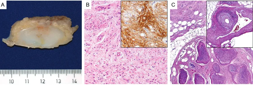
cm-nerve encased in the center. The tumor was lobulated and ill defined, partly covered by fas-cia, and displayed a pale fleshy, solid, white to yellow cut-surface (Figure 2A). The tumor sur-rounded the sural nerve but it was not embed-ded in the nerve substance as intraneural peri-neurioma. Histopathological evaluation reveal- ed a moderately cellular mesenchymal tumor composed of slender bipolar spindle cells with slightly hyperchromatic, occasionally wavy nuclei with fine chromatin, inconspicuous nucleoli and pale eosinophilic cytoplasm with indistinct cell borders. The tumor cells formed short parallel oriented or irregular fascicles with storiform lamellar and focally whorled growth pattern (Figure 2B). Intervening small capillaries and variably prominent interspersed delicate collagen fibers were observed between tumor cells. In the central portion of the tumor, small twigs of peripheral nerves were seen encased by tumor tissue. Furthermore, a promi-nent involvement and entrapment of surround-ing fatty tissue was seen that occasionally deceptively mimicked an integral lipomatous component of the tumor. The resection margins were involved by tumor tissue. No mitotic fig-ures were detected. At the tumor margins sev-eral venous vessels showed remarkable vascu-lopathic changes with significant myointimal spindle cell proliferation, some accompanied by small venule-like capillary vessels (Figure 2C). Immunohistochemical staining for CD34, EMA, alpha-smooth-muscle actin (ASMA), des-min, GLUT-1, claudin-1, S-100 protein, glial
[image:3.612.92.522.71.215.2]fibrillary acidic protein (GFAP), pan-cytokeratin, and Ki67 was performed. The tumor cells expressed CD34, EMA (Figure 2B, inset), GLUT-1, and claudin-GLUT-1, whereas protein S-100, ASMA, desmin, GFAP, and pan-cytokeratin were not expressed. The proliferative activity assessed by Ki67-staining was estimated at 2%. Sections of the sural nerve showed prominent perineuri-al proliferation of similar tumor cells coexpress-ing EMA, claudin-1 and GLUT-1 and extendcoexpress-ing into adjacent fatty tissue. Nests of perineurial cells were seen infiltrating along small venous vessels between fascicles of the nerve trunk. Interestingly, perineurial cell markers highlight-ed an abnormal network of intraneural perineu-rial cells that were arranged in a reticular pat-tern lacking the onion skin appearance of intra-neural perineurioma. The latter involved only a few nerve fascicles of the sural nerve. Trans- mission electron microscopy (EM) performed on 3% glutaraldehyde-fixed, uranyl acetate and lead citrate stained ultrathin sections revealed spindled tumor cells with long tapering nuclei, inconspicuous nucleoli, bipolar cytoplasmic processes embedded in a collagen-rich matrix and foci of numerous intracytoplasmatic globu-lar structures compatible with pinocytotic vesi-cles as well as a discontinuous external lamina – features previously reported for perineurio-mas [2]. Mutation analysis of NF1 (from patient’s blood) revealed a known 4-bp dele-tion in the NF1 gene (c.449_502delTGTT) [7], confirming the germline mutation and diagno-sis of NF-1. Chromosomal analydiagno-sis from blood Figure 2.The resected tumor had ill-defined margins and a pale fleshy, solid cut-surface (A). On microscopic view,
cells revealed a normal karyotype. Furthermore, mutation analysis of NF2 exon 1-15 was per-formed as described previously [5] and revealed no NF2 mutation in the perineurioma. Compa- rative genomic hybridization (CGH) performed as described before [8] detected no chromo-somal imbalances.
Discussion
Soft tissue perineurioma is a rare distinctive tumor entity with exclusive perineurial cell dif-ferentiation as evident from regular expression of perineurial cell markers and ultrastructural features of perineurial cells [2]. Recognition of perineurioma and its distinction from potential low-grade malignant mimics is of clinical impor-tance as this tumor usual behaves in a benign fashion with a low recurrence rate (5%) [2]. In the present case, the tumor was recognizably originating from the sural nerve. It is very unusual for perineurioma to arise from a gross-ly identifiable nerve in contrast to several malig-nant peripheral nerve sheath tumors (MPNST). Thus, our case is unique and it is likely that this unusual gross presentation is related to the background NF-1 disease in this patient. NF-1 (von Recklinghausen disease) is an auto-somal-dominant inherited disorder with an esti-mated worldwide incidence of approximately 1:3,000 individuals which makes NF-1 the most common hereditary multitumor syndrome [9, 10]. The NF1 gene (OMIM *613113) is locat-ed at 17q11.2 [9]. NF1 gene mutations leads to increased cell growth, unopposed Ras activity and constitutive downstream signaling [9]. As a phacomatosis, NF-1 presents various lesions and/or neoplasms of the skin and neural sys-tem, including café-au-lait spots, axillary or inguinal freckling, Lisch-nodules and cutane-ous neurofibromas (60%). An increased sus-ceptibility to other malignances in patients with NF-1 has been observed, including sarcomas (MPNST and others), but squamous cell and other types of carcinomas have been also observed occasionally [11]. Also gastrointesti-nal tumor manifestation has been described, such as small bowel and/or stomach neurofi-bromas and undifferentiated sarcomas of the upper gastrointestinal tract [10]. GISTs arising in the setting of NF-1 are well documented and represent 1.5% of all GISTs [10]. These tumors typically present as small asymptomatic lesions, histologically display spindle cell
mor-phology and so-called skeinoid fibres, a low mitotic rate, and KIT/PDGFRA wild type. In patients with NF-1, a positive family history is reported in approximately 50% of cases [9]. Increased morbidity of disease in individuals born to affected mothers than in those born to affected fathers or those who have new muta-tions suggested a possible maternal impact on the severity of NF-1 [12]. This mechanism may also play a role in the family reported herein. An association of perineuriomas with NF-1, NF-2 or other syndromes has not been observed in larger series. To our knowledge, only a single case of perineurioma associated with NF-1 has been documented [2, 6]. However, in the pres-ent case, the patipres-ent, his brother, and mother had NF-1 and displayed associated lipomas, neurofibromas, a ganglioneuroma, a jejunal GIST, and a colonic juvenile polyp. Of note, an association of NF-1 and juvenile polyps, vascu-lar anomalies (including “prominent veins” on the trunk), and horseshoe kidney have previ-ously been reported by Oktenli et al in a patient with NF-1 and his relatives [13]. Although in our case the presence of horseshoe kidneys was not examined in the patient or his relatives, the patient and his mother suffered from nephroli-thiasis, and a sister underwent renal surgery for unknown reason. Furthermore, our patient dis-played cutaneous angiomatous nodules (Figure 1), a striking vasculopathy in the soft tissue of the lower extremity surrounding the perineurio-ma, a lytic bone lesion (consistent with non-ossifying fibroma), and his brother had a juve-nile polyp, indicating that this family might have a similar phenotype as the family reported by Oktenli et al. [13]. Since an NF1 mutation was not detected in the other family reported by Oktenli et al, a phenotype-genotype correlation can not yet be made. Based on the National Institutes of Health Consensus Conference cri-teria developed in 1988, the diagnosis was made in our patient due to presence of multiple major criteria and positive family history [9]. In the present family, multiple neurofibromas and an affected relative were present as diagnostic criteria for all three patients.
Although chromosomal aberrations at chromo-some 22 have been reported in perineuriomas previously, the perineurioma in this case dis-played no chromosomal imbalances [3, 4]. As previously reported by Lasota et al, the NF2
mutated in sporadic perineuriomas [5]. In that study, point mutations in the 5’-untranslated region and in exons 3, 6, and 8 were detected in 4 of 8 perineuriomas examined [5]. In the present case the perineurioma did not harbor an NF2 mutation in all 15 examined exons and also lacked chromosomal aberrations at chro-mosome 22, thus supporting our hypothesis that the tumor is likely driven by NF1 inactiva-tion in the setting of his genetic disease. Given the facts that the precise subtyping of many benign peripheral nerve sheath tumors might be difficult or even some times impossible with-out the use of immunohistochemistry, and that perineurial markers are not always applied for diagnosing benign neurogenic tumors in pati- ents with a known NF-1, it is not excluded that perineuriomas be under-recognized in NF-1 set-ting. A recent study showed significant over-representation of hybrid peripheral nerve sheath tumors (61% hybrid schwannoma-neu-rofibroma) in patients with schwannomatosis, NF-2, and NF-1 [14]. Retrospective analysis of archival cases revealed that at least 9% of hybrid schwannoma-neurofibromas affected patients with NF-1 in the same study [14]. Although no clear-cut perineurial cell differenti-ation was stated in that study, the authors reported a mosaic GLUT-1 immunopositivity in the schwannoma-like areas in several cases which was associated with CD34 expression suggesting at least partial perineurial differen-tiation. During preparation of this manuscript, another study was published by Kacerovska et al. [15] describing 5 cases of hybrid nerve sheath tumors composed of plexiform neurofi-broma with considerable perineurial compo-nent in patients with NF-1 (three from the same family). All 4 lesions were evident on immuno-histochemistry. The fifth lesion was evident in HE stained sections and showed evidence of malignancy in the S100 positive neurofibroma-tous component. These recent studies suggest under-recognition of perineurial differentiation in benign peripheral nerve sheath tumors tradi-tionally classified as neurofibromas in NF-1. In conclusion, the present case highlights the rare occurrence of soft tissue perineurioma in a background of NF-1. This uncommon entity might be underreported in NF-1 patients. We therefore recommend using perineurial cell markers in classifying benign peripheral nerve sheath tumors in patients with NF-1 in order to assess the possibility of under-recognized NF-1-associated perineurial lesions.
Acknowledgements
Inga-Marie Schaefer is supported by a research grant from the Dr. Mildred Scheel Stiftung für Krebsforschung (No. 110822).
Written informed consent was obtained from the patient and his brother for the publication of this case report and any accompanying images. As next of kin they provided informed consent for their late mother.
Disclosure of conflict of interest
The authors declare that they have no conflict of interest.
Address correspondence to: Dr. Inga-Marie Schaefer, Department of Pathology, Brigham and Women’s Hospital, Harvard Medical School, 75
Francis Street, Boston, MA 02115, USA. Tel: +1-617-732-7070; Fax: +1-617-278-6921; E-mail:
ischaef-er@partners.org
References
[1] Lazarus SS and Trombetta LD. Ultrastructural identification of a benign perineurial cell tu -mor. Cancer 1978; 41: 1823-1829.
[2] Hornick JL and Fletcher CD. Soft tissue peri -neurioma: clinicopathologic analysis of 81 cases including those with atypical histologic features. Am J Surg Pathol 2005; 29: 845-858.
[3] Lasota J, Wozniak A and Debiec-Rychter M. Loss of chromosome 22q and lack of NF2 mu
-tations in perineuriomas [abstract 46]. Mod
Pathol 2000; 13: 11A.
[4] Giannini C, Scheithauer BW, Jenkins RB, Er-landson RA, Perry A, Borell TJ, Hoda RS and Woodruff JM. Soft-tissue perineurioma. Evi-dence for an abnormality of chromosome 22, criteria for diagnosis, and review of the litera-ture. Am J Surg Pathol 1997; 21: 164-173.
[5] Lasota J, Fetsch JF, Wozniak A, Wasag B, Sciot R and Miettinen M. The neurofibromatosis type
2 gene is mutated in perineurial cell tumors: a molecular genetic study of eight cases. Am J Pathol 2001; 158: 1223-1229.
[6] Ausmus GG, Piliang MP, Bergfeld WF and Gold-blum JR. Soft-tissue perineurioma in a
20-year-old patient with neurofibromatosis type 1
(NF1): report of a case and review of the litera-ture. J Cutan Pathol 2007; 34: 726-730.
muta-tional spectrum of the entire NF1 gene does not explain its high mutability but points to a functional domain upstream of the GAP-relat-ed domain. Am J Hum Genet 2000; 66: 790-818.
[8] Gunawan B, Schulten HJ, von Heydebreck A,
Schmidt B, Enders C, Höer J, Langer C, Schüler P, Schindler CG, Kuhlgatz J, Füzesi L. Site-inde
-pendent prognostic value of chromosome 9q
loss in primary gastrointestinal stromal tu-mours. J Pathol 2004; 202: 421-429.
[9] Williams VC, Lucas J, Babcock MA, Gutmann DH, Korf B and Maria BL. Neurofibromatosis
type 1 revisited. Pediatrics 2009; 123: 124-133.
[10] Agaimy A, Vassos N and Croner RS.
Gastroin-testinal manifestations of neurofibromatosis
type 1 (Recklinghausen's disease): clinico-pathological spectrum with pathogenetic con-siderations. Int J Clin Exp Pathol 2012; 5: 852-862.
[11] Knight WA III, Murphy WK and Gottlieb JA.
Neu-rofibromatosis associated with malignant neu
-rofibromas. Arch Dermatol 1973; 107:
747-750.
[12] Miller M and Hall JG. Possible maternal effect
on severity of neurofibromatosis. Lancet 1978;
2: 1071-1073.
[13] Oktenli C, Gul D, Deveci MS, Saglam M, Upad -hyaya M, Thompson P, Consoli C, Kocar IH,
Pi-larski R, Zhou XP and Eng C. Unusual features in a patient with neurofibromatosis type 1:
multiple subcutaneous lipomas, a juvenile polyp in ascending colon, congenital intrahe-patic portosystemic venous shunt, and horse-shoe kidney. Am J Med Genet A 2004; 127A: 298-301.
[14] Harder A, Wesemann M, Hagel C, Schitten-helm J, Fischer S, Tatagiba M, Nagel C, Jeib-mann A, Bohring A, Mautner VF and Paulus W.
Hybrid neurofibroma/schwannoma is overrep -resented among schwannomatosis and
neuro-fibromatosis patients. Am J Surg Pathol 2012;
36: 702-709.
[15] Kacerovska D, Michal M, Kuroda N, Tanaka A,
Sima R, Denisjuk N, Kreuzberg B, Ricarova R and Kazakov DV. Hybrid peripheral nerve
sheath tumors, including a malignant variant