Int J Clin Exp Pathol (2008) 1, 124-133
www.ijcep.com
/IJCEP708010
Original Article
Prognostic Significance of Flow Cytometric
Immunophenotyping in Acute Myeloid Leukemia
Brian A. Webber, Melissa M. Cushing and Shiyong Li
Department of Pathology and Laboratory Medicine, Emory University School of Medicine, Atlanta, GA, USA
Received 2 August 2007; Accepted 20 August 2007; Available online 1 January 2008
Abstract: The prognostic significance of flow cytometric immunophenotyping (FCI) in acute myeloid leukemia (AML) has been controversial. In this study, we re-investigated the possible role of FCI in the prediction of AML relapse following standard chemotherapy. A total of 209 AML cases with follow-up information were analyzed. Among those, 78 cases were in remission (M:F=44/34; mean age of 48.9 years) and 131 had relapse (M:F=71/60; mean age of 51.3 years). The expression of CD34, HLA-DR or a combination of both was significantly different between the remission and relapse groups for all AML as well as AML without t(15;17). None of the pammyeloid markers or their combinations analyzed was found to correlate with treatment outcomes. Complex cytogenetic abnormalities were more likely associated with relapse group than with remission group, but were not statistically significant after excluding AML with t(15;17). In conclusion, FCI is useful in predicting treatment outcome and disease relapse in AML.
Key Words: Flow cytometric immunophenotyping, acute myeloid leukemia, acute promyelocytic leukemia, chromosome translocation, cytogenetics, prognosis
Introduction
Flow cytometric immunophenotyping (FCI) has a well established role as a diagnostic modality in acute leukemias, particularly as a tool for assigning lineage and facilitating further pathologic classifications [1-14]. However, cytogenetic evaluation constitutes the predominant method for assessing prognosis in acute leukemias [15-18]. Although several authors have investigated the prognostic implications of immunophenotype in acute myeloid leukemia (AML) [17, 19-27], no clear consensus has emerged regarding the role of FCI in predicting treatment response, relapse, or overall survival.
While several studies have failed to demonstrate any significant association between FCI and prognosis, others have reported significant correlation between several immunophenotypic markers and clinical outcomes, albeit without universal reproducibility [2, 28-37]. Immunophenotypic markers that, in various studies, have been implicated as predictive of adverse outcomes
include CD7, CD9, CD11b, CD13, CD14, CD33, CD34, CD56, and terminal deoxynucleotidyl transferase (TdT) [35, 38-44]. In addition, co-expression of CD34 and HLA-DR has been shown to be an independent predictor of failure to achieve complete remission (CR) [19, 39, 45-49]. Another study ascribed a more favorable prognosis to cases in which myeloblasts demonstrate a panmyeloid phenotype, co-expressing myeloperoxidase (MPO), CD13, CD33, CDw65 and CD117 [22].
In this report, we retrospectively analyze FCI and cytogenetic findings in 209 cases of AML with or without relapse. Our aim was to determine whether any independent correlation exists between immunophenotype and probability of disease relapse.
Materials and Methods
Case Selection
2003 were retrieved from our pathology electronic database. Cases meeting World Health Organization criteria for AML with available immunophenotyping results and appropriate follow-up information were included and corresponding cytogenetic results were obtained. All AML subtypes were included in the sample population, without exclusion of secondary AML or subtypes with specific cytogenetic abnormalities. Outcomes were defined as complete remission (CR, no evidence of disease at least 10 weeks after induction therapy) or relapse (persistent disease or recurrent disease 10 weeks or greater following induction chemotherapy). Any recurrence of disease occurring before October 2003 was recorded, along with the immunophenotypic and cytogenetic profiles at relapse.
Flow Cytometric Immunophenotyping
Flow cytometric immunophenotyping was performed on bone marrow aspirate or peripheral blood samples collected in RPMI 1640 culture medium. Specimen processing was performed according to a routine red cell lysis protocol. Single cell suspensions were stained with various 4 fluorochrome-conjugated antibody combinations and analyzed in reference to isotype-matched fluorochrome-conjugated control antibodies. Samples were stained with monoclonal antibodies for the following antigens: CD2, CD3, CD4, CD5, CD7, CD8, CD10, CD11b, CD11c, CD13, CD14, CD15, CD16, CD19, CD20, CD22, CD25, CD33, CD34, CD36, CD38, CD45, CD56, CD103, CD117 and HLA-DR (Becton Dickinson Biosciences, San Diego, CA). Although intracytoplasmic staining on permeabilized samples for myeloperoxidase (MPO) and terminal deoxynucleotidyl transferase (TdT) was performed on selected samples when deemed necessary for clinical classification purposes at the time of diagnosis, these results were not included in the analysis due to insufficient numbers. Samples were acquired on a dual-laser FACSCalibur flow cytometer (Becton Dickinson Biosciences) and subsequently analyzed using the CellQuest computer software program (Becton Dickinson Biosciences). Myeloblast immunophenotype was determined with an antigen defined as positive when at least 20% of the myeloblasts expressed the marker at a fluorescence intensity above cutoffs established using the corresponding
isotype-matched control antibody.
Cytogenetic Studies
All cytogenetic studies were performed at Emory Medical Laboratories according to standard protocols with results reported in accordance with the International System for Human Cytogenetic Nomenclature (ISCN) guidelines. Karyotype and/or fluorescence in situ hybridization results at initial presentation and at relapse were retrieved and recorded along with the corresponding FCI results.
Statistical Analysis
Bivariate and multivariate analyses were performed on the total sample, both including and excluding acute promyelocytic leukemia (APL) cases, for correlation between immunophenotype and remission status with endpoints defined as maintained CR or relapse. Multivariate analysis was adjusted for impact of age (less than or greater than 60 years old) and cytogenetic prognostication categories (favorable, intermediate, or unfavorable prognoses). Odds ratios (with corresponding 95% confidence intervals) for relapse were calculated with results less than 1 indicating less likelihood of relapse and results greater than 1 indicating greater likelihood of relapse. Significant results were defined as those with p<0.05.
Results
Clinicopathologic Features
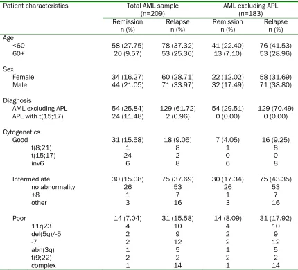
A total of 209 AML cases were retrieved. 78 of the patients were in CR and 131 patients had documented persistent disease or disease relapse. The cohort included 26 cases of APL with documented t(15;17) and all statistical analyses were performed in tandem on the entire patient sample and on the sample excluding these APL cases. Clinical and biological characteristics of the patients are presented in Table 1.
Immunophenotypic Findings
Table 1 Patient characteristics by outcome for both total sample (n=209) and AML only (n=183) Total AML sample
(n=209) AML excluding APL (n=183)
Remission Relapse Remission Relapse Patient characteristics
n (%) n (%) n (%) n (%)
Age
<60 58 (27.75) 78 (37.32) 41 (22.40) 76 (41.53)
60+ 20 (9.57) 53 (25.36) 13 (7.10) 53 (28.96)
Sex
Female 34 (16.27) 60 (28.71) 22 (12.02) 58 (31.69)
Male 44 (21.05) 71 (33.97) 32 (17.49) 71 (38.80)
Diagnosis
AML excluding APL 54 (25.84) 129 (61.72) 54 (29.51) 129 (70.49) APL with t(15;17) 24 (11.48) 2 (0.96) 0 (0.00) 0 (0.00)
Cytogenetics
Good 31 (15.58) 18 (9.05) 7 (4.05) 16 (9.25)
t(8;21) 1 8 1 8
t(15;17) 24 2 0 0
inv6 6 8 6 8
Intermediate 30 (15.08) 75 (37.69) 30 (17.34) 75 (43.35)
no abnormality 26 53 26 53
+8 1 7 1 7
other 3 16 3 16
Poor 14 (7.04) 31 (15.58) 14 (8.09) 31 (17.92)
11q23 4 10 4 10
del(5q)/-5 2 9 2 9
-7 2 12 2 12
abn(3q) 1 5 1 5
t(9;22) 2 2 2 2
complex 1 14 1 14
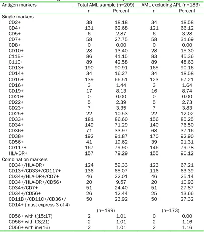
The most frequently expressed antigen was the non-specific lymphoid progenitor marker CD38 (92% of all cases).This was followed by the myeloid lineage markers CD13 (91%), CD33 (87%), and CD117 (80%). Also demonstrating relatively high prevalence rates were the hematopoietic progenitor cell markers CD34 (71%) and HLA-DR (79%). Monocytic markers were moderately frequent: including CD4 (63%), CD11b (41%), CD11c (43%), CD14 (16%), and CD36 (34%). CD7, a T cell antigen known to show aberrant expression in a subset of AML cases, was positive in 28% of all cases. Of note, another T cell antigen, CD2, was expressed in a considerable number of cases (18%). B cell markers CD10, CD19, and CD22 were present in 13%, 8%, and 2% of all cases, respectively.
Not surprisingly, CD34 and HLA-DR were expressed in a higher percentage of cases when APL patients were excluded from the group. This increase was seen for these markers when assessed individually (CD34+ in 71% of total sample and 77% of AML excluding APL sample; HLA-DR in 79% and 90%, respectively) as well as when both markers were co-expressed (59% and 67%, respectively).
Table 2 Prevalence of antigen markers for both total sample and AML excluding APL
Total AML sample (n=209) AML excluding APL (n=183) Antigen markers
n Percent n Percent Single markers
CD2+ 38 18.18 34 18.58
CD4+ 131 62.68 121 66.12
CD5+ 6 2.87 6 3.28
CD7+ 58 27.75 58 31.69
CD8+ 0 0.00 0 0.00
CD10+ 28 13.40 28 15.30
C11B+ 86 41.15 83 45.36
C11C+ 89 42.58 89 48.63
CD13+ 190 90.91 165 90.16
CD14+ 34 16.27 34 18.58
CD15+ 139 66.51 123 67.21
CD16+ 3 1.44 3 1.64
CD19+ 17 8.13 16 8.74
CD20+ 0 0.00 0 0.00
CD22+ 5 2.39 5 2.73
CD23+ 7 3.35 7 3.83
CD25+ 22 10.53 22 12.02
CD33+ 181 86.60 156 85.25
CD34+ 149 71.29 140 76.50
CD36+ 71 33.97 68 37.16
CD38+ 192 91.87 170 92.90
CD56+ 41 19.62 39 21.31
CD117+ 167 79.90 146 79.78
HLA-DR+ 157 79.29 155 90.12
Combination markers
CD34+/HLA-DR+ 124 59.33 123 67.21
CD13+/CD33+/CD117+ 136 65.07 116 63.39
CD34+/HLA-DR+/CD7+ 46 22.01 46 25.14
CD34+/HLA-DR+/CD56+ 20 9.57 20 10.93
CD34+/CD7+ 51 24.40 51 27.87
CD34+/CD56+ 26 12.44 25 13.66
CD11B+/CD11C+/CD36+/
CD14+ (must express 3 of 4) 50 23.92 50 27.32
(n=199) (n=173)
CD56+ with t(15;17) 2 1.01 0 0.00
CD56+ with t(8;21) 2 1.01 2 1.16
CD56+ with inv(16) 2 1.01 2 1.16
least 3 of CD11b, CD11c, CD36, and CD14) comprised 24% of all cases. Only 2 cases of AML with t(8;21) demonstrated CD56 expression. Similarly, CD56 expression was only seen in 6 of the cases with favorable cytogenetic findings.
Cytogenetic Features
Cytogenetic findings were available for 199 of 209 cases. Of the translocations that have been associated with a favorable prognosis—
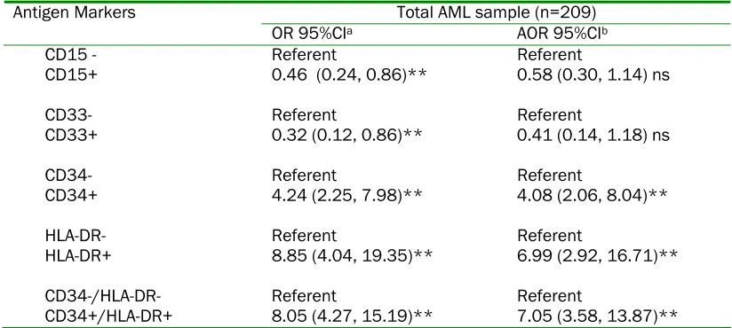
Table 3 Antigen marker as a predictor of relapse for total AML
Total AML sample (n=209) Antigen Markers
OR 95%CIa AOR 95%CIb
CD15 - Referent Referent
CD15+ 0.46 (0.24, 0.86)** 0.58 (0.30, 1.14) ns
CD33- Referent Referent
CD33+ 0.32 (0.12, 0.86)** 0.41 (0.14, 1.18) ns
CD34- Referent Referent
CD34+ 4.24 (2.25, 7.98)** 4.08 (2.06, 8.04)**
HLA-DR- Referent Referent
HLA-DR+ 8.85 (4.04, 19.35)** 6.99 (2.92, 16.71)**
CD34-/HLA-DR- Referent Referent
CD34+/HLA-DR+ 8.05 (4.27, 15.19)** 7.05 (3.58, 13.87)**
aUnadjusted Odds Ratio and 95% Confidence Intervals; bAdjusted Odds Ratio and 95% Confidence Intervals – adjusted for age (less than 60 years or 60+ years) and cytogenetics (good, intermediate or poor); **statistically significant at p<0.05; ns: statistically not significant.
trisomy 8, normal karyotype, or cytogenetic abnormalities other than the above--75 (71%) had relapsed and 30 (29%) were in remission.
Correlation of Immunophenotype with Clinical Outcomes
In the entire sample, 78 (37%) patients were in CR and 131 (63%) had relapsed. When APL cases were excluded from the analysis, 54 (30%) maintained CR status, while 129 (70%) had relapsed. Analysis of the individual markers in the entire sample demonstrated significantly increased likelihood of relapse (p<0.05) in cases expressing CD34 (OR=4.24; 95% CI=2.25-7.98) and HLA-DR (OR=8.85; 95% CI=4.04-19.35). Co-expression of CD34 and HLA-DR was also significantly associated with relapse (OR=8.05; 95% CI=4.27-15.19). Alternatively, CD15 (OR=0.46; 95% CI=0.24-0.86) and CD33 (OR=0.32; 95% CI=0.12-0.86) correlated with maintenance of CR (Table 3).
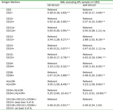
When APL cases were excluded from the analysis sample, cases showing CD34 (OR=3.10; 95% CI=1.52-6.32) or HLA-DR (OR=3.05; 95% CI=1.09-8.49) expression, in addition to those co-expressing CD34 and HLA-DR (OR=5.25; 95% CI=2.64-10.41), remained significantly associated with relapse in the bivariate analysis. Also, CD13 expression in the APL-excluded group correlated with relapse (OR=2.88; 95% CI=1.02-8.19). CD2 (OR=0.39; 95% CI=0.18-0.83), CD10
(OR=0.42; 95% CI=0.18-0.95), CD11b (OR=0.50; 95% CI=0.26-0.95), CD14 (OR=0.45; 95%CI=0.21-0.97), CD15 (OR=0.36; 95% CI=0.17-0.78), and CD36 (OR=0.47; 95% CI=0.24-0.98) all correlated with maintenance of CR. In addition, cases expressing 3 of 4 of the monocytic markers CD11b, CD11c, CD36, and CD14 were also associated with CR (OR=0.46; 95% CI=0.23-0.91) (Table 4).
In the multivariate analysis, with adjustment for age and cytogenetic results, CD34 (OR=4.08; 95% CI=2.06-8.04) and HLA-DR (OR=6.99; 95% CI=2.92-16.71) expression, as well as co-expression of these markers (OR=7.05; 95% CI=3.58-13.87), remained significantly associated with increased relapse rate when all cases were included in the analysis. Importantly, these continued to show significance when APL cases were excluded. The multivariate analysis in the APL-excluded group also showed continued correlation with relapse for CD13 (OR=2.88; 95% CI=1.02-8.19) and with CR for CD2 (OR=0.40; 95% CI=0.17-0.94), CD10 (OR=0.37; 95% CI=0.15-0.90), CD15 (OR=0.42; 95% CI=0.19-0.94), and CD36 (OR=0.48; 95% CI=0.24-0.95) (Table 5).
Table 4 Antigen marker as a predictor of relapse for AML excluding APL
AML excluding APL sample (n=183) Antigen Markers
OR 95%CIa AOR 95%CIb
CD2 - Referent Referent
CD2+ 0.39 (0.18, 0.83)** 0.40 (0.17, 0.94)**
CD10 - Referent Referent
CD10+ 0.42 (0.18, 0.95)** 0.37 (0.15, 0.90)**
CD11b - Referent Referent
CD11b+ 0.50 (0.26, 0.95)** 0.55 (0.28, 1.11) ns
CD13 - Referent Referent
CD13+ 3.44 (1.28, 9.27)** 2.88 (1.02, 8.19)**
CD14 - Referent Referent
CD14+ 0.45 (0.21, 0.97)** 0.47 (0.20, 1.11) ns
CD15 - Referent Referent
CD15+ 0.36 (0.17, 0.78)** 0.42 (0.19, 0.94) **
CD34- Referent Referent
CD34+ 3.10 (1.52, 6.32)** 3.06 (1.43, 6.55)**
CD36- Referent Referent
CD36+ 0.47 (0.24, 0.89)** 0.48 (0.24, 0.95)**
HLA-DR- Referent Referent
HLA-DR+ 3.05 (1.09, 8.49)** 3.23 (1.09, 9.60)**
CD34-/HLA-DR- Referent Referent
CD34+/HLA-DR+ 5.25 (2.64, 10.41)** 5.21 (2.51, 10.84)**
CD11B+/CD11C+/CD36+/
CD14+ (less than 3 of 4) Referent Referent
CD11B+/CD11C+/CD36+/
CD14+ (at least 3 of 4) 0.46 (0.23, 0.91)** 0.49 (0.24, 1.02)
aUnadjusted Odds Ratio and 95% Confidence Intervals; bAdjusted Odds Ratio and 95% Confidence Intervals – adjusted for age (less than 60 years or 60+ years) and cytogenetics (good, intermediate or poor); **statistically significant at p<0.05; ns: statistically not significant.
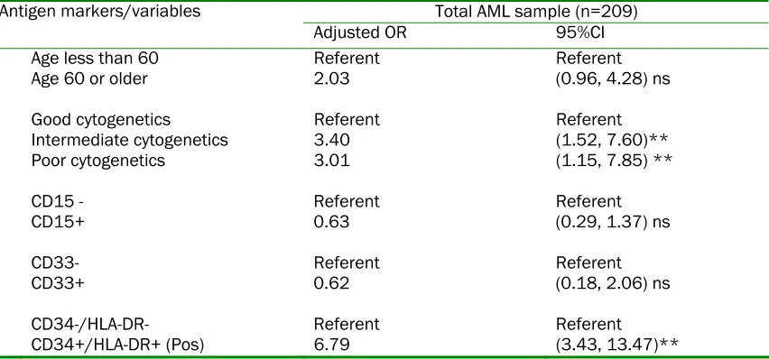
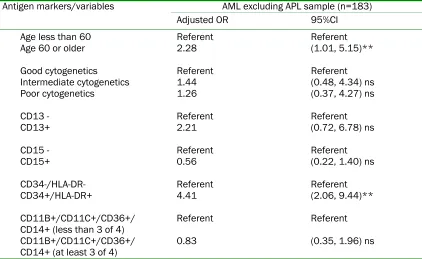
CI=3.43-13.47) and when they were excluded (OR=4.41; 95% CI=2.06-9.44) from the test population. In the former analysis, intermediate (OR=3.40; 95% CI=1.52-7.60) and unfavorable (OR=3.01; 95%CI=1.15-7.85) cytogenetic features were also associated with an increased relapse rate, as was age greater than 60 years old in the latter. In both analyses, however, co-expression of CD34 and HLA-DR was the strongest and independent predictor of relapse (Table 6).
Discussion
single institution. In order to allow comparison with these other studies, some of which excluded APL cases from their test populations, we performed all statistical analyses twice: once including all AML cases (n=209) and the other including only those cases failing to demonstrate t(15;17) (n=183).
Our findings support previous reports ascribing poor prognosis to AML cases with myeloblasts expressing the hematopoietic progenitor cell markers CD34 and HLA-DR [19; 39, 45-49]. Although we identified several antigenic markers associated with prognostic outcomes on initial bivariate analysis, multivariate analysis confirmed only independent CD34 and HLA-DR expression, along with CD34/HLA-DR co-expression, as having significant predictive value in the all inclusive population. While inclusion of cases demonstrating the prognostically favorable t(15;17), almost by definition negative for CD34 and HLA-DR, clearly confounds these results, these markers remained significantly associated with increased relapse risk even when APL cases were excluded from the test population.
In fact, excluding the t(15;17) cases yielded additional significant associations on simple multivariate analysis with CD2, CD10, CD15, and CD36 correlating with CR and CD13 correlating with relapse. While these additional findings admittedly simply add to the complicated landscape of potentially
prognostic markers already described, application of the more stringent multivariate logistic regression model of analysis again revealed only CD34 and HLA-DR co-expression to be significant among the immunophenotypic markers. In fact, co-expression of these markers proved more predictive of poor outcome than advanced age and absence of favorable cytogenetic features.
Given the reportedly favorable prognosis ascribed to cases with a panmyeloid phenotype [22], we investigated and failed to demonstrate any prognostic implications for cases co-expressing the myeloid-exclusive markers present in our panel--namely CD13, CD33, and CD117. Unfortunately, we were unable to implement the more stringent panmyeloid criteria established by Legrand et al (requiring co-expression of CD13, CD33, CDw65, CD117, and MPO) due to insufficient numbers of cases evaluated for MPO and exclusion of CDw65 from routine testing at our institution. Similarly, the reported poor prognosis associated with CD56 expression in AML could not be adequately assessed because of limited numbers of these cases present in our sample population.
[image:7.612.96.522.492.691.2]In conclusion, the results of this study support the findings of previous investigations in ascribing poor prognostic implications to AML cases with myeloblasts co-expressing the hematopoietic progenitor markers CD34 and
Table 5 Multivariate logistic regression results – predictors of relapse for total AML Total AML sample (n=209) Antigen markers/variables
Adjusted OR 95%CI
Age less than 60 Referent Referent
Age 60 or older 2.03 (0.96, 4.28) ns
Good cytogenetics Referent Referent
Intermediate cytogenetics 3.40 (1.52, 7.60)**
Poor cytogenetics 3.01 (1.15, 7.85) **
CD15 - Referent Referent
CD15+ 0.63 (0.29, 1.37) ns
CD33- Referent Referent
CD33+ 0.62 (0.18, 2.06) ns
CD34-/HLA-DR- Referent Referent
CD34+/HLA-DR+ (Pos) 6.79 (3.43, 13.47)**
Table 6 Multivariate logistic regression results – predictors of relapse for AML excluding APL AML excluding APL sample (n=183) Antigen markers/variables
Adjusted OR 95%CI
Age less than 60 Referent Referent
Age 60 or older 2.28 (1.01, 5.15)**
Good cytogenetics Referent Referent
Intermediate cytogenetics 1.44 (0.48, 4.34) ns
Poor cytogenetics 1.26 (0.37, 4.27) ns
CD13 - Referent Referent
CD13+ 2.21 (0.72, 6.78) ns
CD15 - Referent Referent
CD15+ 0.56 (0.22, 1.40) ns
CD34-/HLA-DR- Referent Referent
CD34+/HLA-DR+ 4.41 (2.06, 9.44)**
CD11B+/CD11C+/CD36+/
CD14+ (less than 3 of 4) Referent Referent
CD11B+/CD11C+/CD36+/
CD14+ (at least 3 of 4) 0.83 (0.35, 1.96) ns
OR: odds ratio; CI: confidence interval; **statistically significant at p<0.05; ns: statistically not significant.
HLA-DR. Since almost all AML cases undergo immunophenotyping and given the relative speed and availability of testing, any significant prognostic information to be gleaned from flow cytometric analysis, in conjunction with cytogenetic and other clinical findings, may be helpful in influencing treatment strategies.
Acknowledgements
The authors would like to thank Ms. Laura Zapata for assistance with statistical analysis.
Please address all correspondences to Shiyong Li, MD, PhD, Department of Pathology and Laboratory Medicine, Emory University Hospital F143D, 1364 Clifton Road NE, Atlanta, GA 30322. Tel: 404-712-5456; Fax: 404-712-4140; Email: [email protected]
References
[1] Belov L, de la Vega O, dos Remedios CG, Mulligan SP and Christopherson RI. Immunophenotyping of leukemias using a cluster of differentiation antibody microarray.
Cancer Res 2001;61:4483-4489.
[2] Bradstock K, Matthews J, Benson E, Page F and Bishop J. Prognostic value of immunophenotyping in acute myeloid
leukemia. Australian Leukaemia Study Group.
Blood 1994;84:1220-1225.
[3] Elghetany MT, Sullivan AK, Kurec AS, MacCallum JM, Bloomfield CD, Sobol RE and Davey FR. The use of monoclonal antibodies against primary myeloid granules in normal and leukemic cells. Am J Clin Pathol 1992; 98:430-436.
[4] Haferlach T, Bacher U, Kern W, Schnittger S and Haferlach C. Diagnostic pathways in acute leukemias: a proposal for a multimodal approach. Ann Hematol 2007;86:311-327. [5] Harada N, Okamura S, Kubota A, Shimoda K,
Ikematsu W, Kondo S, Harada M and Niho Y. Analysis of acute myeloid leukemia cells by flow cytometry, introducing a new light-scattering classification. J Cancer Res Clin Oncol 1994;120:553-557.
[6] Kaleem Z, Crawford E, Pathan MH, Jasper L, Covinsky MA, Johnson LR and White G. Flow cytometric analysis of acute leukemias. Diagnostic utility and critical analysis of data.
Arch Pathol Lab Med 2003;127:42-48. [7] Kawada H, Ichikawa Y, Watanabe S, Nagao T
and Arimori S. Flow cytometric analysis of cell-surface antigen expressions on acute myeloid leukemia cell populations according to their cell-size. Leuk Res 1994;18:29-35.
and comparison of immunophenotype, French-American-British classification, and karyotypic abnormalities. Am J Clin Pathol 1998;109:211-220.
[9] Kotylo PK, Seo IS, Smith FO, Heerema NA, Fineberg NS, Miller K, Greene ME, Chou P and Orazi A. Flow cytometric immunophenotypic characterization of pediatric and adult minimally differentiated acute myeloid leukemia (AML-M0). Am J Clin Pathol 2000; 113:193-200.
[10] Lacombe F, Durrieu F, Briais A, Dumain P, Belloc F, Bascans E, Reiffers J, Boisseau MR and Bernard P. Flow cytometry CD45 gating for immunophenotyping of acute myeloid leukemia. Leukemia 1997;11:1878-1886. [11] Paredes-Aguilera R, Romero-Guzman L,
Lopez-Santiago N, Burbano-Ceron L, Camacho-Del Monte O and Nieto-Martinez S. Flow cytometric analysis of cell-surface and intracellular antigens in the diagnosis of acute leukemia.
Am J Hematol 2001;68:69-74.
[12] Ratei R, Karawajew L, Lacombe F, Jagoda K, Del Poeta G, Kraan J, De Santiago M, Kappelmayer J, Bjorklund E, Ludwig WD, Gratama JW and Orfao A. European Working Group of Clinical Cell A: Discriminant function analysis as decision support system for the diagnosis of acute leukemia with a minimal four color screening panel and multiparameter flow cytometry immunophenotyping. Leukemia
2007;21:1204-1211.
[13] Schwonzen M, Diehl V, Dellanna M and Staib P. Immunophenotyping of surface antigens in acute myeloid leukemia by flow cytometry after red blood cell lysis. Leuk Res 2007;31:113-116.
[14] Weir EG and Borowitz MJ. Flow cytometry in the diagnosis of acute leukemia. Semin Hematol
2001;38:124-138.
[15] de Nully Brown P JJ, Pedersen-Bjergaard J, Victor MA and Geisler CH. The prognostic significance of chromosomal analysis and immunophenotyping in 117 patients with de novo acute myeloid leukemia. Leuk Res 1997; 21:985-995.
[16] Girimwade DWH, Oliver F, Wheatley K, Harrison C, Harrison G, Rees J, Hann I, Stevens R, Burnett A and Goldstone A. The importance of diagnostic cytogenetics on outcome in AML: analysis of 1,612 patients entered into the MRC AML 10 trial. The Medical Research Council Adult and Children's Leukaemia Working Parties. Blood 1998;92:2322-2333. [17] Del Poeta G, Stasi R, Venditti A, Suppo G,
Aronica G, Bruno A, Masi M, Tabilio A and Papa G. Prognostic value of cell marker analysis in de novo acute myeloid leukemia. Leukemia
1994;8:388-394.
[18] Rowe JL. Treatment and prognostic factors in acute myeloid leukemia. Baillieres Clin Haematol 1996;9:87-105.
[19] Chang HSF, Yi QL, Patterson B, Brien B and
Minden MD. Prognostic relevance of immuno-phenotyping in 379 patients with acute myeloid leukemia. Leuk Res 2004;28:43-48. [20] Creutzig U, Harbott J, Sperling C, Ritter J,
Zimmermann M, Loffler H, Riehm H, Schellong G and Ludwig WD. Clinical significance of surface antigen expression in children with acute myeloid leukemia. Blood 1995;86:3097-3108.
[21] Kern W, Voskova D, Schoch C, Schnittger S, Hiddemann W and Haferlach T. Prognostic impact of early response to induction therapy as assessed by multiparameter flow cytometry in acute myeloid leukemia. Haematologica
2004;89:528-540.
[22] Legrand O PJ, Baudard M, Cordier A, Lautier R, Simonin G, Zittoun R, Casadevall N and Marie jP. The immunophenotype of 177 adults with acute myeloid leukemia: proposal of a prognostic score. Blood 2000;96:870-877. [23] Perea G, Domingo A, Villamor N, Palacios C,
Junca J, Torres P, Llorente A, Fernandez C, Tormo M, Queipo de Llano MP, Bargay J, Gallart M, Florensa L, Vivancos P, Marti JM, Font L, Berlanga J, Esteve J, Bueno J, Ribera JM, Brunet S, Sierra J, Nomdedeu JF and GroupSpain C. Adverse prognostic impact of CD36 and CD2 expression in adult de novo acute myeloid leukemia patients, Leuk Res
2005;29:1109-1116.
[24] Pereira FG, Metze K, Costa FP, Lima CSP and Lorand-Metze I. Phenotypic quantitative features of patients with acute myeloid leukemia. Neoplasma 2006;53:155-160. [25] Tucker J, Dorey E, Gregory WM, Simpson AP,
Amess JA, Lister TA and Horton MA. Immunophenotype of blast cells in acute myeloid leukemia may be a useful predictive factor for outcome. Hematol Oncol 1990;8:47-58.
[26] Wetzler M, McElwain BK, Stewart CC, Blumenson L, Mortazavi A, Ford LA, Slack JL, Barcos M, Ferrone S and Baer MR. HLA-DR antigen-negative acute myeloid leukemia.
Leukemia 2003;17:707-715.
[27] Zangrando A, Luchini A, Buldini B, Rondelli R, Pession A, Bicciato S, te Kronnie G and Basso G. Immunophenotype signature as a tool to define prognostic subgroups in childhood acute myeloid leukemia. Leukemia 2006;20:888-891.
[28] Ball ED, Davis RB, Griffin JD, Mayer RJ, Davey FR, Arthur DC, Wurster-Hill D, Noll W, Elghetany MT and Allen SL et al. Prognostic value of lymphocyte surface markers in acute myeloid leukemia. Blood 1991;77:2242-2250.
leukemia. Cancer 2006;106:839-847.
[30] Baer MR, Stewart CC, Lawrence D, Arthur DC, Byrd JC, Davey FR, Schiffer CA and Bloomfield CD. Expression of the neural cell adhesion molecule CD56 is associated with short remission duration and survival in acute myeloid leukemia with t(8;21)(q22;q22). Blood
1997;90:1643-1648.
[31] Bahia DM, Yamamoto M, Chauffaille MdL, Kimura EY, Bordin JO, Filgueiras MA and Kerbauy J. Aberrant phenotypes in acute myeloid leukemia: a high frequency and its clinical significance. Haematologica 2001; 86:801-806.
[32] Campos L, Guyotat D, Archimbaud E, Devaux Y, Treille D, Larese A, Maupas J, Gentilhomme O, Ehrsam A and Fiere D. Surface marker expression in adult acute myeloid leukaemia: correlations with initial characteristics, morphology and response to therapy. Br J Haematol 1989;72:161-166.
[33] Ciolli S, Leoni F, Caporale R, Pascarella A, Salti F and Rossi-Ferrini P. CD34 expression fails to predict the outcome in adult acute myeloid leukemia. Haematologica 1993;78:151-155. [34] Jilani I, Estey E, Huh Y, Joe Y, Manshouri T,
Yared M, Giles F, Kantarjian H, Cortes J, Thomas D, Keating M, Freireich E and Albitar M. Differences in CD33 intensity between various myeloid neoplasms. Am J Clin Pathol
2002;118:560-566.
[35] Lee E, Yang J, Leavitt RD, Testa JR, Civin CI, Forrest A and Schiffer CA. The significance of CD34 and TdT determinations in patients with untreated de novo acute myeloid leukemia.
Leukemia 1992;6:1203-1209.
[36] Legras S, Gunthert U, Stauder R, Curt F, Oliferenko S, Kluin-Nelemans HC, Marie JP, Proctor S, Jasmin C and Smadja-Joffe F. A strong expression of CD44-6v correlates with shorter survival of patients with acute myeloid leukemia. Blood 1998;91:3401-3413.
[37] Schwarzinger I, Valent P, Koller U, Marosi C, Schneider B, Haas O, Knapp W, Lechner K and Bettelheim P. Prognostic significance of surface marker expression on blasts of patients with de novo acute myeloblastic leukemia. J Clin Oncol 1990;8:423-430. [38] Del Poeta G, Stasi R, Venditti A, Cox C, Aronica
G, Masi M, Bruno A, Simone MD, Buccisano F and Papa G. CD7 expression in acute myeloid leukemia. Leuk Lymphoma 1995;17:111-119. [39] Graf M, Reif S, Kroll T, Hecht K, Nuessler V and
Schmetzer H. Expression of MAC-1 (CD11b) in acute myeloid leukemia (AML) is associated with an unfavorable prognosis. Am J Hematol
2006;81:227-235.
[40] Kita K, Miwa H, Nakase K, Kawakami K, Kobayashi T, Shirakawa S, Tanaka I, Ohta C, Tsutani H and Oguma S et al. Clinical importance of CD7 expression in acute
myelocytic leukemia. the Japan Cooperative Group of Leukemia/Lymphoma. Blood 1993; 81:2399-2405.
[41] Raspadori D, Damiani D, Michieli M, Stocchi R, Gentili S, Gozzetti A, Masolini P, Michelutti A, Geromin A, Fanin R and Lauria F. CD56 and PGP expression in acute myeloid leukemia: impact on clinical outcome. Haematologica
2002;87:1135-1140.
[42] Saxena A, Sheridan DP, Card RT, McPeek AM, Mewdell CC and Skinnider LF. Biologic and clinical significance of CD7 expression in acute myeloid leukemia. Am J Hematol 1998; 58:278-284.
[43] Seymour JF, Pierce SA, Kantarjian HM, Keating MJ and Estey EH. Investigation of karyotypic, morphologic and clinical features in patients with acute myeloid leukemia blast cells expressing the neural cell adhesion molecule (CD56). Leukemia 1994;8:823-826.
[44] Venditti A, Del Poeta G, Buccisano F, Tamburini A, Cox-Froncillo MC, Aronica G, Bruno A, Del Moro B, Epiceno AM, Battaglia A, Forte L, Postorino M, Cordero V, Santinelli S and Amadori S. Prognostic relevance of the expression of Tdt and CD7 in 335 cases of acute myeloid leukemia. Leukemia 1998; 12:1056-1063.
[45] Guerci A, Merlin JL, Missoum N, Feldmann L, Marchal S, Witz F, Rose C and Guerci O. Predictive value for treatment outcome in acute myeloid leukemia of cellular daunorubicin accumulation and P-glycoprotein expression simultaneously determined by flow cytometry. Blood 1995;85:2147-2153.
[46] Junghanss C, Waak M, Knopp A, Kleine HD, Kundt G, Leithauser M, Hilgendorf I, Wolff D, Casper J and Freund M. Multivariate analyses of prognostic factors in acute myeloid leukemia: relevance of cytogenetic abnormalities and CD34 expression.
Neoplasma 2005;52:402-410.
[47] Lanza F, Rigolin GM, Moretti S, Latorraca A and Castoldi G. Prognostic value of immunophenotypic characteristics of blast cells in acute myeloid leukemia. Leuk Lymphoma 1994;13(Suppl 1):81-85.
[48] Raspadori D, Lauria F, Ventura MA, Rondelli D, Visani G, de Vivo A and Tura S. Incidence and prognostic relevance of CD34 expression in acute myeloblastic leukemia: analysis of 141 cases. Leuk Res 1997;21:603-607.
[49] Solary E, Casasnovas RO, Campos L, BeneMC, Faure G, Maingon P, Falkenrodt A, Lenormand B and Genetet N. Surface markers in adult acute myeloblastic leukemia: correlation of CD19+, CD34+ and CD14+/DR- phenotypes with short survival. Groupe d'Teude Immunologique des Leucemies (GEIL).