Original Article
Detection of
EGFR
mutation in supernatant, cell pellets
of pleural effusion and tumor tissues from non-small
cell lung cancer patients by high resolution melting
analysis and sequencing
Jie Lin1,2*, Ye Gu1,2*, Rui Du1,2, Min Deng1, Yaodan Lu1, Yanqing Ding1,2
1Department of Pathology, Nanfang Hospital, Southern Medical University, Guangzhou 510515, PR China; 2Department of Pathology, School of Basic Medicine, Southern Medical University, Guangzhou 510515, PR China. *Equal contributors.
Received October 12, 2014; Accepted December 1, 2014; Epub December 1, 2014; Published December 15, 2014
Abstract: To determine epidermal growth factor receptor (EGFR) mutation in advanced non-small cell lung cancer
(NSCLC) patients and compare the detection efficiency between different sample resources, both high resolution melting (HRM) analysis and direct sequencing method were used to analyze 36 pleural effusion samples and 22 matched biopsy tumor tissues collected from NSCLC patients. For each pleural effusion sample, the supernatant and the cell pellets were examined separately. Among all the 36 cases of pleural effusion samples, 18 mutations
of EGFR were found in cell-free supernatant while 13 mutations were found in the cell pellets as detected by HRM analysis. In the 22 matched samples, 13 cases of EGFR mutations were identified in paraffin-embedded biopsy
tissue samples, 12 cases in the cell-free supernatant and 9 cases in the cell pellets of pleural effusion. EGFR
muta-tions in 15 cases out of the total 36 pleural effusion samples detected by direct sequencing were also identified by HRM analysis, giving 100% efficiency for HRM method. The results established the important role of HRM as a reliable and efficient method to determine EGFR mutation status and indicated the feasibility of using pleural effu
-sion in replacement of biopsy tissues in particular clinical cases. Furthermore, the cell-free supernatant of pleural
effusion might be a better resource for mutation detection than cell pellets.
Keywords: EGFR, NSCLC, pleural effusion, supernatant, HRM
Introduction
Lung cancer is the main cause of cancer-relat-ed death all over the world. Non-small cell lung cancer (NSCLC) is the most common form of lung cancer and accounts for about 80% of lung cancer [1, 2]. The traditional first-line treat -ment of advanced NSCLC often involves opera-tive treatment and platinum-based combina-tion chemotherapy [2]. However, due to the lack of overt symptoms, approximately 60-85% of patients are diagnosed in advanced lung can-cer period when operative treatment is no more viable and only combination chemotherapy can be applied to inhibit tumor growth, under which circumstance, conventional chemotherapy nor -mally fails to realize long-term therapeutic effect.
EGFR mutation spectrum of early NSCLC patients can normally be evaluated using sam -ples of surgical removed cancer tissue. However, advanced NSCLC patients who have missed the appropriate time of operative treat-ment may lose the opportunity to receive drug sensitivity test attributing to unobtainable tumor tissue. Thus the availability of noninva -sive diagnostic specimens is of great impor-tance. Pleural effusion is a convenient clinical sample with important clinical diagnostic sig-nificance. It may be an alternative source sup -plying useful information about the mutation status of the EGFR gene. If EGFR gene muta-tion determinamuta-tion can be achieved with more attainable pleural effusion samples, then tar-geted drug therapy will be possible for advanced NSCLC patients, which will contribute to vital clinical and practical value [8].
Up to now, different methods have been recruit-ed to detect EGFR mutation and their suitability for EGFR mutation analysis has also been more and more valued [2]. Direct sequencing is a pre-dominant criterion since it is an economical method and can successfully detect all muta -tions [9, 10]. However, direct sequencing is time-consuming and low-sensitivity, limiting its widely application. Hence some faster and more sensitive testing methods are needed. The high resolution melting (HRM) technology has become a hot-spot in the field of life sci -ence in recent years [11]. HRM analysis is an attractive screening method based on the physical property of nucleic acid, which adopts saturable dye to monitor the variation of nucle -ic acid melting curve. New instruments com-bined with DNA intercalating dyes that can be used at saturating concentrations allow the dis-crimination of sequence changes in PCR ampli-cons without manual handling of PCR products [12], making it an ideal candidate to detect DNA sequence changes with advantages of low cost, high throughput, high sensitivity, high specificity and convenience [13].
In this study, we investigate the concordance of
EGFR mutations in pleural effusion including cell-free pleural fluid and cellular pellets, and tumor tissue samples from biopsy of the same patients in order to verify the application of pleural effusion in EGFR mutation detection. Both HRM analysis and direct sequencing method were applied and their efficiency is also compared.
Materials and methods
Patients and tumor samples
This study was approved by the Institutional Review Board of Nanfang Hospital, Southern Medical University, and written informed con -sent was obtained. A total number of 36 cases of malignant pleural effusion and 22 cases of matched tumor tissue samples obtained by thoracoscopic lung biopsy were recruited. The diagnosis of NSCLC was based on cytological or histological findings. The pathological diagno -sis was adenocarcinoma in 35 patients and large cell carcinoma in the other patients. There are 16 males and 20 females altogether. The age range was from 31 to 82 ages (median 61.5 years). All individuals in this manuscript have been given written informed consent to publish these case details.
DNA extraction
The pleural effusion samples were centrifugat-ed, then cell-free supernatant and cell pellets were collected respectively. Genomic DNA was extracted by the use of QIAamp DNA Midi Kit according to the manufacturer’s protocols. The tumor tissue samples were embedded by par -affin, and then genomic DNA was extracted by the used of QIAamp DNA FFPE Tissue Kit according to the manufacturer’s protocols.
PCR amplification and HRM for EGFR mutation
detection
HRM analysis was carried out by using Human
EGFR Gene Mutation Test Kit (HNME-01, He-lixgen (Guangzhou) Co. Ltd.) on a LightCyclerTM 480 PCR (Roche Diagnostics) according to the manufacturer’s protocol. The reaction mixture consists of 20 ng of genomic DNA, 300 nM of each primer, 0.5 mM MgCl2 and 1× Master Mix containing LC Green® Plus+ Melting Dye (Biofire Diagnostics) with PCR-grade water adjusted to a final volume of 10 µl. Sample loading was conducted strictly following the manufacturer’s instructions.
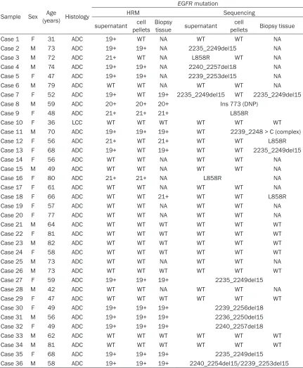
95°C for 30 s, which declined to 40°C for 30 s, 75°C for 1 s, and risen up to 95°C again for fluorescence signal captured 20 times per 1°C resulting on a ramp rate of 0.2°C/s. Nucleotide variation was detected based on HRM curve acquisition using the LightCyclerTM 480 Gene Scanning Software (version 1.5). All PCR
[image:3.612.92.523.87.610.2]sam-ples were plotted according to their melting profiles. The normalized graph shows the degree of reduction in fluorescence over a tem -perature range (75°C to 95°C). Under the dif-ference graph, melting profiles of the samples were compared to that of significant deviations from the horizontal line; those with aberrant Table 1. Summary of EGFR mutations detected by HRM and sequencing from the 36 NSCLC samples
Sample Sex (years) HistologyAge
EGFR mutation
HRM Sequencing
supernatant pelletscell Biopsy tissue supernatant pelletscell Biopsy tissue
Case 1 F 31 ADC 19+ WT NA WT WT NA Case 2 M 73 ADC 19+ 19+ NA 2235_2249del15 NA Case 3 M 72 ADC 21+ WT NA L858R WT NA Case 4 M 74 ADC 19+ 19+ NA 2240_2257del18 NA Case 5 F 47 ADC 19+ 19+ NA 2239_2253del15 NA
Case 6 M 79 ADC WT WT NA WT WT NA
Case 7 F 52 ADC 19+ WT 19+ 2235_2249del15 WT 2235_2249del15 Case 8 M 59 ADC 20+ 20+ 20+ Ins 773 (DNP)
Case 9 F 48 ADC 21+ 21+ 21+ L858R
Case 10 F 36 LCC WT WT WT WT WT WT Case 11 M 70 ADC 19+ 19+ 19+ WT 2239_2248 > C (complex) Case 12 F 56 ADC 21+ WT 21+ WT WT L858R Case 13 F 68 ADC 19+ WT 19+ WT WT 2235_2249del15 Case 14 F 56 ADC WT WT NA WT WT NA Case 15 M 49 ADC WT WT NA WT WT NA Case 16 F 80 ADC 21+ 21+ NA L858R NA Case 17 F 61 ADC WT WT NA WT WT NA Case 18 F 66 ADC WT WT 21+ WT WT L858R Case 19 F 57 ADC WT WT NA WT WT NA Case 20 F 77 ADC WT WT NA WT WT NA Case 21 M 64 ADC WT WT WT WT WT WT Case 22 F 81 ADC WT WT WT WT WT WT Case 23 M 82 ADC WT WT WT WT WT WT Case 24 F 58 ADC WT WT WT WT WT WT Case 25 M 73 ADC WT WT NA WT WT NA Case 26 M 73 ADC WT WT WT WT WT WT Case 27 F 59 ADC 19+ 19+ 19+ 2235_2249del15 Case 28 M 42 ADC WT WT NA WT WT NA Case 29 F 47 ADC WT WT WT WT WT WT Case 30 F 49 ADC 19+ 19+ 19+ 2239_2256del18 Case 31 M 56 ADC 19+ 19+ 19+ 2236_2250del15 Case 32 F 49 ADC 19+ 19+ 19+ 2240_2257del18 Case 33 M 62 ADC WT WT WT WT WT WT Case 34 M 81 ADC WT WT WT WT WT WT Case 35 F 68 ADC 19+ 19+ 19+ 2235_2249del15
melting curves implying the presence of muta -tion were all recorded as HRM positive. All PCR reactions were performed in duplicate.
The obtained PCR products were purified using QIAquick PCR purification kit and directly sequenced by BGI-Shenzhen to further confirm the genetic variants as detected in HRM analysis.
Statistical analysis
Chi-square test (SPSS Statistics 20.0) was per-formed to compare the detection efficiency of
EGFR mutation between pleural effusion and tumor tissue samples. A P value < 0.05 was considered statistically different. In addition, an inter-rater reliability analysis using the Kappa statistic was performed to determine consistency between the pleural effusion and biopsy samples.
Results
Detection of EGFR mutations by HRM analysis
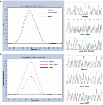
[image:4.612.92.522.73.511.2]HRM analysis. About 18 (18/36, 50%) muta -tions were detected in the cell-free supernatant and 13 (13/36, 36.1%) mutations in cell pellets (Table 1). EGFR mutations in exon 19 were the most common (13/36, 36.1%), followed by those in exon 21 (5/36; 13.9 %) and exon 20 (1/36; 2.8%). The different plots and sequenc -ing traces of wild type for EGFR exon 19 and exon 21 are shown in Figure 1. No point muta-tion of exon 18 was detected in this study. The total mutation rate of exon 19 and exon 21 was 50% (18/36). There were discrepancies in the detected mutations between pleural effusion
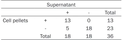
cell pellets and cell-free supernatant. 13 cases of mutation were detected in both cell-free supernatant and cell pellets, while 5 cases of mutation were detected only in cell-free super -natant (Table 1; Figure 2A). Thus, cell-free supernatant is more suitable than cell pellets in
EGFR mutation assessment.
[image:5.612.90.522.73.401.2]In the 22 tumor tissue samples, 13 mutations was detected by HRM, including 9 cases of exon 19 deletion, 3 cases of exon 21 point mutation and 1 case of exon 20 mutation. Among these mutations, 9 mutations were
Figure 2. Comparison of EGFR mutation status among different sample resources detected by HRM analysis and
sequencing. A. HRM results from the supernatant and cell pellets of pleural effusion in 36 samples; B. HRM results from the supernatant, cell pellets of pleural effusion and tumor tissues in the paired 22 samples; C. Sequencing results from the supernatant and cell pellets of pleural effusions in 36 samples; D. Sequencing results from the supernatant, cell pellets and tumor tissues in the paired 22 samples. The vertical coordinate shows the number of positive mutation samples detected in different EGFR exons or mutation types listed on the horizontal coordi -nate (exon 18 was not included since no mutation was detected). Overall, 12 cases were detected in both pleural
effusion and tumor tissues using HRM method, 1 case of mutation was only detected in tumor tissue samples. Different efficiency degree was revealed in mutation type A, B and C for the three different sample sources. The pie chart displays the total ratio of positive mutation samples detected in all exons or mutation types using differ
-ent sample sources. Mutations types repres-ented by each capital letter are: A. 2235_2249del15; B. L858R; C. 2239_2248>C (complex); D. 2240_2257del18; E. 2239_2253del15; F. 2240_2254del15; G. 2239_2256del18;
Table 2. Comparison of detection efficiency of
EGFR mutation between 22 pleural effusion samples and matched biopsy tumor tissues by Kappa statistic analysis
Pleural effusion
+ - Total Tumor tissue + 12 1 13
- 0 9 9
Total 12 10 22
+: EGFR positive mutation; -: EGFR negative mutation.
detected in cell pellets and 3 more were detect-ed in the supernatant of pleural effusion, adddetect-ed up to a total of 12 positive mutations detected with pleural effusion. An extra case of mutation was only detected in tumor tissue samples (Table 1; Figure 2B). The consistency of hydro -thorax samples and biopsy specimens subjects to 95.5% (21/22) according to HRM results.
Detection of EGFR mutations by sequencing
The PCR products of HRM were all purified and sequenced (Figure 1). EGFR mutations were found in 15 of the 36 pleural effusions. Mutation types encompass deletion mutation in exon 19, L858R in exon 21 and Ins773 (DNP) in exon 20. No missense mutation in exon 18 was found. There are two kinds of deletion mutation in case 36. In total, 15 kinds of muta -tion were detected in the supernatant of pleu-ral effusion, 14 of which were also detected in cell pellets (Figure 2C). No sample was found to have EGFR mutations by direct sequencing alone (Table 1). Among all these mutations, 2235_2249del15 was observed in 4 cases of tumor tissues, 3 cases of the supernatant of pleural effusion, and only 2 cases of the cell pellets. Mutation L858R was observed in 3 cases of tumor tissues and only 1 case of pleu -ral effusion (Figure 2D). In the 22 cases of
paired samples, 3 cases of mutation detected in tumor tissue samples were not successfully detected in pleural effusion, indicating a con-sistency of 86.4% (19/22). The efficiency of hydrothorax sample resulted in 78.6% (11/14) for both the supernatant and cell pellets.
Comparison of detection efficiency of EGFR
mutation status between biopsy and cytologi-cal samples
Statistical analysis showed that the kappa val -ues reflecting detection efficiency of EGFR
mutation between tumor tissue samples and hydrothorax samples, supernatants of pleural effusion and cell pellets were 0.908 and 0.722 (P < 0.05; Tables 2, 3), respectively. No signifi -cant difference was detected between tumor tissue samples and pleural effusion (P > 0.05). The high consistency between these two differ -ent sample sources indicates the effectiveness of using cytological samples as a potential sub -stitute of biopsy specimen.
Discussion
Nowadays, lung cancer ranks the highest mor -bidity and mortality among different malignant tumor types and is responsible for approxi -mately 1.38 million deaths each year world -wide [14]. EGFR mutation analysis in NSCLC is a pivotal process during clinic treatment, which can be utilized to predict the patient’s response to EGFR-TKIs [15-17]. Previous studies have proved the therapeutic effect of EGFR-TKI on patients carrying positive EGFR mutations [6, 10, 18, 19]. Therefore, examination of the mutation status of EGFR can provide crucial suggestions on which treatment protocol might be chosen and which patient will be benefit from EGFR targeted therapy along with the prognostic evaluation afterwards [20, 21]. However, advanced NSCLC patients heretofore must take biopsy or needle core biopsy to get samples to obtain unequivocal classification diagnosis and genetic detection for targeted drug screening. Concurrently, although paraffin embedded tumor tissue is still the gold-stan-dard sample for EGFR mutation detection and results from molecular analyses using the tradi -tional tumor tissue samples were validated by clinical outcomes [22], it has some restrictions such as inadequate tissue acquisition and non-ideal tissue positions [23]. To overcome the Table 3. Comparison of detection efficiency of
EGFR mutation between the supernatant and cell pellets of pleural effusion samples by Kappa statistic analysis
Supernatant
+ - Total Cell pellets + 13 0 13
- 5 18 23
Total 18 18 36
[image:6.612.88.289.270.337.2]limitation of sample collection in EGFR targeted therapy, numerous studies have employed cytological samples to assess gene mutation, which is in the ascendant currently and has gained favorable clinical effects. In this study, we have selected an atraumatic tumor cell type for patients to whom only adjuvant chemother -apy is available instead of conventional opera -tive treatment, namely, deciduous hydrothorax tumor cell as DNA samples for clinical EGFR
mutation detection to conduct targeted drug screening. These cytological samples were added on the basis of tumor tissues as supple-ment for mutation assesssupple-ment. Our results show a coherence of 95.5% (21/22) between mutation analyses of 22 paired biopsies and hydrothorax samples from 22 patients. The interrater reliability was found to be statistically significant with a value of Kappa =0.908 (P < 0.05) which indicates fair agreement between these two materials [24]. It offers a kind of tan -talizing possibility for advanced NSCLC patients to get the appropriate treatment inferred from
EGFR mutation conditions. Now that fresh tis-sue is not easily accessible for advanced NSCLC patients, cytological samples can serve as a supplement to biopsy specimen as have been suggested by other studies [25-31]. Furthermore, sensitivity methods were also applied in their studies [25-31].
In our study, there was one case of mutation that detected only in tumor tissues but not in cytological sample, showing an efficiency of 92.3% (12/13) for hydrothorax samples. Several explanations might be responsible for this scenario. Firstly, even though HRM analysis was proved to be a suitable methodology to test samples with a low level of DNA content [12], the tumor cells collected in hydrothorax samples were normally far less than surgical samples. Secondly, since malignant mutations do not occur in every single tumor cell, we can -not rule out the possibility of omitting some malignant tumor cells during the acquisition of pleural effusion. In addition, a relative small region or focal distribution of pathological change may lead to failed sampling in a non-optimal puncture site as well. These factors can largely reduced the DNA quantity in pleural fluid to a degree even lower than the resolution capability of HRM analysis. Finally, even though hydrothorax exfoliative cytologic examination is an easily accessible source of specimen with
convenient manipulation and comparatively high sensitivity, the veracity of hydrothorax samples has always been labile in different studies. The positive mutation rate detected in pleural fluid ranges from 23% to 70% as report -ed before [32]. Bas-ed on our analyses, the pos -itive mutation rate detected was 92.3% (12/13) by HRM analysis and 78.6% (11/14) by gene sequencing. From the above, we believe that the pleural effusion samples is an ideal substi-tute of tumor tissues which can provide pivotal evidence for malignance diagnosis under spe-cial circumstances, yet commonly, surgical tumor tissue should still be the main sample source. A combined test incorporating cytologi -cal samples with biopsy tissue samples can be developed to guarantee the accuracy of EGFR
mutation detection.
It is also noteworthy that the positive mutation rate detected from cell-free supernatant of pleural effusions was higher than that detected from cell pellets using either HRM analysis or direct sequencing method in our study. This is probably correlated with a higher content of dissolved tumor DNA in the supernatant. Under high speed of centrifugation, the tumor cells were damaged and DNA molecules were released from the nucleus, making up the dom -inant components of the supernatant. Even though the pleural effusion often has low tumor cell content than fresh tissues, we can more or less offset this defect by extracting the super -natant part for detection as far as possible. The conformity extrapolates to 83.3% (31/36) between the top phase and the sediment cells in pleural effusions based on HRM results. Our results are in accord with previous study report -ed by Liu et al [33], suggesting the superna -tants of pleural effusion could better reflect the real status of EGFR mutation. Thus, it may pro -vide a more suitable alternative of biopsy tumor tissues than cell pellets.
In the current study, both the direct sequencing and HRM method were recruited to detect
detected mutations, giving a congruence of 91.7% (33/36). More specifically, these three mutations all came from the supernatant part of pleural effusions with probably higher DNA content than the cell pellets. This evident vari-ance reflected the different efficiency of the two methods and proved that gene sequencing does not support investigations of most cytolo -gy samples with low and insufficient tumor cell content. Generally, a threshold of 20% mutant tumor cells was required to be detected by gene sequencing reliably [3, 34].
Up to now, high-sensitivity, high-throughput and low-cost technologies are making an impact on genomic research by providing new strategies to fulfill gene mutation determination and scan -ning, among which the HRM analysis has been specifically noted giving the prominent superi -orities of easy operationand wide range of utili-zation [35, 36]. Previous studies have proved that mutant alleles at levels of 1% to 10% can be detected by HRM [3, 28]. As a result, it has been widely used in analysis of different can -cer-related genes. Gonzalez-Bosquet et al. per -formed HRM analysis in exons of candidate genes like PIK3CA, ERBB2, KRAS, TP53, EGFR, BRAF, GATA3 and FGFR3 known to harbor established commonly observed mutations [13]. Their results indicating that HRM analysis is a rapid, sensitive and economical method with enormous potential for the detection of DNA sequence changes. Meanwhile, the intrin-sic characteristics of HRM endows it with the ability to purify and directly sequence target DNA after analysis without damaging DNA structure, making it a perfect tool of SNP pre-screening before sequencing. However, leaving aside all the advantages, HRM is a robust method that only judges the existence of muta -tions while no detailed mutation information can be dissected. It cannot completely replace the sequencing method despite of its much higher sensitivity and efficiency. On the con -trary, direct sequencing has long been used as a historical standard to detect all known or unknown mutations and provides detailed mutation information. So far, the status of genomic sequence in mutation analysis is still remarkable and positive samples of HRM must be analyzed by direct sequencing to ensure the accuracy. It is expected to be a serious obsta -cle for HRM method to further develop resulting from its imperfection in clarifying the mutation type and gene sequencing has to be followed up to determine this issue integrally.
In conclusion, our study showed EGFR muta-tion assessment in pleural effusion samples from NSCLC patients is basically consistent with that in tumor tissue samples obtained by traditional biopsy, indicating that cytological sample is worth to be generalized and adopted clinically. The results also suggested that cell-free supernatant of pleural effusion is a more suitable alternative than cell pellets, but an increased sample size in individual level should be included in further research to confirm this finding. Meanwhile, as a sensitive and reliable method, HRM analysis can be well applied in
EGFR mutation detection in somatic cells and mutation screening before sequencing.
Acknowledgements
National Natural Science Foundation of China (Grant No. 30800414) and Pearl River Science & Technology New Star Foundation of Guang-zhou City (Grant No. 2012J2200044).
Disclosure of conflict of interest
None.
Address correspondence to: Dr. Jie Lin, Department
of Pathology, Nanfang Hospital, Southern Medical University, 1838 Guangzhou Avenue, Guangzhou 510515, PR China. E-mail: [email protected]
References
[1] Zhang X, Zhao Y, Wang M, Yap WS and Chang AY. Detection and comparison of epidermal growth factor receptor mutations in cells and
fluid of malignant pleural effusion in non-small
cell lung cancer. Lung Cancer 2008; 60: 175-182.
[2] Ellison G, Zhu G, Moulis A, Dearden S, Speake G and McCormack R. EGFR mutation testing in
lung cancer: a review of available methods and
their use for analysis of tumour tissue and cy
-tology samples. J Clin Pathol 2013; 66: 79-89.
[3] Krypuy M, Newnham GM, Thomas DM, Conron
M and Dobrovis A. High resolution melting
analysis for the rapid and sensitive detection of mutations in clinical samples: KRAS codon
12 and 13 mutations in non-mall cell lung can-cer. BMC Cancer 2006; 6: 295.
[4] Erali M, Voelkerding KV and Wittwer CT. High
resolution melting applications for clinical
lab-oratory medicine. Exp Mol Pathol 2008; 85:
50-58.
accurate high resolution melting (HRM)
tech-nology-based assay to screen for common K-ras mutations. Cell Oncol 2009; 31:
161-167.
[6] Guo H, Wan Y, Tian G, Liu Q, Kang Y, Li Y, Yao Z and Lin D. EGFR mutations predict a favorable
outcome for malignant leural effusion of lung
adenocarcinoma with Tarceva therapy. Oncol
Rep 2012; 27: 880-890.
[7] Ikeda T, Nakamura Y, Yamaguchi H, Tomonaga N, Doi S, Nakatomi K, Iida T, Motoshima K, Mizoguchi K, Nagayasu T, Tsukamoto K and Kohno S. Direct Comparison of 3 PCR Methods in Detecting EGFR Mutations in Patients with
advanced Non–Small-Cell Lung Cancer. Clin Lung Cancer 2012; 13: 369-374.
[8] Yeo CD, Kim JW, Kim KH, Ha JH, Rhee CK, Kim SJ, Kim YK, Park CK, Lee SH, Park MS and Yim HW. Detection and comparison of EGFR muta
-tions in matched tumor tissues, cell blocks,
pleural effusions, and sera from patients with
NSCLC with malignant pleural effusion, by PNA
clamping and direct sequencing. Lung Cancer 2013; 81: 207-212.
[9] Harlé A, Busser B, Rouyer M, Harter V, Genin P,
Leroux A and Merlin JL. Comparison of COBAS
4800 KRAS, TaqMan PCR and High Resolution Melting PCR assays for the detection of KRAS somatic mutations in formalin-fixed paraffin
embedded colorectal carcinomas. Virchows Arch 2013; 462: 329-335.
[10] Shi Yeen TN, Pathmanathan R, Shiran MS,
Ahmad Zaid FA, Cheah YK. Detection of epider -mal growth factor receptor mutations in
forma-lin fixed paraffin embedded biopsies in Malaysian non-small cell lung cancer patients.
J Biomed Sci 2013; 20: 1-7.
[11] Fassina A, Gazziero A, Zardo D, Corradin M, Aldighieri E and Rossi GP. Detection of EGFR and KRAS mutations on transthoracic needle aspiration of lung nodules by high resolution melting analysis. J Clin Pathol 2009; 62:
1096-1102.
[12] Do H, Krypuy M, Mitchell PL, Fox SB and Dobrovic A. High resolution melting analysis for rapid and sensitive EGFR and KRAS mutation detection in formalin fixed paraffin embedded
biopsies. BMC Cancer 2008; 8: 142.
[13] Gonzalez-Bosquet J, Calcei J, Wei JS, Garcia-Closas M, Sherman ME, Hewitt S, Vockley J, Lissowska J, Yang HP, Khan J and Chanock S. Detection of Somatic Mutations by High-Resolution DNA Melting (HRM) Analysis in
Multiple Cancers. PLoS One 2011; 6: e14522. [14] Ferlay J, Shin HR, Bray F, Forman D, Mathers C
and Parkin DM. Estimates of worldwide burden
of cancer in 2008: GLOBOCAN 2008. Int J Cancer 2010; 127: 2893-2917.
[15] Lynch TJ, Bell DW, Sordella R, Gurubhagavatula S, Okimoto RA, Brannigan BW, Harris PL,
Haserlat SM, Supko JG, Haluska FG, Louis DN,
Christiani DC, Settleman J and Haber DA. Activating mutations in the epidermal growth
factor receptor underlying responsiveness of non–small-cell lung cancer to gefitinib. N Engl
J Med 2004; 350: 2129-2139.
[16] Paez JG, Jänne PA, Lee JC, Tracy S, Greulich H, Gabriel S, Herman P, Kaye FJ, Lindeman N, Boggon TJ, Naoki K, Sasaki H, Fujii Y, Eck MJ, Sellers WR, Johnson BE and Meyerson M. EGFR mutations in lung cancer: correlation with clinical response to gefitinib therapy.
Science 2004; 304: 1497-1500.
[17] Pao W, Miller V, Zakowski M, Doherty J, Politi K, Sarkaria I, Singh B, Heelan R, Rusch V, Fulton L, Mardis E, Kupfer D, Wilson R, Kris M and Varmus H. EGFR receptor gene mutations are common in lung cancers from “never smok
-ers” and are associated with sensitivity of tu
-mors to gefitinib and erlotinib. PNAS 2004;
101: 13306-13311.
[18] Miller VA, Kris MG, Shah N, Patel J, Azzoli C, Gomez J, Krug LM, Pao W, Rizvi N, Pizzo B, Tyson L, Venkatraman E, Ben-Porat L, Memoli N, Zakowski M, Rusch V and Heelan RT. Bronchioloalveolar pathologic subtype and smoking history predict sensitivity to gefitinib
in advanced non-small-cell lung cancer. J Clin Oncol 2004; 22: 1103-1109.
[19] Ando M, Okamoto I, Yamamoto N, Takeda K, Tamura K, Seto T, Ariyoshi Y and Fukuoka M.
Predictive factors for interstitial lung disease, antitumor response, and survival in non-small
cell lung cancer patients treated with gefitinib.
J Clin Oncol 2006; 24: 2549-2556.
[20] He M, Capelletti M, Nafa K, Yun CH, Arcila ME, Miller VA, Ginsberg MS, Zhao B, Kris MG, Eck MJ, Jänne PA, Ladanyi M and Oxnard GR. EGFR exon 19 insertions: a new family of sensitizing EGFR mutations in lung adenocarcinoma. Clin
Cancer Res 2012; 18: 1790-1797.
[21] Brevet M, Johnson ML, Azzoli CG and Ladanyi M. Detection of EGFR mutations in plasam DNA from lung cancer patients by mass spec
-trometry genotyping is predictive of tumor EGFR status and response to EGFR inhibitors.
Lung cancer 2011; 73: 96-102.
[22] Lozano MD, Zulueta JJ, Cheveste JI, Gúrpide A,
Seijo LM, Martín-Algarra S, Del Barrio A, Pio R,
Idoate MA, Labiano T, Perez-Gracia JL.
Assessment of epidermal growth factor
recep-tor and K-ras mutation status in cytological
stained smears of non-small cell lung cancer patients: correlation with clinical outcomes. Oncologist 2011; 16: 877-885.
[23] Simone G, Mangia A, Malfettone A, Rubini V
and Siciliano M. Chromogenic in situ hybridiza
cell lung carcinomas and lung metastases from colo-rectal cancer. J Exp Clin Cancer Res 2010; 29: 125.
[24] Landis JR and Koch GG. The measurement of
observer agreement for Galegorical data. Biometrics 1997; 34: 209-212.
[25] da Cunha Santos G, Saieg MA, Geddie W and
Leighl N. EGFR gene status in cytological sam -ples of nonsmall cell lung carcinoma: contro-versies and opportunities. Ann Oncol 2011; 119: 80-91.
[26] Goto K, Satouchi M, Ishii G, Nishio K, Hagiwara K, Mitsudomi T, Whiteley J, Donald E, McCormack R and Todo T. An evaluation study of EGFR mutation tests utilized for
non-small-cell lung cancer in the diagnostic setting. Ann Oncol 2012; 23: 2914-2919.
[27] Boldrini L, Gisfredi S, Ursino S, Camacci T,
Baldini E, Melfi F and Fontanini G. Mutational analysis in cytological specimens of advanced
lung adenocarcinoma: A sensitive method for molecular diagnosis. J Thorac Oncol 2007; 2: 1086-1090.
[28] Nomoto K, Tsuta K, Takano T, Fukui T and Yokozawa K. Detection of EGFR mutations in archived cytologic specimens of non-small cell
lung cancer using high resolution melting
anal-ysis. Am J Clin Pathol 2006; 126: 608-615.
[29] Smith GD, Chadwick BE, Willmore-Payne C and Bentz JS. Detection of epidermal growth factor receptor gene mutations in cytology speci -mens from patients with non-small cell lung cancer utilising high-resolution melting
ampli-con analysis. J Clin Pathol 2008; 61: 487-493.
[30] Oshita F, Matsukuma S, Yoshihara M, Sakuma Y, Ohgane N, Kameda Y, Saito H, Yamada K, Tsuchiya E and Miyagi Y. Novel heteroduplex method using small cytology specimens with a remarkably high success rate for analysing EGFR gene mutations with a significant corre
-lation to gefitinib efficacy in non-small-cell lung
cancer. Br J Cancer 2006; 95: 1070-1075.
[31] Billah S, Stewart J, Staerkel G, Chen S, Gong Y and Guo M. EGFR and KRAS mutations in lung carcinoma. Cancer Cytopathol 2011; 119:
111-117.
[32] Attanoos RL and Gibbs AR. The comparative
accuracy of different pleural biopsy techniques
in the diagnosis of malignant mesothelioma.
Histopathology 2008; 53: 340-344.
[33] Liu D, Lu Y, Hu Z, Wu N, Nie X, Xia Y, Han Y, Li Q,
Zhu G and Bai C. Malignant Pleural Effusion Supernatants Are Substitutes for Metastatic
Pleural Tumor Tissues in EGFR Mutation Test
in Patients with Advanced Lung Adenoca- rcinoma. PLoS One 2014; 9: e89946.
[34] Ogino S, Kawasaki T, Brahmandam M, Yan L, Cantor M, Namgyal C, Mino-Kenudson M, Lauwers GY, Loda M and Fuchs CS. Sensitive sequencing method for KRAS mutation detec
-tion by Pyrosequencing. J Mol Diagn 2005; 7:
413-421.
[35] Guedes JG, Veiga I, Rocha P, Pinto P, Pinto C,
Pinheiro M, Peixoto A, Fragoso M, Raimundo A, Ferreira P, Machado M, Sousa N, Lopes P,
Araújo A, Macedo J, Alves F, Coutinho C,
Henrique R, Santos LL and Teixeira MR. High
resolution melting analysis of KRAS, BRAF and PIK3CA in KRAS exon 2 wild-type metastatic
colorectal cancer. BMC Cancer 2013; 13: 169. [36] Obul J, Itoga S, Abliz M, Sato K, Ishige T, Utsuno
E, Matsushita K, Matsubara H and Nomura F. High-resolution melting analyses for gene
scanning of APC, MLH1, MSH2, and MSH6
as-sociated with hereditary colorectal cancer.