1302
https://doi.org/10.1107/S2056989017011379 Acta Cryst.(2017). E73, 1302–1304research communications
Received 18 July 2017 Accepted 2 August 2017
Edited by M. Weil, Vienna University of Technology, Austria
Keywords:crystal structure; theophylline; cobalt; hydrogen bonding.
CCDC reference:1566195
Supporting information:this article has supporting information at journals.iucr.org/e
Crystal structure of
tetraaquabis(1,3-dimethyl-2,6-dioxo-7
H
-purin-7-ido-
jN
7)cobalt(II)
Hicham El Hamdani,aMohammed El Amaneaand Carine Duhayonb,c*
aEquipe Metallation, Complexes Moleculaires et Applications, Universite Moulay, Ismail, Faculte des Sciences, Meknes,
BP 11201 Zitoune, 50000 Meknes, Morocco,bLaboratoire de Chimie de Coordination du CNRS, 205, route de Narbonne, BP 44099, F-31077 Toulouse Cedex 4, France, andcUniversite´ de Toulouse, UPS, INPT, F-31077 Toulouse Cedex 4, France. *Correspondence e-mail: elhamdanihicham41@gmail.com
The title complex, [Co(C7H7N4O2)2(H2O)4], comprises mononuclear molecules
consisting of a CoII ion, two deprotonated theophylline ligands (systematic name: 1,3-dimethyl-7H-purine-2,6-dione) and four coordinating water mol-ecules. The CoIIatom lies on an inversion centre and has a slightly distorted octahedral coordination environment, with two N atoms of two trans-oriented theophylline ligands and the O atoms of four water molecules. An intra-molecular hydrogen bond stabilizes this conformation. A three-dimensional supramolecular network structure is formed by intermolecular O—H O and O—H N hydrogen bonds.
1. Chemical context
Theophylline (systematic name: 1,3-dimethyl-7H -purine-2,6-dione) belongs to the family of xanthines, which are purine derivatives. It is related to dietary xanthines caffeine and theobromine and is an important pharmacologic compound (Shukla & Mishra, 1994). Usually, synthetic drugs of theo-phylline are used for the treatment of disorder in the physiological functions of the pulmonary system (Childs, 2004) because theophylline is a bronchodilator that is given against asthma and bronchospasm in adults (Chenet al., 2007).
The complexing ability of theophylline has been studied towards modelling metal interactions with the guanine base of nucleic acids (Orbell et al., 1988). Theophylline can be deprotonated in basic or neutral media. In the majority of cases, the resulting anionic ligand is monodentate and coor-dinates through the N7 atom of theophylline (Marzilli et al., 1973; Begum & Manohar, 1994; Bombiczet al., 1997; Buncelet al., 1985), while only in a few cases has a different coordina-tion behaviour been reported,e.g.through the N9 atom of the imidazole ring (Aoki & Yamazaki, 1980). In addition, depro-tonated theophylline may act as a bidentate ligand, where the
initial metal bonding to N7 is supplemented by coordination to the O6 atom, forming an N7/O6 chelate (Cozaket al., 1986). In this study, we reacted theophylline with the CoIIion to yield the title complex, [Co(C7H7N4O2)2(H2O)4].
2. Structural commentary
The molecular structure of the title complex is shown in Fig. 1. The complex lies across an inversion centre, with the CoII atom being coordinated in a slightly distorted octahedral environment by four aqua ligands in the equatorial sites and the imidazole ring N atoms of two 1,3-dimethyl-2,6-dioxo-7H -purin-7-ide ligands [N1 and N1i; see Table 1 for symmetry code], in the axial sites. The Co—O bond lengths are shorter than the Co—N bond length (Table 1). The purine ring system is essentially planar, with a maximum deviation of 0.029 A˚ for N5; methyl atoms C10 and C12 deviate from this mean plane by 0.117 and 0.12 A˚ , respectively. The molecular confor-mation is stabilized by an intramolecular O—H O hydrogen bond between a water molecule (O15) and a carbonyl O atom (O13) (Table 2), leading to anS(7) graph-set motif (Bernstein
et al., 1995).
3. Supramolecular features
In the crystal, the mononuclear units are connected into a layered arrangement parallel to (010). The coordinating water molecules are involved in various hydrogen-bonding inter-actions (Table 2), including R2
4(8) graph-set motifs that are
formed through (O14 O11ii= 2.817 (2) A˚ and O14 O11iii= 2.773 (2) A˚ ; see Table 2 for symmetry codes) between a coordinating water molecule and the carbonyl groups of symmetry-related theophylline ligands (Fig. 2). In addition, water molecule O15 is hydrogen bonded to the nonmethylated N atom of the imidazole group (O15 N3iv= 2.799 (2) A˚ ; see
Table 2 for symmetry code), leading to an overall three-dimensional network.
4. Synthesis and crystallization
Theophylline (360 mg, 2 mmol) was dissolved in water (20 ml). An aqueous solution (10 ml) of NaOH (80 mg, 2 mmol) was added slowly. CoCl26H2O (237 mg, 1 mmol) in
water (10 ml) was then added. Pink single crystals of the title compound suitable for X-ray analysis were obtained after several months by slow evaporation of the solvent at room temperature.
5. Refinement
Details of data collection and structure refinement are summarized in Table 3. The calculated strategy was based on monoclinic chiral symmetry, with a completeness of 100%, an average multiplicity of 11.4 and no missing reflections. However, some reflections were still missing after data collection, thus reducing the completeness to less than 100%. All H atoms were located in a difference map, but those attached to C atoms were repositioned geometrically. The H atoms were refined with soft restraints on bond lengths and angles to regularize their geometry (C—H = 0.93–0.98 A˚ , N—H = 0.86–0.89 A˚ , N—H = 0.86 A˚ and O—H = 0.82 A˚) and
research communications
Acta Cryst.(2017). E73, 1302–1304 El Hamdaniet al. [Co(C7H7N4O2)2(H2O)4]
1303
Table 1
Selected geometric parameters (A˚ ,).
N1—Co1 2.1847 (12) O15—Co1 2.0756 (10)
O14—Co1 2.1022 (11)
N1—Co1—N1i 180 O14—Co1—O15i 91.42 (4)
N1—Co1—O14 88.00 (4) N1—Co1—O15 90.47 (4)
[image:2.610.44.297.94.142.2]Symmetry code: (i)x;yþ1;zþ1.
Table 2
Hydrogen-bond geometry (A˚ ,).
D—H A D—H H A D A D—H A
O14—H142 O11ii 0.81 1.99 2.773 (2) 164
O15—H151 O13 0.83 1.82 2.638 (2) 173
O14—H141 O11iii 0.81 2.01 2.817 (2) 174
O15—H152 N3iv 0.82 2.01 2.799 (2) 162
Symmetry codes: (ii)xþ1;yþ1;zþ2; (iii)x;y;z1; (iv)x;yþ1 2;zþ
3 2.
Figure 2
[image:2.610.315.563.521.721.2]The crystal structure, showing the overall three-dimensional hydrogen-bonded network (hydrogen bonds as dashed lines).
Figure 1
Uiso(H) values (in the range 1.2–1.5 times Ueqof the parent
atom), after which the positions were refined with riding constraints (Cooperet al., 2010).
Acknowledgements
The authors would like to thank the LCC CNRS (Laboratory of Chemistry of Coordination) for their help.
References
Aoki, K. & Yamazaki, H. (1980).Chem. Commun.pp. 186–188. Begum, N. S. & Manohar, H. (1994).Polyhedron,13, 307–312. Bernstein, J., Davis, R. E., Shimoni, L. & Chang, N.-L. (1995).Angew.
Chem. Int. Ed. Engl.34, 1555–1573.
Betteridge, P. W., Carruthers, J. R., Cooper, R. I., Prout, K. & Watkin, D. J. (2003).J. Appl. Cryst.36, 1487.
Bombicz, P., Madarasz, J., Forizs, E. & Foch, I. (1997).Polyhedron,16, 3601–3607.
Bruker (2006).APEX2. Bruker AXS Inc., Madison, Wisconsin, USA. Buncel, E., Kumar, R., Norris, A. R. & Beauchamp, A. L. (1985).Can.
J. Chem.63, 2575–2581.
Chen, A. M., Ellison, M. E., Peresypkin, A., Wenslow, R. M., Variankaval, N., Savarin, C. G., Natishan, T. K., Mathre, D. J., Dormer, P. G., Euler, D. H., Ball, R. G., Ye, Z., Wang, Y. & Santos, I. (2007).Chem. Commun.pp. 419–421.
Childs, S. L. (2004).J. Am. Chem. Soc.126, 13335–13342.
Cooper, R. I., Thompson, A. L. & Watkin, D. J. (2010).J. Appl. Cryst.
43, 1100–1107.
Cozak, D., Mardhy, A., Olivier, M. J. & Beauchamp, A. L. (1986).
Inorg. Chem.25, 2600–2606.
Marzilli, L. G., Kistenmacher, T. J. & Chang, C. H. (1973). J. Am. Chem. Soc.95, 7507–7508.
Nonius (2001).COLLECT. Nonius BV, Delft, The Netherlands. Orbell, J. D., Taylor, M. R., Birch, S. L., Lowton, S. E., Vilkins, L. M. &
Keefe, L. J. (1988).Inorg. Chim. Acta,152, 125–134.
Otwinowski, Z. & Minor, W. (1997).Methods in Enzymology, Vol. 276, Macromolecular Crystallography, Part A, edited by C. W. Carter Jr & R. M. Sweet, pp. 307–326. New York: Academic Press.
Palatinus, L. & Chapuis, G. (2007).J. Appl. Cryst.40, 786–790. Shukla, M. K. & Mishra, P. C. (1994).J. Mol. Struct.324, 241–249. Watkin, D. J., Prout, C. K. & Pearce, L. J. (1996). CAMERON.
Chemical Crystallography Laboratory, Oxford, UK.
1304
El Hamdaniet al. [Co(C7H7N4O2)2(H2O)4] Acta Cryst.(2017). E73, 1302–1304 [image:3.610.312.562.88.367.2]research communications
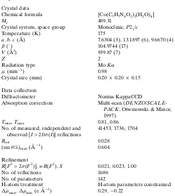
Table 3
Experimental details.
Crystal data
Chemical formula [Co(C7H7N4O2)2(H2O)4]
Mr 489.31
Crystal system, space group Monoclinic,P21/c
Temperature (K) 175
a,b,c(A˚ ) 7.6304 (3), 13.1897 (6), 9.6670 (4)
() 104.9744 (17)
V(A˚3) 939.87 (7)
Z 2
Radiation type MoK
(mm1) 0.98
Crystal size (mm) 0.200.200.15
Data collection
Diffractometer Nonius KappaCCD
Absorption correction Multi-scan (DENZO/
SCALE-PACK; Otwinowski & Minor, 1997)
Tmin,Tmax 0.81, 0.86
No. of measured, independent and observed [I> 2.0(I)] reflections
41453, 1736, 1704
Rint 0.028
(sin/)max(A˚ 1
) 0.604
Refinement
R[F2> 2(F2)],wR(F2),S 0.021, 0.023, 1.00
No. of reflections 1686
No. of parameters 142
H-atom treatment H-atom parameters constrained
max,min(e A˚ 3
) 0.29,0.22
supporting information
sup-1
Acta Cryst. (2017). E73, 1302-1304
supporting information
Acta Cryst. (2017). E73, 1302-1304 [https://doi.org/10.1107/S2056989017011379]
Crystal structure of tetraaquabis(1,3-dimethyl-2,6-dioxo-7
H
-purin-7-ido-κ
N
7)cobalt(II)
Hicham El Hamdani, Mohammed El Amane and Carine Duhayon
Computing details
Data collection: COLLECT (Nonius, 2001).; cell refinement: DENZO/SCALEPACK (Otwinowski & Minor, 1997); data
reduction: APEX2 (Bruker, 2006); program(s) used to solve structure: SUPERFLIP (Palatinus & Chapuis, 2007);
program(s) used to refine structure: CRYSTALS (Betteridge et al., 2003); molecular graphics: CAMERON (Watkin et al.,
1996); software used to prepare material for publication: CRYSTALS (Betteridge et al., 2003).
Tetraaquabis(1,3-dimethyl-2,6-dioxo-7H-purin-7-ido-κN7)cobalt(II)
Crystal data
[Co(C7H7N4O2)2(H2O)4]
Mr = 489.31
Monoclinic, P21/c
Hall symbol: -P 2ybc
a = 7.6304 (3) Å
b = 13.1897 (6) Å
c = 9.6670 (4) Å
β = 104.9744 (17)°
V = 939.87 (7) Å3
Z = 2
F(000) = 506
Dx = 1.729 Mg m−3
Mo Kα radiation, λ = 0.71073 Å
Cell parameters from 9823 reflections
θ = 3–25°
µ = 0.98 mm−1
T = 175 K
Block, pale pink 0.20 × 0.20 × 0.15 mm
Data collection
Nonius KappaCCD diffractometer
Graphite monochromator
φ & ω scans
Absorption correction: multi-scan
(DENZO/SCALEPACK; Otwinowski & Minor, 1997)
Tmin = 0.81, Tmax = 0.86
41453 measured reflections 1736 independent reflections 1704 reflections with I > 2.0σ(I)
Rint = 0.028
θmax = 25.4°, θmin = 2.7°
h = −9→9
k = −15→15
l = −11→11
Refinement
Refinement on F
Least-squares matrix: full
R[F2 > 2σ(F2)] = 0.021
wR(F2) = 0.023
S = 1.00
1686 reflections 142 parameters 0 restraints
Primary atom site location: other
Hydrogen site location: difference Fourier map H-atom parameters constrained
Method = Quasi-Unit weights W = 1.0 or
1./4Fsq
(Δ/σ)max = 0.0003458
Δρmax = 0.29 e Å−3
supporting information
sup-2
Acta Cryst. (2017). E73, 1302-1304
Special details
Experimental. The crystal was placed in the cold stream of an Oxford Cryosystems open-flow nitrogen cryostat (Cosier & Glazer, 1986) with a nominal stability of 0.1 K.
Cosier, J. & Glazer, A.M., 1986. J. Appl. Cryst. 105-107.
Fractional atomic coordinates and isotropic or equivalent isotropic displacement parameters (Å2)
x y z Uiso*/Ueq
N1 0.05846 (17) 0.45527 (9) 0.72491 (13) 0.0126
N3 0.04606 (18) 0.33836 (10) 0.89795 (13) 0.0151
N5 0.24314 (17) 0.42364 (10) 1.10036 (13) 0.0143
N7 0.34987 (17) 0.58201 (10) 1.04872 (13) 0.0139
C2 −0.0068 (2) 0.36737 (12) 0.75802 (16) 0.0143
C4 0.1522 (2) 0.41609 (11) 0.95762 (15) 0.0124
C6 0.3386 (2) 0.50912 (12) 1.14880 (16) 0.0145
C8 0.2695 (2) 0.57724 (11) 0.89958 (16) 0.0136
C9 0.16404 (19) 0.48879 (11) 0.85772 (15) 0.0119
C10 0.2355 (2) 0.34042 (13) 1.19853 (17) 0.0218
C12 0.4475 (2) 0.67528 (12) 1.10615 (17) 0.0196
O11 0.41386 (15) 0.52183 (9) 1.27770 (11) 0.0188
O13 0.29724 (16) 0.64754 (8) 0.82313 (12) 0.0206
O14 0.26896 (14) 0.46163 (9) 0.50496 (11) 0.0197
O15 0.08751 (15) 0.64646 (8) 0.55927 (11) 0.0172
Co1 0.0000 0.5000 0.5000 0.0103
H21 −0.0864 0.3279 0.6895 0.0161*
H103 0.3447 0.3423 1.2769 0.0336*
H101 0.1296 0.3467 1.2352 0.0345*
H102 0.2316 0.2770 1.1486 0.0330*
H121 0.4345 0.7242 1.0325 0.0318*
H122 0.4018 0.7025 1.1814 0.0314*
H123 0.5741 0.6614 1.1459 0.0311*
H142 0.3487 0.4654 0.5786 0.0298*
H151 0.1601 0.6467 0.6392 0.0273*
H141 0.3056 0.4824 0.4383 0.0306*
H152 0.0280 0.6987 0.5575 0.0275*
Atomic displacement parameters (Å2)
U11 U22 U33 U12 U13 U23
N1 0.0148 (6) 0.0118 (6) 0.0100 (6) −0.0004 (5) 0.0012 (5) −0.0006 (5)
N3 0.0188 (7) 0.0135 (6) 0.0124 (6) −0.0009 (5) 0.0031 (5) 0.0011 (5)
N5 0.0164 (6) 0.0158 (6) 0.0094 (6) 0.0006 (5) 0.0010 (5) 0.0024 (5)
N7 0.0138 (6) 0.0148 (6) 0.0114 (6) −0.0019 (5) 0.0003 (5) −0.0019 (5)
C2 0.0163 (7) 0.0135 (7) 0.0122 (7) −0.0016 (6) 0.0019 (6) −0.0014 (6)
C4 0.0125 (7) 0.0136 (7) 0.0109 (7) 0.0021 (6) 0.0026 (6) 0.0001 (6)
C6 0.0112 (7) 0.0191 (8) 0.0129 (7) 0.0028 (6) 0.0026 (6) −0.0008 (6)
C8 0.0127 (7) 0.0150 (7) 0.0122 (7) 0.0020 (6) 0.0015 (6) −0.0008 (6)
supporting information
sup-3
Acta Cryst. (2017). E73, 1302-1304
C10 0.0312 (9) 0.0187 (8) 0.0139 (8) 0.0009 (7) 0.0030 (7) 0.0065 (6)
C12 0.0216 (8) 0.0177 (8) 0.0171 (8) −0.0042 (7) 0.0004 (6) −0.0049 (6)
O11 0.0170 (5) 0.0280 (6) 0.0089 (5) −0.0012 (5) −0.0008 (4) −0.0012 (5)
O13 0.0272 (6) 0.0171 (6) 0.0141 (5) −0.0082 (5) −0.0008 (5) 0.0032 (5)
O14 0.0146 (5) 0.0329 (7) 0.0105 (5) 0.0010 (5) 0.0012 (4) 0.0005 (5)
O15 0.0236 (6) 0.0119 (5) 0.0125 (5) 0.0007 (5) −0.0021 (4) −0.0016 (4)
Co1 0.01218 (14) 0.00993 (14) 0.00793 (14) −0.00056 (11) 0.00085 (10) −0.00020 (11)
Geometric parameters (Å, º)
N1—C2 1.333 (2) C8—C9 1.416 (2)
N1—C9 1.3994 (18) C8—O13 1.2375 (19)
N1—Co1 2.1847 (12) C10—H103 0.971
N3—C2 1.363 (2) C10—H101 0.966
N3—C4 1.341 (2) C10—H102 0.962
N5—C4 1.3786 (19) C12—H121 0.947
N5—C6 1.358 (2) C12—H122 0.955
N5—C10 1.4620 (19) C12—H123 0.961
N7—C6 1.382 (2) O14—Co1 2.1022 (11)
N7—C8 1.4149 (19) O14—H142 0.810
N7—C12 1.4706 (19) O14—H141 0.813
C2—H21 0.933 O15—Co1 2.0756 (10)
C4—C9 1.380 (2) O15—H151 0.826
C6—O11 1.2414 (18) O15—H152 0.823
C2—N1—C9 102.52 (12) H103—C10—H102 108.8
C2—N1—Co1 118.74 (10) H101—C10—H102 109.7
C9—N1—Co1 138.49 (10) N7—C12—H121 110.0
C2—N3—C4 101.73 (12) N7—C12—H122 110.7
C4—N5—C6 119.43 (13) H121—C12—H122 109.2
C4—N5—C10 120.08 (13) N7—C12—H123 110.6
C6—N5—C10 120.48 (13) H121—C12—H123 109.2
C6—N7—C8 126.39 (13) H122—C12—H123 107.1
C6—N7—C12 115.76 (12) Co1—O14—H142 120.9
C8—N7—C12 117.76 (13) Co1—O14—H141 115.7
N3—C2—N1 116.71 (13) H142—O14—H141 110.0
N3—C2—H21 121.3 Co1—O15—H151 110.6
N1—C2—H21 122.0 Co1—O15—H152 129.6
N5—C4—N3 125.28 (14) H151—O15—H152 104.5
N5—C4—C9 122.90 (14) N1—Co1—N1i 180
N3—C4—C9 111.81 (13) N1—Co1—O14i 92.00 (4)
N7—C6—N5 117.44 (13) N1i—Co1—O14i 88.00 (4)
N7—C6—O11 120.82 (14) N1—Co1—O14 88.00 (4)
N5—C6—O11 121.74 (14) N1i—Co1—O14 92.00 (4)
N7—C8—C9 113.12 (13) O14i—Co1—O14 180
N7—C8—O13 118.63 (13) N1—Co1—O15i 89.53 (4)
C9—C8—O13 128.25 (14) N1i—Co1—O15i 90.47 (4)
supporting information
sup-4
Acta Cryst. (2017). E73, 1302-1304
C8—C9—C4 120.50 (13) O14—Co1—O15i 91.42 (4)
N1—C9—C4 107.23 (13) N1—Co1—O15 90.47 (4)
N5—C10—H103 108.5 N1i—Co1—O15 89.53 (4)
N5—C10—H101 110.6 O14i—Co1—O15 91.42 (4)
H103—C10—H101 110.0 O14—Co1—O15 88.58 (4)
N5—C10—H102 109.2 O15i—Co1—O15 180
Symmetry code: (i) −x, −y+1, −z+1.
Hydrogen-bond geometry (Å, º)
D—H···A D—H H···A D···A D—H···A
O14—H142···O11ii 0.81 1.99 2.773 (2) 164
O15—H151···O13 0.83 1.82 2.638 (2) 173
O14—H141···O11iii 0.81 2.01 2.817 (2) 174
O15—H152···N3iv 0.82 2.01 2.799 (2) 162