Optimisation of the PCR Method for the Detection
of
Campylobacter jejuni
and
Campylobacter coli
in Samples
of Ready-to-Eat Chicken Meals
Zdeňka ŠabatkoVá
1,2, kateřina DemNeroVá
1and Jarmila PaZlaroVá
11
Department of biochemistry and microbiology, Faculty of Food and biochemical
technology, Institute of Chemical technology in Prague, Prague, Czech republic;
2Department of biomagnetic techniques, Institute of Systems biology and ecology,
academy of Science of the Czech republic, České budějovice, Czech republic
Abstract
Šabatková Z., Demnerová K., Pazlarová J. (2008): Optimisation of the PCR method for the detec-tion of Campylobacter jejuniandCampylobacter coliin samples of ready-to-eat chicken meals.Czech J. Food Sci., 26: 291–297.
This work compared the use of polymerase chain reaction (PCR) and the conventional CSN/ISO/10272 culture-based methods in the detection of Campylobacter species in ready-to-eat meals made from chicken meat. PCR was carried out with the primers specific to C. jejuni, C. coli, C. lari, and was modified with an internal control. The detection of campylobacters by PCR was performed on both untreated and spiked samples of real food purchased in local stores. For PCR, the detection limit was 2 CFU/g after 48 h enrichment in Park and Sanders broth. Duplex PCR proved to be highly reliable in the detection of campylobacters in different food types. Without extra spiking, samples from a global fast food chain exhibited positive amplification of the PCR product while but negative results were obtained from the cultivation of the same samples.
Keywords: polymerase chain reaction; internal control; Campylobacter spp.
In the last few decades, new food-borne patho-gens have been identified. Campylobacter, a food-borne bacteria, is one of the leading causes of diarrhea illness throughout the world (Friedman
et al. 2000). The genus Campylobacter comprises 16 closely related species and 6 sub-species of gram-negative bacteria, all of which are capable of colonising the gastrointestinal tracts of a wide variety of host species (Volokhov et al. 2003). Epidemiological datashow that the most
signifi-cant of these Campylobacter pathogen species are the thermotolerant C. jejuni and C. coli (Mead et al. 1999). As these bacteria are currently part of the microflora in farmed animals (poultry, pigs, cattle), contaminated foods and water appear to be the most common vehicles of transmission to humans. However, Campylobacter is also hosted in wild birds (Glunder et al. 1992).
The conventional methods for the detection and differentiation of Campylobacter species are
tedious and time consuming, usually taking five days to produce a negative result and up to seven days to confirm a positive result. In recent years, numerous molecular diagnostic approaches for the detection and identification of Campylobacter spp. have been developed, including various PCR-based assays (Chuma et al. 1997; Fermer & Engvall 1999; Lübeck et al. 2003a; Sabatkova et al. 2004). PCR methods have several advantages because they are faster and more sensitive and specific than the cultivation-based procedures. However, thus far few PCR-based studies have aimed at dif-ferentiating between species.
Our goal was to develop a PCR-based rapid screening method for the detection of campy-lobacters in ready-to-eat foods. The method consisted of two steps: (i) elimination of false negatives obtained in the detection of thermotol-erant species of Campylobacter spp.; (ii) species identification.
MAtERiAlS AnD MEthODS
Bacterial strains.Campylobacter jejuni subsp.
jejuni CCM 6212 and Campylobacter coli CCM 6211 (Czech Collection of Microorganisms, Ma-saryk University Brno, Czech Republic), were used for testing PCR detection limits and for the spiking of food samples.
Food samples. Four samples of ready-to-eat
meals (chicken pieces in jelly; chicken sausage with cheese; chicken baguette; fried chicken pieces)
were purchased from local stores.
Cultivation and enumeration of bacteria.
The Campylobacter strains were grown either on Karmali agar (Hi-media, Mumbai, India) or in Park and Sanders broth (Hi-media, Mumbai, India) to which sheep blood was added. They were incubated in a microaerophilic atmosphere at 42°C for 24–48 hours. For the cell enumera-tion, cell suspensions were serially diluted 1:10 in 0.85% NaCl solution. For each dilution, the cell number (CFU/ml) was determined by plating on Karmali agar.
Preparation of food samples. 25-g portions
of each food matrix were homogenised at 1:10 (225 ml) with Park and Sanders broth in a stom-acher for 1 min to macerate them. To contami-nate the samples, the first parts of the respective mixtures after homogenisation were spiked with approximately 101 CFU of C. jejuni or C. coli per g of food. The second parts of the mixtures were
spiked with approximately 100 of C. jejuni or C.
coli per g of food. The third parts of the mixtures were incubated unspiked as controls. All mixtures were incubated in a microaerophilic atmosphere at 37°C for 4 h, and then at 42°C for further 48 hours. After 24 h of enrichment, 1 ml aliquots of each mixture were extracted for PCR analysis only. After 48 h, 1 ml aliquots of each mixture were extracted for both PCR and standard micro-biological analyses (CSN ISO 10272). Following the enrichment, the standard microbiological approach (CSN ISO 10272) was followed to de-termine whether or not the spiked and unspiked samples contained Campylobacter spp. All 1-ml portions (24 h, 48 h) were centrifuged at 10 000 g for 5 minutes. The resulting pellets were kept at –20°C for later DNA analysis; DNA was extracted from the food samples by the use of three rapid methods: (i) extraction by boiling, (ii) extraction by treatment with proteinase K, (iii) resin based extraction, details in Sabatkova et al. (2004). Prior to the use of each extraction method, the sample pellet was allowed to thaw at 4°C, washed with 0.1M Tris buffer (pH 8) and centrifuged at 7000 rpm for 5 min.
Preparation of PCR internal control. With
a LightCycler® 2.0 Instrument (F. Hoffmann-La Roche Ltd., Basel, Switzerland).
PCR with internal control. To identify the thermo-
tolerant species of Campylobacter spp. present in the food samples (24 h, 48 h), PCR was carried out following the procedure described by Sabatkova
et al. (2004), with some modifications to the primer concentrations and with the addition of the internal control. The primers used were specific to C. jejuni,
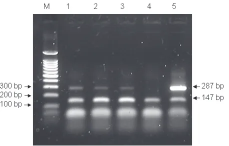
C. coli, and C. lari. The primer concentrations selected were 0.88µM for the forward primer and 0.96µM for the reverse primer. The concentration of the internal control was selected to be 0.05 pg of IC per reaction. The determination of PCR sensitivity was performed using DNA extracted by three rapid methods (Sabatkova et al. 2004) from the serial dilutions (in the range of 100 to 102 CFU/ml) of the strain C. jejuni CCM 6212. The presence of the thermotolerant species of Campy-lobacter spp. (Figure 1) is demonstrated by 287 bp product, and the presence of the internal control is demonstrated by 147 bp product on agarose gel (Bio-Rad, New Orleans, USA) stained with ethid-ium bromide (Fluka, Buchs, Switzerland).
Duplex PCR. Duplex PCR was used to
differenti-ate between the thermotolerant species, C. jejuni,
C. coli.
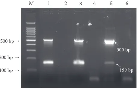
It has been shown that the primers based on the sequence for putative oxidoreductase enable the specific detection of C. jejuni (Winters & Slavik 1995). In terms of the specific detection of C. coli, Linton et al. (1997) have described the use of primers containing the 3 end of the putative aspartokinase gene and a downstream short open reading frame (ORF) encoding a gene of unknown function. Optimal PCR conditions were estab-lished by testing various parameters, including: different annealing temperatures; different DNA polymerases (in different concentrations); and different concentrations of MgCl2. A final reac-tion volume of 25 μl was created by the addireac-tion of the following components: 2.5 μl of sample; 0.4μM of each primer (Generi Biotech, Hradec Králové, Czech Republic); 2mM MgCl2 (Invitrogen, Carlsbad, USA); 0.2mM of each deoxynucleotide (Promega, Madison, USA); the reaction buffer (Invitrogen, Carlsbad, USA); and 0.65U of Plati-num taq DNA polymerase (Invitrogen, USA). The amplification was initiated with DNA denaturation at 95°C for 3 min, followed by a 40-cycle reaction (95°C for 1 min; 57°C for 1 min; 72°C for 1 min), and extension at 72°C for 3 min. Amplicons were
detected in 1% (w/v) agarose gel electrophoresis stained by ethidium bromide. The presence of
C. jejuni was demonstrated by 159 bp product and the presence of C. coli by 500 bp product (Fig-ure 2). For the determination of sensitivity duplex PCR were carried out with all the described DNA extractions from the serial dilutions of the strain
C. jejuni subsp. jejuni CCM 6212 and e. coli CCM 6211 ranging from 100 to 104 CFU/ml.
RESultS AnD DiSCuSSiOn Detection of campylobacters using PCR
with internal control (iC)
It is necessary to select an such IC concentration that is able to produce a visible band on agarose gel while, at the same time, not reducing the intensity of the target product. On the basis of IC titration, 0.05 pg of IC per reaction was chosen for the detec-tion of Campylobacter (data not shown). Because the original primer concentrations (0.44μM and 0.48μM) produced only weak bands of PCR prod-ucts in the presence of the internal control, it was necessary to double them. After this modification, the detection limit for the DNA extracted from the suspension of the pure CCM 6212 strain was found to be the same as the detection limit previ-ously found by Sabatkova et al. (2004) using PCR. The detection limit ranged from 100–101 CFU/ml for DNA extracted with proteinase K (Figure 1)
Lane M– marker: 100 bp DNA ladder, lanes 1 to 5, DNA extracted by treatment with proteinase K from different amounts of C. jejuni in CFU/ml, 102 CFU/ml (lane 1),
101 CFU/ml (lane 2), 100 CFU/ml (lane 3), DNA free (lane 4),
[image:3.595.306.535.497.643.2]105 CFU/ml (lane 5)
and resin-based extraction (data not shown). The detection limit for DNA extracted with boiling lyses, a less sophisticated method, was 102 CFU/ml (data not shown).
Differentiation between C. jejuni and C. coli
using duplex PCR
The sensitivity of the use of duplex PCR with pure cultures was determined using the minimum number of Campylobacter cells that could be am-plified. The results of the optimised procedure are shown in Figure 2; the detection limit of C. jejuni
and C. coli being 102 CFU/ml for DNA extracted with proteinase K, resin-based extraction (data not shown), and 103 CFU/ml (data not shown) when extracted with boiling lyses.
Examination of spiked food samples
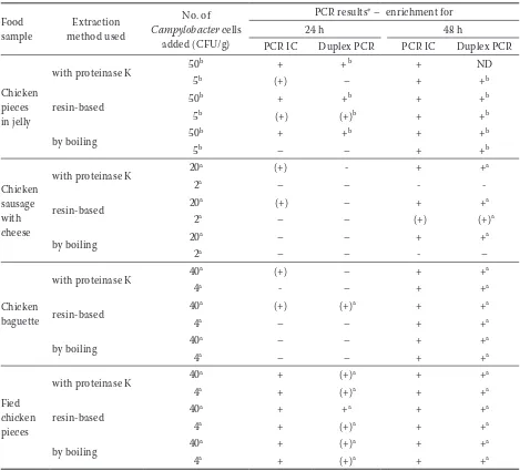
All Campylobacter detections were qualitative. The results summarised in Table 1 were obtained by screening PCR from four independent spiked food matrices. The occurrence of Campylobacter
spp. in all spiked food samples was proven using the standard microbiological method of plating on Karmali agar.
(A) The first analysis was conducted after 24 h of the enrichment cultivation. At this stage, diverse results were obtained by the extraction methods used. For the spiked matrices of approximately 101 CFU/g, negative PCR results were obtained during the analyses of the two food matrices
extracted by boiling (Table 1). However, using duplex PCR, C. jejuni and C. coli were detected in three of the four food matrices extracted by the resin-based method. After 24 h enrichment, we were able to detect at least 20 CFU/g in the chicken products, which is similar to the results published by Magistrado et al. (2001). For spiked food matrices of approximately 100 CFU/g, positive PCR results were obtained in the analyses of the two food matrices extracted by the resin-based method.
The poorest results were obtained with DNA extracted by boiling. With respect to this find-ing, Mohran et al. (1998) suggested that within
Campylobacter populations is a subset that does not release PCR-detectable DNA upon boiling in water, which could explain why no amplicons were obtained for the two food samples extracted by boiling (Table 1). We suggest that the negative results obtained using duplex PCR were caused by the lower sensitivity of the method, as well as by the complex nature of the food matrix after only 24 h enrichment.
(B) The second analysis was conducted after 48 h cultivation. Positive PCR results, indicat-ing the presence of the thermotolerant group of
Campylobacter spp., were obtained for all spiked samples. C. jejuni and/or C. coli. were identified in all four samples using duplex PCR (Table 1). In the case of the spiked chicken pieces in jelly of approximately 101 CFU/g, duplex PCR was not carried out for the DNA extracted by the treat-ment with proteinase K because these species had previously been satisfactorily identifed after 24 h of enrichment. In the case of the spiked chicken sausage with cheese of approximately 100 CFU/g, positive PCR results were obtained only when using the extraction by the resin-based method. The species determined using duplex PCR on the DNA extracted from the food samples cor-responded to the species used for spiking the samples.
The detection limit prescribed by the cultivation method, of 1 cell per 25 g of food (0.04 CFU/g) for either 24 h or 48 h enrichment, was not achieved in our experiments. The best detection limit achieved was 2 CFU/g after 48 h enrichment (Table 1), which was obtained for DNA extracted by the resin-based method.
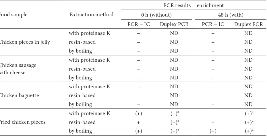
(C) In parallel to the analytical experiments described above (A, B), the control analysis was also conducted on the unspiked food samples with
Lane M– marker: 100 bp DNA ladder, CCM 6211 C. coli
(lane 1), CCM 6212 C. jejuni (lane 2), mixed culture of C. je- juni and C. coli (lane 3), DNA free (lane 4)
Figure 2.Results of optimised Duplex PCR protocol detecting C. jejuni and C. coli
500 bp→
200 bp →
100 bp →
[image:4.595.64.292.83.231.2]mixed results (Table 2). With the exception of the fried chicken pieces, negative PCR results were obtained with all food samples, and were confirmed by the cultivation method used. In the case of the positive PCR results obtained with the fried chicken pieces, no variance was found between the extraction methods used, and this positive finding was not confirmed by the cultivation method used. This suggests that, while the bacterial cells were destroyed during the food preparation process,
a sufficient amount of DNA remained that could be amplified by PCR.
[image:5.595.63.532.114.539.2]The main limitation of PCR methods in the ex-amination of food samples is the frequent presence of inhibiting compounds that can interfere with the amplification reaction and, consequently, result in either a negative or a false negative analyses. An internal control (IC) should be used in PCR proce-dures to prevent false negative results, particularly when food samples are to be examined. As evident
Table 1. Results obtained by the use of PCR on spiked ready-to-eat chicken meal samples extracted by three different methods after enrichment
Food
sample method usedExtraction
No. of
Campylobacter cells added (CFU/g)
PCR results* – enrichment for
24 h 48 h
PCR IC Duplex PCR PCR IC Duplex PCR
Chicken pieces in jelly
with proteinase K 50b + + b + ND
5b (+) – + +b
resin-based 50b + +b + +b
5b (+) (+)b + +b
by boiling 50b + +b + +b
5b – – + +b
Chicken sausage with cheese
with proteinase K 20a (+) - + +a
2a – – -
-resin-based 20a (+) – + +a
2a – – (+) (+)a
by boiling 20a – – + +a
2a – – - –
Chicken baguette
with proteinase K 40a (+) – + +a
4a - – + +a
resin-based 40a (+) (+)a + +a
4a – – + +a
by boiling 40a – – + +a
4a – – + +a
Fied chicken pieces
with proteinase K 40a + (+)a + +a
4a + (+)a + +a
resin-based 40a + +a + +a
4a + (+)a + +a
by boiling 40a + (+)a + +a
4a + (+)a + +a
aC. jejuni; bC. coli; + strong band, (+) weak band; – negative band, ND not done
from previous studies, the IC can be developed in several different ways (Sachadyn & Kur 1998; Cubero et al. 2002; Lübeck et al. 2003b). The ad-vantage of the IC used in this study is its simplicity, accessibility, and universality. As the amplification of one product may influence that of another, and as the band intensity depends on the amounts of the target DNA and control DNA, it is necessary to find the appropriate ratio of IC DNA to target DNA experimentally. To obtain reliable results, it is necessary to store the IC in a highly concentrated form, because when stored at low concentrations, it may be degraded and lead to irreproducible results (Sachadyn & Kur 1998).
PCR inhibition can be partially overcome by the use of a suitable DNA extraction protocol (Cubero
et al. 1999). The rapid extraction methods used in this study are cheap, fast, and undemanding, but their capacity to remove inhibitors is not efficient enough for all types of food matrix. This is why it is important to use an IC in the application of PCR methods.
Our detection limit (2 CFU/g after 48h enrich-ment) did not reach the level of the ISO norm (0.04 CFU/g). After 24h enrichment, our detection limit was comparable with the limit obtained by Magistrado et al. (2001), who, after 17 h en-richment of chicken rinse, detected 31.7 CFU/g.
As our detection limit was determined in highly complex matrices, such as chicken sausage with cheese, we can assume that the use of a simpler matrix, in which inhibiting compounds are not present, would enable us to achieve a detection limit similar to the ISO norm. We plan to inves-tigate this in a future study.
References
Chuma T., Yano K., Omori K., Yugi H. (1997): Direct detection of Campylobacter jejuni in chicken cecal contents by PCR. Journal of Veterinary Medical Sci-ence, 59: 85–87.
CSN ISO 10272 Microbiology of food and animal feeding stuffs – Horizontal method for detection of thermo-tolerant Campylobacter.
Cubero J., Martınez M.C., Llop P., Lopez M.M. (1999): A simple and efficient PCR method for the detection of
agrobacterium tumefaciens in plant tumours. Journal of Applied Microbiology, 86: 591–602.
Cubero J., van der Wolf J., van Beckhoven J., Lopt M.M. (2002): An internal control for the diagnosis of crown gall by PCR. Journal of Microbiological Meth-ods, 51: 387–392.
[image:6.595.67.532.115.353.2]Fermer C., Engvall E.O. (1999): Specific PCR identifi-cation and differentiation of the thermophilic campy-lobacters, Campylobacter jejuni, C. coli, C. lari and Table 2. Results obtained by the use of PCR on unspiked ready-to-eat chicken meal samples (both enriched and nonenriched) extracted by three different methods
Food sample Extraction method
PCR results – enrichment
0 h (without) 48 h (with)
PCR – IC Duplex PCR PCR – IC Duplex PCR
Chicken pieces in jelly
with proteinase K – ND – ND
resin-based – ND – ND
by boiling – ND – ND
Chicken sausage with cheese
with proteinase K – ND – ND
resin-based – ND – ND
by boiling – ND – ND
Chicken baguette
with proteinase K –- ND – ND
resin-based – ND – ND
by boiling – ND - ND
Fried chicken pieces
with proteinase K (+) (+)a + (+)a
resin-based + (+)a + (+)a
by boiling (+) (+)a (+) (+)a
C. upsaliensis. Journal of Clinical Microbiology, 37: 3370–3373.
Friedman C.R., Neimann J., Wegener H.C., Tauxe R.V. (2000): Epidemiology of Campylobacter jejuni infections in the United States and other industrialized nations. In: Nachamkin I., Blaser M.J. (eds): Campylobacter. 2nd Ed. ASM Press, Washington: 121–138.
Glunder G., Neumann U., Braune S. (1992): Occur-rence of Campylobacter spp. in young gulls, duration of Campylobacter infection and reinfection by contact. Journal of Veterinary Medicine series B – Infectious Diseases and Veterinary Public Health, 39: 119–122. Linton D., Lawson A.J., Owen R.J., Stanley J. (1997):
PCR detection, identification to species level, and fin-gerprinting of Campylobacter jejuni andCampylobacter coli direct from diarrheic samples. Journal of Clinical Microbiology, 35: 2568–2572.
LübeckP.S., Cook N., Wagner M., Fach P., Hoor-far J. (2003a): Towards an international standard for PCR-based detection of food-borne thermotolerant campylobacters: Validation in a multicenter collabo-rative trial. Applied Environmental Microbiology, 69: 5670–5672.
Lübeck P.S., Wolffs P., On S.L.W., Agens P., Radström P., Hoorfar J. (2003b): Towards an international standard for PCR-based detection of food-borne ther-motolerant campylobacters: Assay development and analytical validation. Applied Environmental Micro-biology, 69: 5664–5669.
Magistrado P.A., Garcia M.M., Raymundo A.K. (2001): Isolation and polymerase chain reaction-based
detection of Campylobacter jejuni and Campylobacter coli from poultry in the Philippines. International Journal of Food Microbiology, 70: 197–206.
Mead P.S., Slutsker L., Dietz V., McCaig L.F., Bresee J.S., Shapiro C., Griffin P.M., Tauxe R.V. (1999): Food-related illness and death in the United States. Emergency Infectious Diseases, 5: 607–625.
Mohran Z.S., Arthur R.R., Oyofo B.A., Peruski L.F., Wasfy M.O., Ismail T.F., Murphy J.R. (1998): Differentiation of Campylobacter isolates on the basis of sensitivity to boiling in water as measured by PCR-detectable DNA. Applied Environmental Microbiology,
64: 363–365.
Sabatkova Z., Pazlarova J., Demnerova K. (2004): Sample processing effect on polymerase chain reaction used for identification of Campylobacter jejuni. Folia Microbiologica, 49: 693–697.
Sachadyn P., Kur J. (1998): The construction and use of PCR internal control. Molecular and Cellular Probes,
12: 259–262.
Volokhov D., Chizhikov V., Chumakov K., Ra-sooly A. (2003): Microarray-based identification of thermophilic Campylobacter jejuni, C. coli, C. lari
and C. upsaliensis. Journal of Clinical Microbiology,
41: 4071–4080.
Winters D.K., Slavik M.F. (1995): Evaluation of a PCR based assay for specific detection of Campylobacter jejuni in chicken washes. Molecular and Cellular Probes,
9: 307–310.
Received for publication January 14, 2008 Accepted after corrections March 27, 2008
Corresponding author:
Prof. Ing. Kateřina Demnerová, CSc., Vysoká škola chemicko-technologická, Fakulta potravinářské a biochemické technologie, Ústav biochemie a mikrobiologie, Technická 3, 166 28 Praha 6, Česká republika