4-Chloro-2-[(
E
)-(4-fluorophenyl)imino-methyl]phenol
Tian-Jun Feng
College of Mathematics and Physics, Lanzhou Jiaotong University, Lanzhou 730070, People’s Republic of China
Correspondence e-mail: [email protected]
Received 4 December 2013; accepted 9 December 2013
Key indicators: single-crystal X-ray study;T= 293 K; mean(C–C) = 0.005 A˚;
Rfactor = 0.056;wRfactor = 0.153; data-to-parameter ratio = 13.0.
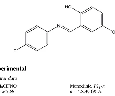
In the title Schiff base molecule, C13H9ClFNO, the benzene rings are twisted slightly with respect to each other, making a dihedral angle of 7.92 (2). An intramolecular O—H N
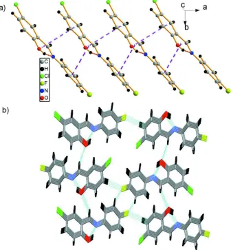
hydrogen bond occurs. In the crystal, an infinite chain is formed along thec-axis direction by–stacking interactions between the phenyl rings and the six-membered hydrogen-bonded ring of neighboring Schiff base ligands [centroid– centroid distances of 3.698 (2) and 3.660 (3) A˚ ]. Neighboring chains are linked into a three-dimensional supramolecular structure by C—H O and C—H F hydrogen bonds.
Related literature
For the coordination modes of Schiff base ligands with tran-sition metals, see: Ebrahimipouret al.(2012); Guoet al.(2013). For the biological activity of Schiff base ligands, see: Sawadaet al.(2001); Maet al.(2013); Siddiquiet al.(2006).
Experimental
Crystal data
C13H9ClFNO
Mr= 249.66
Monoclinic,P21=n a= 4.5140 (9) A˚
b= 20.560 (4) A˚
c= 12.0712 (19) A˚
= 94.153 (16)
V= 1117.4 (3) A˚3
Z= 4
MoKradiation
= 0.34 mm1
T= 293 K
0.340.270.22 mm
Data collection
Agilent Xcalibur (Eos, Gemini) diffractometer
Absorption correction: multi-scan (CrysAlis PRO; Agilent, 2011)
Tmin= 0.908,Tmax= 0.942
6481 measured reflections 2016 independent reflections 1143 reflections withI> 2(I)
Rint= 0.065
Refinement
R[F2> 2(F2)] = 0.056
wR(F2) = 0.153
S= 1.04 2016 reflections
155 parameters
H-atom parameters constrained
max= 0.19 e A˚
3
min=0.23 e A˚
[image:1.610.60.257.523.687.2]3
Table 1
Hydrogen-bond geometry (A˚ ,).
D—H A D—H H A D A D—H A
C7—H7 O1i
0.93 2.69 3.569 (4) 158
C10—H10 F1ii
0.93 2.67 3.481 (4) 147
O1—H1 N1 0.82 1.88 2.613 (3) 148
Symmetry codes: (i)xþ1 2;yþ
1 2;zþ
1
2; (ii)xþ3;yþ1;zþ2.
Data collection: CrysAlis PRO(Agilent, 2011); cell refinement: CrysAlis PRO; data reduction: CrysAlis PRO; program(s) used to solve structure: SHELXS97(Sheldrick, 2008); program(s) used to refine structure:SHELXL97(Sheldrick, 2008); molecular graphics: ORTEPIII (Burnett & Johnson, 1996); software used to prepare material for publication:SHELXL97.
The author acknowledges Lanzhou Jiaotong University for supporting this work.
Supplementary data and figures for this paper are available from the IUCr electronic archives (Reference: ZL2572).
References
Agilent (2011). CrysAlis PRO and CrysAlis RED. Agilent Technologies, Yarnton, England.
Burnett, M. N. & Johnson, C. K. (1996).ORTEPIII. Report ORNL-6895. Oak Ridge National Laboratory, Tennessee, USA.
Ebrahimipour, S. Y., Mague, J. T., Akbari, A. & Takjoo, R. (2012).J. Mol. Struct.1028, 148–155.
Guo, H. F., Zhao, X., Ma, D. Y., Xie, A. P. & Shen, W. B. (2013).Transition Met. Chem.38, 299–305.
Ma, D. Y., Zhang, L. X., Rao, X. Y., Wu, T. L., Li, D. H. & Xie, X. Q. (2013).J. Coord. Chem.66, 1486–1496.
Sawada, H., Yanagida, K., Inaba, Y., Sugiya, M., Kawase, T. & Tomita, T. (2001).Eur. Polym. J.37, 1433–1439.
Sheldrick, G. M. (2008).Acta Cryst.A64, 112–122.
Siddiqui, J. I., Iqbal, A., Ahmad, S. & Weaver, G. W. (2006).Molecules,11, 206–211.
Acta Crystallographica Section E
Structure Reports
Online
supporting information
Acta Cryst. (2014). E70, o42 [https://doi.org/10.1107/S1600536813033278]
4-Chloro-2-[(
E
)-(4-fluorophenyl)iminomethyl]phenol
Tian-Jun Feng
S1. Comment
Schiff bases are considered important compounds because of their wide range of biological activities, and also because of
their use as ligands in conjunction with transition metals. Schiff base ligands usually coordinate to a metal ion through the
imine nitrogen atom, but coordination via other functional groups, e.g. through oxygen or carbon, has also been reported
(Ebrahimipour et al., 2012; Guo et al., 2013). Schiff bases derived from salicyladehyde and fluoroaniline, specifically,
have been considered as potential pharmaceutically interesting compounds as several of the members of this family of
molecules have shown antitumor, antimicrobial or antiviral activities (Sawada et al., 2001; Ma et al., 2013; Siddiqui et
al., 2006). As an extension of our work on the structural characterization of Schiff base compounds, the solid state
structure of the title compound is reported.
The molecular structure of the title compound shows an E configuration, with a C8—N1=C7—C1 torsion angle of
178.33 (2) °. The bond distance of N1=C7 at 1.276 (3) Å is a typical double bond. It is noteworthy that the H1 atom
bonded to O1 is involved in an O1—H1···N1 intramolecular hydrogen bond, which results in the formation of a
six-membered ring (Table 1). The dihedral angle between the two planes of the chlorophenol ring and fluorphenyl ring is
7.92 (2) °. An infinite chain is formed by two types of π-π stacking interactions between the phenyl rings (C1—C6 and
C8—C13) and the six-membered hydrogen bonded ring (C1/C2/O1/H1/N1/C7) of neighboring Schiff base ligands, with
centroid–centroid distances of 3.698 (2) and 3.660 (3) Å, respectively and interplanar spacings of 3.395 (2) Å (Fig. 2a).
Finally, neighboring chains are linked into a three-dimensional supramolecular structure by weak C—H···O and C—H···F
hydrogen bonding interactions (Fig. 2 b, Table 1).
S2. Experimental
Title compound was prepared by the condensation of 5-chlorosalicylaldehyde (0.783 g, 5 mmol) and 4-fluoroaniline
(0.556 g, 5 mmol) in ethanol (15 ml) as the reaction medium. Glacial acetic acid (0.4 ml) was added and the solution was
heated under reflux for 5 h and then allowed to cool to room temperature. The yellow precipitate was recrystallized from
ethanol to give the title compound as straw yellow crystals. Yield 0.20 g (80%). [m.p. 361–363 K; IR (KBr, cm-1):
1637(s), 1560(m), 1508(w), 1460(w), 1392(w), 1324(w), 1288(w), 1210(w), 1120(w), 1054(w), 982(w), 932(w), 876(w),
810(w), 747(w), 709(w), 675(w), 564(w), 511(w); 1H NMR (CDCl
3, δ, p.p.m.) 13.11 (s, 1H), 8.55 (s, 1H), 6.99–7.39 (m,
7H); 13C NMR (CDCl
3, δ, p.p.m.) 161.1, 161.0, 160.7, 159.6, 144.2, 144.1, 133.0, 131.2, 123.8, 122.7, 122.6, 119.9,
118.9, 116.5, 116.2].
S3. Refinement
H atoms were fixed geometrically and treated as riding with O—H = 0.82 Å (hydroxy) and C—H = 0.93 Å, Uiso(H) = 1.2
Ueq(C) for aromatic H atoms and Uiso(H) = 1.5 Ueq(O) for the hydroxy H atom. The hightest residual electron density peak
Figure 1
The molecular structure of the title compound, showing 30% probability displacement ellipsoids.
Figure 2
(a) View of an infinite chain of the title compound formed by π-π stacking interactions (purple dashed lines); (b) View of
the three-dimensional supramolecular structure of the title compound formed by C—H···O and C—H···F hydrogen bonds
[image:3.610.138.474.266.629.2]4-Chloro-2-[(E)-(4-fluorophenyl)iminomethyl]phenol
Crystal data
C13H9ClFNO
Mr = 249.66
Monoclinic, P21/n
Hall symbol: -P 2yn
a = 4.5140 (9) Å
b = 20.560 (4) Å
c = 12.0712 (19) Å
β = 94.153 (16)°
V = 1117.4 (3) Å3
Z = 4
F(000) = 512
Dx = 1.484 Mg m−3
Mo Kα radiation, λ = 0.71073 Å
Cell parameters from 3100 reflections
θ = 1.7–26.0°
µ = 0.34 mm−1
T = 293 K
Block, yellow
0.34 × 0.27 × 0.22 mm
Data collection
Agilent Xcalibur (Eos, Gemini) diffractometer
Radiation source: fine-focus sealed tube Graphite monochromator
ω scans
Absorption correction: multi-scan (CrysAlis PRO; Agilent, 2011)
Tmin = 0.908, Tmax = 0.942
6481 measured reflections 2016 independent reflections 1143 reflections with I > 2σ(I)
Rint = 0.065
θmax = 25.2°, θmin = 2.6°
h = −5→5
k = −23→24
l = −14→14
Refinement
Refinement on F2
Least-squares matrix: full
R[F2 > 2σ(F2)] = 0.056
wR(F2) = 0.153
S = 1.04
2016 reflections 155 parameters 0 restraints
Primary atom site location: structure-invariant direct methods
Secondary atom site location: difference Fourier map
Hydrogen site location: inferred from neighbouring sites
H-atom parameters constrained
w = 1/[σ2(F
o2) + (0.0516P)2] where P = (Fo2 + 2Fc2)/3 (Δ/σ)max < 0.001
Δρmax = 0.19 e Å−3
Δρmin = −0.23 e Å−3
Special details
Geometry. All esds (except the esd in the dihedral angle between two l.s. planes) are estimated using the full covariance matrix. The cell esds are taken into account individually in the estimation of esds in distances, angles and torsion angles; correlations between esds in cell parameters are only used when they are defined by crystal symmetry. An approximate (isotropic) treatment of cell esds is used for estimating esds involving l.s. planes.
Refinement. Refinement of F2 against ALL reflections. The weighted R-factor wR and goodness of fit S are based on F2,
conventional R-factors R are based on F, with F set to zero for negative F2. The threshold expression of F2 > 2sigma(F2) is
used only for calculating R-factors(gt) etc. and is not relevant to the choice of reflections for refinement. R-factors based
on F2 are statistically about twice as large as those based on F, and R- factors based on ALL data will be even larger.
Fractional atomic coordinates and isotropic or equivalent isotropic displacement parameters (Å2)
x y z Uiso*/Ueq
C1 0.2601 (7) 0.22941 (16) 0.7977 (3) 0.0364 (8)
C2 0.1287 (7) 0.22602 (17) 0.6887 (3) 0.0409 (8)
C3 −0.0847 (8) 0.17908 (18) 0.6617 (3) 0.0485 (10)
C4 −0.1685 (8) 0.13523 (18) 0.7395 (3) 0.0480 (9)
H4 −0.3112 0.1038 0.7202 0.058*
C5 −0.0384 (8) 0.13834 (17) 0.8465 (3) 0.0462 (9)
C6 0.1714 (7) 0.18464 (16) 0.8761 (3) 0.0456 (9)
H6 0.2552 0.1863 0.9488 0.055*
C7 0.4848 (7) 0.27750 (17) 0.8303 (3) 0.0420 (9)
H7 0.5699 0.2772 0.9028 0.050*
C8 0.7834 (7) 0.36833 (16) 0.7949 (3) 0.0380 (8)
C9 0.8940 (7) 0.37974 (17) 0.9038 (3) 0.0477 (9)
H9 0.8310 0.3540 0.9610 0.057*
C10 1.0968 (8) 0.42908 (17) 0.9277 (3) 0.0517 (10)
H10 1.1698 0.4370 1.0005 0.062*
C11 1.1880 (8) 0.46601 (17) 0.8424 (3) 0.0487 (9)
C12 1.0866 (8) 0.45657 (18) 0.7349 (3) 0.0524 (10)
H12 1.1526 0.4824 0.6785 0.063*
C13 0.8819 (8) 0.40728 (17) 0.7116 (3) 0.0494 (10)
H13 0.8093 0.4002 0.6385 0.059*
Cl1 −0.1447 (3) 0.08228 (5) 0.94489 (9) 0.0769 (4)
F1 1.3866 (5) 0.51483 (10) 0.86620 (19) 0.0767 (7)
N1 0.5694 (6) 0.32018 (13) 0.7626 (2) 0.0407 (7)
O1 0.2061 (6) 0.26776 (13) 0.60954 (19) 0.0573 (7)
H1 0.3290 0.2937 0.6366 0.086*
Atomic displacement parameters (Å2)
U11 U22 U33 U12 U13 U23
C1 0.0361 (18) 0.039 (2) 0.0332 (19) 0.0018 (16) −0.0011 (15) −0.0019 (16)
C2 0.044 (2) 0.043 (2) 0.035 (2) −0.0010 (17) 0.0013 (16) −0.0047 (17)
C3 0.052 (2) 0.054 (2) 0.038 (2) 0.0012 (19) −0.0047 (18) −0.0142 (19)
C4 0.048 (2) 0.044 (2) 0.051 (2) −0.0036 (18) 0.0013 (19) −0.0134 (19)
C5 0.053 (2) 0.041 (2) 0.044 (2) −0.0051 (18) 0.0023 (18) −0.0012 (17)
C6 0.051 (2) 0.048 (2) 0.037 (2) 0.0005 (18) −0.0034 (17) −0.0033 (18)
C7 0.0398 (19) 0.046 (2) 0.039 (2) 0.0023 (17) −0.0052 (16) −0.0055 (18)
C8 0.0378 (19) 0.036 (2) 0.039 (2) 0.0013 (15) −0.0008 (16) −0.0005 (17)
C9 0.052 (2) 0.048 (2) 0.043 (2) −0.0075 (18) 0.0015 (18) −0.0019 (18)
C10 0.059 (2) 0.049 (2) 0.045 (2) −0.0026 (19) −0.0100 (19) −0.0030 (19)
C11 0.045 (2) 0.042 (2) 0.058 (3) −0.0080 (17) −0.0024 (19) −0.004 (2)
C12 0.056 (2) 0.049 (2) 0.053 (2) −0.005 (2) 0.007 (2) 0.0058 (19)
C13 0.052 (2) 0.053 (2) 0.042 (2) 0.0011 (19) −0.0058 (18) 0.0001 (19)
Cl1 0.0965 (9) 0.0668 (8) 0.0659 (7) −0.0274 (6) −0.0031 (6) 0.0137 (6)
F1 0.0852 (16) 0.0563 (15) 0.0870 (17) −0.0318 (13) −0.0043 (13) −0.0019 (13)
N1 0.0397 (16) 0.0414 (17) 0.0407 (17) −0.0025 (14) 0.0010 (13) −0.0040 (15)
O1 0.0689 (19) 0.0628 (18) 0.0385 (14) −0.0163 (14) −0.0068 (13) 0.0018 (14)
Geometric parameters (Å, º)
C1—C6 1.400 (4) C8—C13 1.384 (4)
C1—C7 1.450 (4) C8—N1 1.418 (4)
C2—O1 1.349 (4) C9—C10 1.383 (5)
C2—C3 1.385 (4) C9—H9 0.9300
C3—C4 1.374 (5) C10—C11 1.366 (5)
C3—H3 0.9300 C10—H10 0.9300
C4—C5 1.381 (5) C11—C12 1.359 (5)
C4—H4 0.9300 C11—F1 1.362 (4)
C5—C6 1.372 (4) C12—C13 1.386 (5)
C5—Cl1 1.747 (4) C12—H12 0.9300
C6—H6 0.9300 C13—H13 0.9300
C7—N1 1.276 (4) O1—H1 0.8200
C7—H7 0.9300
C6—C1—C2 118.6 (3) C13—C8—C9 118.5 (3)
C6—C1—C7 119.6 (3) C13—C8—N1 116.9 (3)
C2—C1—C7 121.8 (3) C9—C8—N1 124.6 (3)
O1—C2—C3 119.2 (3) C10—C9—C8 120.5 (3)
O1—C2—C1 121.2 (3) C10—C9—H9 119.8
C3—C2—C1 119.5 (3) C8—C9—H9 119.8
C4—C3—C2 121.2 (3) C11—C10—C9 118.8 (3)
C4—C3—H3 119.4 C11—C10—H10 120.6
C2—C3—H3 119.4 C9—C10—H10 120.6
C3—C4—C5 119.3 (3) C12—C11—F1 118.5 (3)
C3—C4—H4 120.4 C12—C11—C10 122.8 (4)
C5—C4—H4 120.4 F1—C11—C10 118.7 (3)
C6—C5—C4 121.0 (3) C11—C12—C13 118.1 (4)
C6—C5—Cl1 119.9 (3) C11—C12—H12 120.9
C4—C5—Cl1 119.1 (3) C13—C12—H12 120.9
C5—C6—C1 120.4 (3) C8—C13—C12 121.3 (3)
C5—C6—H6 119.8 C8—C13—H13 119.3
C1—C6—H6 119.8 C12—C13—H13 119.3
N1—C7—C1 122.1 (3) C7—N1—C8 122.3 (3)
N1—C7—H7 119.0 C2—O1—H1 109.5
C1—C7—H7 119.0
Hydrogen-bond geometry (Å, º)
D—H···A D—H H···A D···A D—H···A
C7—H7···O1i 0.93 2.69 3.569 (4) 158
C10—H10···F1ii 0.93 2.67 3.481 (4) 147
O1—H1···N1 0.82 1.88 2.613 (3) 148