research communications
Acta Cryst.(2018). E74, 1735–1740 https://doi.org/10.1107/S2056989018015578
1735
Received 2 November 2018 Accepted 2 November 2018
Edited by W. T. A. Harrison, University of Aberdeen, Scotland
‡ Additional correspondence author, e-mail: [email protected].
Keywords:crystal structure; carboxylate; mol-ecular salt; hydrogen-bonding.
CCDC references:1876928; 1876927; 1876926
Supporting information:this article has supporting information at journals.iucr.org/e
Crystal structures of the 1:1 salts of
2-amino-4-nitrobenzoate with each of
(2-hydroxyethyl)di-methylazanium,
tert
-butyl(2-hydroxyethyl)azanium
and
1,3-dihydroxy-2-(hydroxymethyl)propan-2-aminium
James L. Wardella‡ and Edward R. T. Tiekinkb*
aDepartment of Chemistry, University of Aberdeen, Old Aberdeen, AB24 3UE, Scotland, andbResearch Centre for
Crystalline Materials, School of Science and Technology, Sunway University, 47500 Bandar Sunway, Selangor Darul Ehsan, Malaysia. *Correspondence e-mail: [email protected]
The crystal and molecular structures of the title molecular salts, C4H12NO+C7H5N2O4, (I), C6H16NO+C7H5N2O4, (II), and C4H12NO3+
-C7H5N2O4, (III), are described. The common feature of these salts is the
presence of the 2-amino-4-nitrobenzoate anion, which exhibit non-chemically significant variations in the conformational relationships between the carboxyl-ate and nitro groups, and between these and the benzene rings they are connected to. The number of ammonium-N—H H atoms in the cations increases from one to three in (I) to (III), respectively, and this variation significantly influences the supramolecular aggregation patterns in the respective crystals. Thus, a linear supramolecular chain along [100] sustained by charge-assisted tertiary-ammonium-N—H O(carboxylate), hydroxy-O—H O(carboxylate) and amino-N—H O(carboxylate) hydrogen-bonds is apparent in the crystal of (I). Chains are connected into a three-dimensional architecture by methyl-C— H O(hydroxy) and–interactions, the latter between benzene rings [inter-centroid separation = 3.5796 (10) A˚ ]. In the crystal of (II), a supramolecular tube propagating along [901] arises as a result of charge-assisted secondary-ammonium-N—H O(carboxylate) and hydroxy-O—H O(carboxylate) hydrogen-bonding. These are connected by methylene- and methyl-C— H O(nitro) and–stacking between benzene rings [inter-centroid separa-tion = 3.5226 (10) A˚ ]. Finally, double-layers parallel to (100) sustained by charge-assisted ammonium-N—H O(carboxylate), ammonium-N—H
O(hydroxy) and hydroxy-O—H O(carboxylate) hydrogen-bonds are apparent in the crystal of (III). These are connected in a three-dimensional architecture by amine-N—H O(nitro) hydrogen-bonds.
1. Chemical context
Despite being tetramorphic (Wardell & Tiekink, 2011; Wardell & Wardell, 2016), readily forming co-crystals (Wardell & Tiekink, 2011) and providing systematic series of crystals of alkali metal, e.g. Na+, K+(Smith, 2013), Rb+(Smith, 2014a) and Cs+(Smith & Wermuth, 2011), and ammonium salts, see
below, studies of the relatively small benzoic acid derivative, 2-amino-4-nitrobenzoic acid, are still comparatively limited. Most crystallographic investigations of the acid have focused upon an evaluation of the hydrogen-bonding propensities occurring in derived ammonium salts of the 2-amino-4-nitro-benzoate anion. Thus, studies have been described with a range of salts, starting with the simplest, i.e. N(+)H4
(Smith, 2014b), H2NN (+)
H3 (Wardell et al., 2017) and
(H2N)2C N (+)
H2(Smithet al., 2007) toR2N (+)
H2,i.e. R= Me,
n-Bu (Wardellet al., 2016), cyclohexyl (Smithet al., 2004) and R2= (CH2CH2)2O (Smith & Lynch, 2016), and more
compli-cated ammonium cations such as 4-(4-acetylphenyl)piperazin-1-ium (Jotani et al., 2018) and the dication, H3N(+)CH2CH2N(+)H3(Smithet al., 2002). As a continuation
of on-going interest in this area, the results of co-crystal-lization experiments between 2-amino-4-nitrobenzoic acid (LH) and amines substituted with hydroxy groups,i.e. each of Me2N(CH2CH2OH),
(t-Bu)N(H)CH2CH2OH and (HOCH2)3CNH2 are described
whereupon the anhydrous 1:1 salts, i.e. [Me2N (+)
H(CH
2-CH2OH)]L (I), [(t-Bu)N (+)
H2(CH2CH2OH)]L (II) and
[(HOCH2)3CN (+)
H3]L(III), were isolated. Herein, a
descrip-tion of the crystal and molecular structures of (I)–(III) are presented.
2. Structural commentary
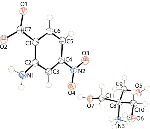
The molecular structures of the constituent ions in (I) are shown in Fig. 1 and selected geometric data for this and for (II) and (III), are collected in Table 1. That proton transfer occurred during co-crystallization is confirmed by the experimental equivalence of the C7. . .O1, O2 bond lengths of 1.270 (2) and 1.258 (2) A˚ , respectively, in the 2-amino-4-nitrobenzoate anion and in the pattern of hydrogen-bonding interactions, as described below inSupramolecular features. In the anion, the carboxylate group is tilted out of the plane of the benzene ring to which it is connected with the dihedral
angle being 6.7 (3). Similarly, the nitro group lies out of the
plane of the benzene ring, forming a dihedral angle of 6.6 (3).
A dis-rotatory relationship between the carboxylate and nitro substituents is indicated by the dihedral angle between them of 11.5 (4). An intramolecular amine-N1—H
O1(carboxylate) hydrogen-bond is noted which closes anS(6) loop, Table 2. In the Me2N
(+)
(H)CH2CH2OH cation, the N3—
C8—C9—O5 torsion angle of 71.15 (19) is indicative of a
syn-clinal conformation.
The anion in (II), Fig. 2, presents essentially the same features as just described for (I), Tables 1 and 3, with the exception of the con-rotatory relationship between the carboxylate and nitro substituents. The (t-Bu)N(+)H2(CH
2-CH2OH) cation is relatively rare, being reported for the first
time in its salt with sulfathiazolate only in 2012 (Armanet al.,
1736
Wardell and Tiekink C4H12NO+, C6H16NO+and C4H12NO3+C7H5N2O4 Acta Cryst.(2018). E74, 1735–1740
[image:2.610.315.565.68.248.2]research communications
Figure 2
[image:2.610.45.297.268.417.2]The molecular structures of the ions comprising the asymmetric unit of (II) showing the atom-labelling scheme and displacement ellipsoids at the 70% probability level. The dashed line indicates a hydrogen bond.
Figure 3
[image:2.610.315.558.500.709.2]The molecular structures of the ions comprising the asymmetric unit of (III) showing the atom-labelling scheme and displacement ellipsoids at the 70% probability level.
Figure 1
[image:2.610.46.292.583.711.2]2012). As for the cation in (I), the N3—C12—C13—O5 torsion angle for the cation in (II) of55.18 (18) is indicative of a
syn-clinal conformation.
The anion in (III), Fig. 3, exhibits the greatest twist between the carboxylate and benzene groups among the series but, a con-rotatory relationship between the carboxylate and nitro substituents means the dihedral angle between them is not as great as in the anion of (I), Tables 1 and 4. The (HOCH2)3CN
(+)
H3 cation exhibits N3—C8—C9—O5, N3—
C8—C10—O6 and N3—C8—C11—O7 torsion angles of
59.01 (18),49.84 (19) and58.12 (18), respectively,
indi-catingsyn-clinal relationships.
3. Supramolecular features
As expected from the chemical compositions of (I)–(III), significant charge-assisted hydrogen-bonding is apparent in their respective crystals. Geometric data characterizing these and other identified interactions are collated in Tables 2–4, respectively.
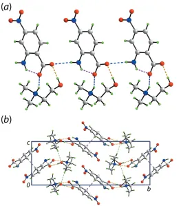
As indicated in Fig. 1, the anion and cation in (I) are linked viacharge-assisted ammonium-N3—H O(carboxylate) and hydroxy-O—H O(carboxylate) hydrogen-bonds to form a nine-membered { OCO HNC2OH} heterosynthon. These
are connected into a linear, supramolecular chain along the a-axis directionviaamino-N—H O(carboxylate) hydrogen-bonds, Fig. 4(a). The chains are linked along thebaxisvia–
interactions between benzene rings [inter-centroid separation = 3.5796 (10) A˚ for symmetry operation:x,y, 1z], and
research communications
Acta Cryst.(2018). E74, 1735–1740 Wardell and Tiekink C
[image:3.610.42.297.91.161.2]4H12NO+, C6H16NO+and C4H12NO3+C7H5N2O4
1737
Figure 4
The molecular packing in (I): (a) linear, supramolecular chain along the a axis sustained by charge-assisted amine-N—H O(carboxylate) and hydroxy-O—H O(carboxylate) hydrogen-bonding interactions shown as blue and orange dashed lines, respectively; intramolecular amine-N— H O(carboxylate) hydrogen bonds are represented by pink dashed lines, and (b) a view of the unit-cell contents in projection down the aaxis. The methyl-C—H O(hydroxy) and–interactions are shown as green and purple dashed lines, respectively.
Table 1
Selected geometric data (A˚ ,) for (I)–(III).
Parameter (I) (II) (III)
C7. . .O1 1.270 (2) 1.259 (2) 1.270 (2)
C7. . .O2 1.258 (2) 1.2678 (19) 1.264 (2)
CO2/C6 6.7 (3) 6.21 (13) 14.80 (17)
NO2/C6 6.6 (3) 3.28 (13) 6.58 (18)
CO2/NO2 11.5 (4) 2.94 (17) 9.7 (3)
Figure 5
[image:3.610.44.307.357.656.2]methyl-C—H O(hydroxy) interactions link molecules along the c-axis to consolidate the three-dimensional packing, Fig. 4(b).
In the crystal of (II), the charge-assisted ammonium-N3— H O(carboxylate) and hydroxy-O—H O(carboxylate) hydrogen-bonds, that lead to the formation of a nine-membered { OCO HNC2OH} heterosynthon, observed in
(I) persist, Fig. 5(a). However, in (II), through the agency of having two ammonium-N—H H atoms, the second H atom bridges a neighbouring carboxylate-O2 atom leading to the formation of a supramolecular tube, as highlighted in Fig. 5(b). As seen from Fig. 5(b), the benzene rings are aligned to be
1738
Wardell and Tiekink C4H12NO+, C6H16NO+and C4H12NO3+C7H5N2O4 Acta Cryst.(2018). E74, 1735–1740
[image:4.610.41.297.91.162.2]research communications
Figure 6
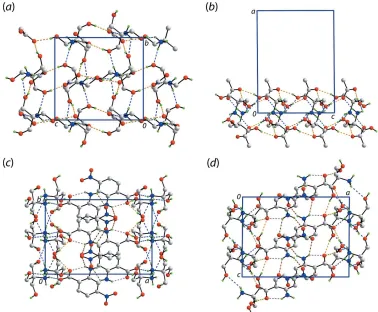
The molecular packing in (III): (a) plan and (b) views of the double-layer sustained by charge-assisted ammonium-N3—H O(carboxylate), ammonium-N3—H O(hydroxy) (blue dashed lines) and hydroxy-O—H O(carboxylate) (orange dashed lines) hydrogen bonds, and views of the unit-cell contents in projection down the (c)caxis and (d)baxis. In (c) and (d), the intramolecular amine-N—H O(carboxylate) and amine-N— H O(nitro) interactions are represented by pink and brown dashed lines, respectively. In each of (a)–(d), non-participating H atoms are omitted.
Table 2
Hydrogen-bond geometry (A˚ ,) for (I).
D—H A D—H H A D A D—H A
N1—H1N O1 0.89 (2) 1.97 (2) 2.6698 (19) 135 (2) N1—H2N O2i 0.88 (1) 2.24 (2) 3.010 (2) 146 (2) N3—H3N O1 0.90 (1) 1.82 (1) 2.6722 (18) 156 (2) O5—H5O O2 0.84 (2) 1.83 (2) 2.6731 (19) 179 (3)
C10—H10B O5ii 0.98 2.48 3.406 (2) 157
Symmetry codes: (i)x1;y;z; (ii)x3 2;y
1 2;z
3 2.
Table 3
Hydrogen-bond geometry (A˚ ,) for (II).
D—H A D—H H A D A D—H A
N1—H1N O1 0.87 (2) 2.00 (2) 2.665 (2) 132 (2) N1—H2N O5 0.89 (2) 2.17 (2) 3.058 (2) 176 (2) N3—H3N O1i 0.90 (2) 1.73 (2) 2.637 (2) 178 (2)
N3—H4N O2ii 0.89 (2) 1.98 (2) 2.849 (2) 166 (2)
O5—H5O O2i 0.84 (2) 1.92 (2) 2.7546 (18) 173 (2) C11—H11A O4iii 0.98 2.49 3.450 (3) 165
C12—H12A O3 0.99 2.50 3.445 (2) 159
Symmetry codes: (i) xþ1 2;yþ
3 2;z
1
2; (ii) xþ1;y;zþ 1 2; (iii)
xþ1 2;yþ
1 2;z
1 2.
Table 4
Hydrogen-bond geometry (A˚ ,) for (III).
D—H A D—H H A D A D—H A
N1—H1N O1 0.88 (2) 2.02 (2) 2.678 (2) 131 (2) N1—H1N O3i 0.88 (2) 2.50 (2) 3.210 (2) 138 (1) N1—H2N O4ii 0.88 (1) 2.56 (2) 3.094 (2) 120 (2)
N3—H3N O6iii 0.89 (2) 2.34 (2) 2.934 (2) 124 (1) N3—H3N O7iv 0.89 (2) 2.44 (2) 3.065 (2) 128 (2)
N3—H4N O5v 0.89 (1) 2.08 (1) 2.945 (2) 165 (2) N3—H5N O2vi 0.89 (2) 1.92 (2) 2.773 (2) 160 (2)
O5—H5O O2vii 0.85 (2) 1.90 (2) 2.7453 (18) 175 (2)
O6—H6O O1viii 0.84 (2) 1.88 (2) 2.6993 (19) 163 (2)
O7—H7O O1ix 0.84 (2) 2.07 (2) 2.8905 (18) 164 (2)
Symmetry codes: (i) xþ1;yþ1 2;zþ
3
2; (ii) x;yþ 1 2;z
1 2; (iii)
xþ2;y1 2;zþ
1
2; (iv) xþ2;yþ1;zþ1; (v) xþ2;yþ 1 2;zþ
1 2; (vi)
xþ1;yþ1 2;z
3
2; (vii) xþ1;y 1 2;zþ
1
2; (viii) xþ1;yþ2;zþ1; (ix)
xþ1;y1 2;zþ
[image:4.610.117.495.388.700.2]proximate and, indeed, they interactvia–stacking with the inter-centroid separation being 3.4944 (9) A˚ (symmetry operation: 1x,y,1
2z). The carboxylate-O2 atom forms two
hydrogen-bonds. The connections between the tubes are of the type methylene- and methyl-C—H O(nitro), involving both nitro-O atoms, as well as–stacking between benzene rings [inter-centroid separation = 3.5226 (10) A˚ for symmetry operation: 1x, 1y,z]. A view of the unit-cell contents is shown in Fig. 5(c), highlighting the intra- and inter-tube–
stacking along thec-axis direction.
In the crystal of (III), supramolecular double-layers in the bc-plane are formed as a result of charge-assisted ammonium-N3—H O(carboxylate), ammonium-N3—H O(hydroxy) and hydroxy-O—H O(carboxylate) hydrogen-bonds. The ammonium-N3—H3N atom is bifurcated, forming two weak ammonium-N3—H O(hydroxy) hydrogen-bonds. A view normal to the plane of the double-layer and a side-on view are shown in Fig. 6(a) and (b), respectively. From the latter, the intra-layer region comprises the ammonium groups, each of which forms four N—H O hydrogen-bonds to carboxylate and hydroxy groups on either side. Each hydroxy group of the cation forms a hydroxy-O—H O(carboxylate) hydrogen-bond with a carboxylate-O atom derived from a different anion, and each accepts an ammonium-N—H atom derived
from a different cation. Each carboxylate-O atom forms two hydrogen-bonds, the O1 accepts hydrogen-bonds from different hydroxy groups, and the O2 atom accept hydrogen-bonds from hydroxy and ammonium groups. Projecting to either side of the double-layer are the nitrobenzene groups, Fig. 6(c) and (d). These provide the links to construct the three-dimensional architecture, i.e. via amine-N—H
O(nitro) interactions, involving both nitro-O atoms.
The obvious trend from the present study is the increase in dimensionality of the supramolecular aggregation pattern,i.e. chain in (I), tube in (II) and double-layer in (III), as the number of acidic ammonium-N—H atoms increases.
4. Database survey
As indicated in theChemical context, a number of ammonium salts of the anion derived from 2-amino-4-nitrobenzoic acid have now been described. The key conformational indicators for the anion are the dihedral angles formed between CO2/C6/
NO2. The smallest dihedral angles between the CO2/C6, C6/
NO2and CO2/NO2pairs of least-squares planes of 3.44 (14),
0.69 (11) and 3.2 (2)are found for the anion in the salt with
H3N (+)
CH2CH2N (+)
H3 (Smith et al., 2002). Conversely, the
greatest CO2/C6, C6/NO2 and CO2/NO2 dihedral angles of
research communications
Acta Cryst.(2018). E74, 1735–1740 Wardell and Tiekink C
[image:5.610.47.563.91.421.2]4H12NO+, C6H16NO+and C4H12NO3+C7H5N2O4
1739
Table 5
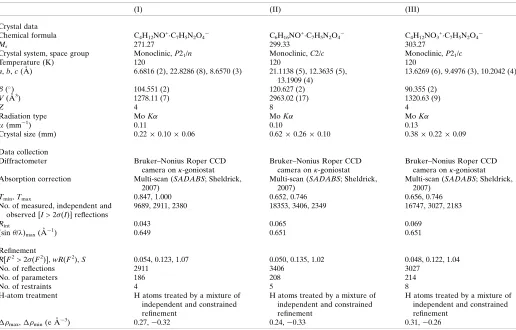
Experimental details.
(I) (II) (III)
Crystal data
Chemical formula C4H12NO+C7H5N2O4
C6H16NO+C7H5N2O4
C4H12NO3+C7H5N2O4
Mr 271.27 299.33 303.27
Crystal system, space group Monoclinic,P21/n Monoclinic,C2/c Monoclinic,P21/c
Temperature (K) 120 120 120
a,b,c(A˚ ) 6.6816 (2), 22.8286 (8), 8.6570 (3) 21.1138 (5), 12.3635 (5), 13.1909 (4)
13.6269 (6), 9.4976 (3), 10.2042 (4)
() 104.551 (2) 120.627 (2) 90.355 (2)
V(A˚3) 1278.11 (7) 2963.02 (17) 1320.63 (9)
Z 4 8 4
Radiation type MoK MoK MoK
(mm1) 0.11 0.10 0.13
Crystal size (mm) 0.220.100.06 0.620.260.10 0.380.220.09
Data collection
Diffractometer Bruker–Nonius Roper CCD
camera on-goniostat
Bruker–Nonius Roper CCD camera on-goniostat
Bruker–Nonius Roper CCD camera on-goniostat Absorption correction Multi-scan (SADABS; Sheldrick,
2007)
Multi-scan (SADABS; Sheldrick, 2007)
Multi-scan (SADABS; Sheldrick, 2007)
Tmin,Tmax 0.847, 1.000 0.652, 0.746 0.656, 0.746
No. of measured, independent and observed [I> 2(I)] reflections
9689, 2911, 2380 18353, 3406, 2349 16747, 3027, 2183
Rint 0.043 0.065 0.069
(sin/ )max(A˚
1
) 0.649 0.651 0.651
Refinement
R[F2> 2(F2)],wR(F2),S 0.054, 0.123, 1.07 0.050, 0.135, 1.02 0.048, 0.122, 1.04
No. of reflections 2911 3406 3027
No. of parameters 186 208 214
No. of restraints 4 5 8
H-atom treatment H atoms treated by a mixture of independent and constrained refinement
H atoms treated by a mixture of independent and constrained refinement
H atoms treated by a mixture of independent and constrained refinement
max,min(e A˚
3) 0.27,0.32 0.24,0.33 0.31,0.26
Computer programs:DENZO(Otwinowski & Minor, 1997),COLLECT(Hooft, 1998),SHELXS97(Sheldrick, 2008),SHELXL2014(Sheldrick, 2015),ORTEP-3 for Windows
26.4 (3), 12.6 (3) and 26.73 (14), respectively, are found in the
N(+)H4(Smith, 2014b),n-Bu2N (+)
H2(Wardellet al., 2016) and
H2NN (+)
H3 (Wardell et al., 2017) salts, respectively. The
respective dihedral angles in (I)–(III), described herein, fall within these ranges.
5. Synthesis and crystallization
Preparation of dimethyl(2-hydroxyethyl)ammonium 2-amino-4-nitrobenzoate (I). To a solution of 2-amino-4-nitrobenzoic acid (1 mmol) in methanol (10 ml) was added a solution of dimethyl(2-hydroxyethyl)amine (1 mmol) in methanol (10 ml). The reaction mixture was refluxed for 15 mins, and then maintained at room temperature. Crystals of (I) were collected after three days. M.p. 444–447 K. Anal. calcd.: C, 48.89; H, 5.97, N, 15.54. Found: C, 48.81; H, 5.89; N, 14.68%. IR (KBr, cm1): 3500–2700 (br, s; with maxima at 3439, 3324, 3219, 2978, 2826), 1632, 1537, 1433, 1381, 1346, 1329, 1279, 1263, 1209, 1140, 1099, 1072, 1022, 918, 858, 823, 785, 731, 692, 684, 577, 513, 486.
Preparation of tert-butyl(2-hydroxyethyl)ammonium nitrobenzoate (II). To a solution of 2-amino-4-nitrobenzoic acid (1 mmol) in methanol (10 ml) was added a solution of tert-butyl(2-hydroxyethyl)amine (1 mmol) in methanol (10 ml). The reaction mixture was refluxed for 15 mins, and then maintained at room temperature. Crystals of (II) were collected after 3 days. M.p. 429–431 K. Anal. calcd.: C, 52.34; H, 6.76, N, 14.09. Found: C, 52.27; H, 6.89; N, 13.99%. IR (KBr, cm1): 3550–2700 (br,s, with maxima at 3430, 3327, 3224, 2968, 2810), 1640, 1446, 1370, 1351, 1329, 1269, 1221 1137, 1085, 1034, 858, 8245, 739, 687, 587.
Preparation of tris(hydroxymethyl)methylammonium nitrobenzoate (III): To a solution of 2-amino-4-nitrobenzoic acid (1 mmol) in ethanol (10 ml) was added a solution of tris(hydroxymethyl)methylamine (1 mmol) in ethanol (10 ml). The reaction mixture was refluxed for 10 mins, and then maintained at room temperature. Crystals of (III) were collected after two days. M.p. 460–463 K. Anal. calcd.: C, 51.06; H, 5.71, N, 14.89. Found: C, 50.94; H, 5.80; N, 14.79% IR (KBr, cm1): 3700–2400 (br, s, with maxima at 3514, 3477, 3398, 3314, 3256, 3078, 2823 and 2538), 1647, 1431, 1350 1250, 1146, 1115, 1063, 1010, 872, 827, 736, 689, 596, 578 511, 484, 1549, 1356.
6. Refinement
Crystal data, data collection and structure refinement details are summarized in Table 5. Carbon-bound H atoms were placed in calculated positions (C—H = 0.95–0.99 A˚ ) and were included in the refinement in the riding-model approximation,
with Uiso(H) set to 1.2–1.5Ueq(C). The O- and N-bound H
atoms were located from difference maps, but refined with O—H = 0.840.01 A˚ andUiso(H) = 1.5Ueq(O), and with N—H
= 0.86–0.880.01 A˚ andUiso(H) = 1.2Ueq(N), respectively. In
the refinement of (II), owing to poor agreement, a reflection, i.e. (0 2 0), was omitted from the final cycles of refinement.
Acknowledgements
The authors thank the National Crystallographic Service, based at the University of Southampton, for collecting the data.
Funding information
JLW thanks CNPq, Brazil, for a grant. The authors are also grateful to Sunway University (INT-RRO-2017-096) for support of this research.
References
Arman, H. D., Kaulgud, T. & Tiekink, E. R. T. (2012).Acta Cryst. E68, o2662–o2663.
Brandenburg, K. (2006).DIAMOND. Crystal Impact GbR, Bonn, Germany.
Farrugia, L. J. (2012).J. Appl. Cryst.45, 849–854.
Hooft, R. W. W. (1998). COLLECT. Nonius BV, Delft, The Netherlands.
Jotani, M. M., Wardell, J. L. & Tiekink, E. R. T. (2018).Z. Kristallogr. Cryst. Mat. doi: https://doi. org/10.1515/zkri-2018-2101.
Otwinowski, Z. & Minor, W. (1997).Methods in Enzymology, Vol. 276, Macromolecular Crystallography, Part A, edited by C. W. Carter Jr & R. M. Sweet, pp. 307–326. New York: Academic Press. Sheldrick, G. M. (2007). SADABS. Bruker AXS Inc., Madison,
Wisconsin, USA.
Sheldrick, G. M. (2008).Acta Cryst.A64, 112–122. Sheldrick, G. M. (2015).Acta Cryst.C71, 3–8. Smith, G. (2013).Acta Cryst.C69, 1472–1477. Smith, G. (2014a).Acta Cryst.E70, m192–m193.
Smith, G. (2014b). Private Communication (Refcode: DOBPIV). CCDC, Cambridge, England.
Smith, G. & Lynch, D. E. (2016).Acta Cryst.C72, 105–111. Smith, G. & Wermuth, U. D. (2011).Acta Cryst.E67, m1047–m1048. Smith, G., Wermuth, U. D. & Healy, P. C. (2004).Acta Cryst.E60,
o684–o686.
Smith, G., Wermuth, U. D., Healy, P. C. & White, J. M. (2007).Acta Cryst.E63, o7–o9.
Smith, G., Wermuth, U. D. & White, J. M. (2002).Acta Cryst.E58, o1088–o1090.
Wardell, J. L., Jotani, M. M. & Tiekink, E. R. T. (2016).Acta Cryst. E72, 1618–1627.
Wardell, J. L., Jotani, M. M. & Tiekink, E. R. T. (2017).Acta Cryst. E73, 579–585.
Wardell, J. L. & Tiekink, E. R. T. (2011).J. Chem. Crystallogr.41, 1418–1424.
Wardell, S. M. S. V. & Wardell, J. L. (2016).J. Chem. Crystallogr.46, 34–43.
Westrip, S. P. (2010).J. Appl. Cryst.43, 920–925.
1740
Wardell and Tiekink C4H12NO+, C6H16NO+and C4H12NO3+C7H5N2O4 Acta Cryst.(2018). E74, 1735–1740
supporting information
sup-1
Acta Cryst. (2018). E74, 1735-1740
supporting information
Acta Cryst. (2018). E74, 1735-1740 [https://doi.org/10.1107/S2056989018015578]
Crystal structures of the 1:1 salts of 2-amino-4-nitrobenzoate with each of
(2-hydroxyethyl)dimethylazanium,
tert
-butyl(2-hydroxyethyl)azanium and
1,3-di-hydroxy-2-(hydroxymethyl)propan-2-aminium
James L. Wardell and Edward R. T. Tiekink
Computing details
For all structures, data collection: COLLECT (Hooft, 1998); cell refinement: DENZO (Otwinowski & Minor, 1997) and
COLLECT (Hooft, 1998); data reduction: DENZO (Otwinowski & Minor, 1997) and COLLECT (Hooft, 1998);
program(s) used to solve structure: SHELXS97 (Sheldrick, 2008); program(s) used to refine structure: SHELXL2014
(Sheldrick, 2015); molecular graphics: ORTEP-3 for Windows (Farrugia, 2012) and DIAMOND (Brandenburg, 2006);
software used to prepare material for publication: publCIF (Westrip, 2010).
2-Amino-4-nitrobenzoate (2-hydroxyethyl)dimethylazanium (I)
Crystal data
C4H12NO+·C7H5N2O4− Mr = 271.27
Monoclinic, P21/n a = 6.6816 (2) Å b = 22.8286 (8) Å c = 8.6570 (3) Å β = 104.551 (2)° V = 1278.11 (7) Å3
Z = 4
F(000) = 576 Dx = 1.410 Mg m−3
Mo Kα radiation, λ = 0.71073 Å Cell parameters from 8678 reflections θ = 2.9–27.5°
µ = 0.11 mm−1 T = 120 K Blade, yellow
0.22 × 0.10 × 0.06 mm
Data collection
Bruker–Nonius Roper CCD camera on κ -goniostat
diffractometer
Radiation source: Bruker–Nonius FR591 rotating anode
Graphite monochromator
Detector resolution: 9.091 pixels mm-1 φ & ω scans
Absorption correction: multi-scan (SADABS; Sheldrick, 2007)
Tmin = 0.847, Tmax = 1.000
9689 measured reflections 2911 independent reflections 2380 reflections with I > 2σ(I) Rint = 0.043
θmax = 27.5°, θmin = 3.0° h = −8→8
k = −29→29 l = −11→10
Refinement
Refinement on F2
Least-squares matrix: full R[F2 > 2σ(F2)] = 0.054 wR(F2) = 0.123 S = 1.07
2911 reflections 186 parameters 4 restraints
supporting information
sup-2
Acta Cryst. (2018). E74, 1735-1740
Hydrogen site location: mixed
H atoms treated by a mixture of independent and constrained refinement
w = 1/[σ2(F
o2) + (0.0305P)2 + 1.1579P]
where P = (Fo2 + 2Fc2)/3
(Δ/σ)max < 0.001
Δρmax = 0.27 e Å−3
Δρmin = −0.31 e Å−3
Special details
Geometry. All esds (except the esd in the dihedral angle between two l.s. planes) are estimated using the full covariance matrix. The cell esds are taken into account individually in the estimation of esds in distances, angles and torsion angles; correlations between esds in cell parameters are only used when they are defined by crystal symmetry. An approximate (isotropic) treatment of cell esds is used for estimating esds involving l.s. planes.
Fractional atomic coordinates and isotropic or equivalent isotropic displacement parameters (Å2)
x y z Uiso*/Ueq
supporting information
sup-3
Acta Cryst. (2018). E74, 1735-1740
H11B 0.1760 0.3208 0.7508 0.038* H11C −0.0481 0.3012 0.6482 0.038*
Atomic displacement parameters (Å2)
U11 U22 U33 U12 U13 U23
O1 0.0236 (6) 0.0214 (6) 0.0258 (7) −0.0023 (5) 0.0074 (5) −0.0044 (5) O2 0.0251 (7) 0.0280 (7) 0.0326 (7) −0.0051 (5) 0.0126 (6) −0.0029 (6) O3 0.0404 (10) 0.0457 (10) 0.0971 (16) −0.0189 (8) 0.0135 (10) −0.0275 (10) O4 0.0809 (13) 0.0412 (9) 0.0422 (10) −0.0246 (9) 0.0125 (9) −0.0190 (8) N1 0.0216 (8) 0.0259 (8) 0.0358 (9) −0.0034 (6) 0.0115 (7) −0.0050 (7) N2 0.0422 (11) 0.0233 (8) 0.0432 (11) −0.0063 (8) −0.0018 (8) −0.0034 (8) C1 0.0206 (8) 0.0158 (8) 0.0182 (8) 0.0004 (6) 0.0015 (6) 0.0023 (6) C2 0.0206 (8) 0.0171 (8) 0.0214 (8) 0.0016 (6) 0.0009 (6) 0.0034 (7) C3 0.0214 (9) 0.0200 (8) 0.0328 (10) −0.0014 (7) 0.0005 (7) 0.0039 (8) C4 0.0332 (10) 0.0169 (8) 0.0266 (9) −0.0031 (7) −0.0028 (8) −0.0002 (7) C5 0.0388 (11) 0.0197 (9) 0.0253 (9) 0.0016 (8) 0.0061 (8) −0.0015 (7) C6 0.0269 (9) 0.0219 (9) 0.0217 (9) 0.0030 (7) 0.0052 (7) 0.0019 (7) C7 0.0196 (8) 0.0177 (8) 0.0185 (8) 0.0005 (6) 0.0025 (6) 0.0046 (6) O5 0.0202 (6) 0.0237 (6) 0.0296 (7) −0.0017 (5) 0.0041 (5) 0.0036 (5) N3 0.0183 (7) 0.0195 (7) 0.0186 (7) −0.0002 (6) 0.0042 (5) −0.0013 (6) C8 0.0218 (9) 0.0229 (9) 0.0304 (10) −0.0054 (7) 0.0067 (7) −0.0026 (7) C9 0.0225 (9) 0.0256 (9) 0.0307 (10) −0.0007 (7) 0.0106 (7) 0.0070 (8) C10 0.0208 (8) 0.0318 (10) 0.0209 (9) −0.0013 (7) 0.0014 (7) −0.0028 (7) C11 0.0288 (9) 0.0273 (9) 0.0211 (9) 0.0027 (8) 0.0099 (7) −0.0034 (7)
Geometric parameters (Å, º)
O1—C7 1.270 (2) O5—C9 1.418 (2) O2—C7 1.258 (2) O5—H5O 0.843 (10) O3—N2 1.229 (3) N3—C10 1.488 (2) O4—N2 1.229 (3) N3—C11 1.493 (2) N1—C2 1.363 (2) N3—C8 1.498 (2) N1—H1N 0.886 (10) N3—H3N 0.902 (9) N1—H2N 0.881 (9) C8—C9 1.512 (3) N2—C4 1.474 (2) C8—H8A 0.9900 C1—C6 1.399 (2) C8—H8B 0.9900 C1—C2 1.413 (2) C9—H9A 0.9900 C1—C7 1.509 (2) C9—H9B 0.9900 C2—C3 1.410 (2) C10—H10A 0.9800 C3—C4 1.374 (3) C10—H10B 0.9800 C3—H3 0.9500 C10—H10C 0.9800 C4—C5 1.387 (3) C11—H11A 0.9800 C5—C6 1.382 (3) C11—H11B 0.9800 C5—H5 0.9500 C11—H11C 0.9800 C6—H6 0.9500
supporting information
sup-4
Acta Cryst. (2018). E74, 1735-1740
C2—N1—H2N 117.5 (15) C11—N3—C8 111.54 (14) H1N—N1—H2N 117 (2) C10—N3—H3N 105.8 (13) O3—N2—O4 123.56 (18) C11—N3—H3N 107.4 (13) O3—N2—C4 118.39 (19) C8—N3—H3N 110.1 (13) O4—N2—C4 118.05 (19) N3—C8—C9 112.70 (14) C6—C1—C2 119.52 (16) N3—C8—H8A 109.1 C6—C1—C7 117.77 (15) C9—C8—H8A 109.1 C2—C1—C7 122.59 (15) N3—C8—H8B 109.1 N1—C2—C3 118.61 (16) C9—C8—H8B 109.1 N1—C2—C1 123.25 (15) H8A—C8—H8B 107.8 C3—C2—C1 118.14 (16) O5—C9—C8 111.72 (15) C4—C3—C2 119.58 (17) O5—C9—H9A 109.3 C4—C3—H3 120.2 C8—C9—H9A 109.3 C2—C3—H3 120.2 O5—C9—H9B 109.3 C3—C4—C5 123.60 (17) C8—C9—H9B 109.3 C3—C4—N2 117.77 (18) H9A—C9—H9B 107.9 C5—C4—N2 118.63 (18) N3—C10—H10A 109.5 C6—C5—C4 116.63 (18) N3—C10—H10B 109.5 C6—C5—H5 121.7 H10A—C10—H10B 109.5 C4—C5—H5 121.7 N3—C10—H10C 109.5 C5—C6—C1 122.49 (18) H10A—C10—H10C 109.5 C5—C6—H6 118.8 H10B—C10—H10C 109.5 C1—C6—H6 118.8 N3—C11—H11A 109.5 O2—C7—O1 123.80 (16) N3—C11—H11B 109.5 O2—C7—C1 117.90 (15) H11A—C11—H11B 109.5 O1—C7—C1 118.28 (15) N3—C11—H11C 109.5 C9—O5—H5O 108.9 (16) H11A—C11—H11C 109.5 C10—N3—C11 110.07 (14) H11B—C11—H11C 109.5
C6—C1—C2—N1 177.60 (16) C3—C4—C5—C6 −1.6 (3) C7—C1—C2—N1 −6.6 (3) N2—C4—C5—C6 177.51 (17) C6—C1—C2—C3 −1.6 (2) C4—C5—C6—C1 0.1 (3) C7—C1—C2—C3 174.24 (15) C2—C1—C6—C5 1.5 (3) N1—C2—C3—C4 −179.05 (17) C7—C1—C6—C5 −174.52 (16) C1—C2—C3—C4 0.2 (2) C6—C1—C7—O2 −3.1 (2) C2—C3—C4—C5 1.5 (3) C2—C1—C7—O2 −178.95 (15) C2—C3—C4—N2 −177.62 (16) C6—C1—C7—O1 175.26 (15) O3—N2—C4—C3 −7.2 (3) C2—C1—C7—O1 −0.6 (2) O4—N2—C4—C3 173.39 (19) C10—N3—C8—C9 −73.87 (19) O3—N2—C4—C5 173.7 (2) C11—N3—C8—C9 162.49 (15) O4—N2—C4—C5 −5.7 (3) N3—C8—C9—O5 −71.15 (19)
Hydrogen-bond geometry (Å, º)
D—H···A D—H H···A D···A D—H···A
N1—H1N···O1 0.89 (2) 1.97 (2) 2.6698 (19) 135 (2) N1—H2N···O2i 0.88 (1) 2.24 (2) 3.010 (2) 146 (2)
supporting information
sup-5
Acta Cryst. (2018). E74, 1735-1740
O5—H5O···O2 0.84 (2) 1.83 (2) 2.6731 (19) 179 (3) C10—H10B···O5ii 0.98 2.48 3.406 (2) 157
Symmetry codes: (i) x−1, y, z; (ii) x−3/2, −y−1/2, z−3/2.
2-Amino-4-nitrobenzoate tert-butyl(2-hydroxyethyl)azanium (II)
Crystal data
C6H16NO+·C7H5N2O4− Mr = 299.33
Monoclinic, C2/c a = 21.1138 (5) Å b = 12.3635 (5) Å c = 13.1909 (4) Å β = 120.627 (2)° V = 2963.02 (17) Å3
Z = 8
F(000) = 1280 Dx = 1.342 Mg m−3
Mo Kα radiation, λ = 0.71073 Å Cell parameters from 8229 reflections θ = 2.9–27.5°
µ = 0.10 mm−1 T = 120 K Slab, orange
0.62 × 0.26 × 0.10 mm
Data collection
Bruker–Nonius Roper CCD camera on κ -goniostat
diffractometer
Radiation source: Bruker–Nonius FR591 rotating anode
Graphite monochromator
Detector resolution: 9.091 pixels mm-1 φ & ω scans
Absorption correction: multi-scan (SADABS; Sheldrick, 2007)
Tmin = 0.652, Tmax = 0.746
18353 measured reflections 3406 independent reflections 2349 reflections with I > 2σ(I) Rint = 0.065
θmax = 27.6°, θmin = 3.0° h = −27→27
k = −16→15 l = −17→17
Refinement
Refinement on F2
Least-squares matrix: full R[F2 > 2σ(F2)] = 0.050 wR(F2) = 0.135 S = 1.02 3406 reflections 208 parameters 5 restraints
Primary atom site location: structure-invariant direct methods
Hydrogen site location: mixed
H atoms treated by a mixture of independent and constrained refinement
w = 1/[σ2(F
o2) + (0.0712P)2 + 0.7685P]
where P = (Fo2 + 2Fc2)/3
(Δ/σ)max < 0.001
Δρmax = 0.24 e Å−3
Δρmin = −0.33 e Å−3
Special details
Geometry. All esds (except the esd in the dihedral angle between two l.s. planes) are estimated using the full covariance matrix. The cell esds are taken into account individually in the estimation of esds in distances, angles and torsion angles; correlations between esds in cell parameters are only used when they are defined by crystal symmetry. An approximate (isotropic) treatment of cell esds is used for estimating esds involving l.s. planes.
Fractional atomic coordinates and isotropic or equivalent isotropic displacement parameters (Å2)
x y z Uiso*/Ueq
supporting information
sup-6
Acta Cryst. (2018). E74, 1735-1740
O4 0.54488 (7) 0.22405 (10) 0.17418 (13) 0.0400 (4) N1 0.52324 (7) 0.69991 (12) 0.14202 (13) 0.0241 (3) N2 0.56872 (7) 0.31665 (12) 0.18823 (12) 0.0241 (3) C1 0.41726 (8) 0.57689 (13) 0.05554 (13) 0.0188 (4) C2 0.49416 (8) 0.59915 (13) 0.12014 (12) 0.0187 (4) C3 0.54245 (8) 0.51022 (13) 0.16275 (13) 0.0192 (4) H3 0.5941 0.5221 0.2073 0.023* C4 0.51565 (8) 0.40665 (13) 0.14053 (13) 0.0195 (4) C5 0.44091 (8) 0.38249 (14) 0.07707 (13) 0.0215 (4) H5 0.4235 0.3101 0.0624 0.026* C6 0.39357 (8) 0.46956 (14) 0.03667 (13) 0.0214 (4) H6 0.3421 0.4558 −0.0063 0.026* C7 0.36039 (8) 0.66455 (14) 0.00382 (13) 0.0216 (4) O5 0.69062 (6) 0.71809 (10) 0.29593 (11) 0.0303 (3) N3 0.79731 (7) 0.56819 (12) 0.45647 (12) 0.0231 (3) C8 0.84793 (9) 0.47891 (15) 0.53503 (16) 0.0292 (4) C9 0.80464 (11) 0.37480 (16) 0.5120 (2) 0.0444 (5) H9A 0.7623 0.3876 0.5219 0.067* H9B 0.8363 0.3192 0.5678 0.067* H9C 0.7873 0.3503 0.4313 0.067* C10 0.87684 (12) 0.51860 (17) 0.66040 (17) 0.0448 (5) H10A 0.8355 0.5298 0.6732 0.067* H10B 0.9032 0.5870 0.6724 0.067* H10C 0.9104 0.4646 0.7163 0.067* C11 0.91074 (10) 0.46495 (17) 0.51089 (19) 0.0402 (5) H11A 0.9478 0.4160 0.5696 0.060* H11B 0.9333 0.5355 0.5155 0.060* H11C 0.8916 0.4344 0.4320 0.060* C12 0.75766 (9) 0.54960 (16) 0.32642 (15) 0.0310 (4) H12A 0.7141 0.5033 0.3028 0.037* H12B 0.7905 0.5118 0.3049 0.037* C13 0.73361 (9) 0.65690 (16) 0.26214 (15) 0.0301 (4) H13A 0.7777 0.6992 0.2789 0.036* H13B 0.7046 0.6434 0.1762 0.036* H1N 0.4925 (9) 0.7539 (12) 0.1137 (17) 0.036* H2N 0.5715 (5) 0.7082 (16) 0.1885 (15) 0.036* H5O 0.7197 (10) 0.7625 (14) 0.3464 (15) 0.045* H3N 0.8263 (9) 0.6272 (11) 0.4739 (16) 0.036* H4N 0.7637 (8) 0.5810 (16) 0.4767 (16) 0.036*
Atomic displacement parameters (Å2)
U11 U22 U33 U12 U13 U23
supporting information
sup-7
Acta Cryst. (2018). E74, 1735-1740
N2 0.0223 (7) 0.0237 (8) 0.0254 (7) 0.0007 (6) 0.0114 (6) 0.0000 (6) C1 0.0181 (8) 0.0232 (9) 0.0162 (7) 0.0006 (7) 0.0096 (6) 0.0000 (6) C2 0.0192 (8) 0.0221 (9) 0.0158 (7) −0.0003 (7) 0.0097 (6) −0.0004 (6) C3 0.0160 (7) 0.0246 (9) 0.0177 (7) −0.0009 (6) 0.0090 (6) −0.0006 (6) C4 0.0206 (8) 0.0194 (9) 0.0194 (7) 0.0028 (7) 0.0110 (6) 0.0018 (6) C5 0.0222 (8) 0.0208 (9) 0.0229 (8) −0.0042 (7) 0.0125 (7) −0.0024 (7) C6 0.0172 (7) 0.0294 (10) 0.0182 (7) −0.0029 (7) 0.0095 (6) −0.0022 (7) C7 0.0200 (8) 0.0271 (10) 0.0174 (7) 0.0036 (7) 0.0093 (6) 0.0022 (7) O5 0.0202 (6) 0.0296 (8) 0.0306 (7) −0.0012 (5) 0.0053 (5) −0.0053 (5) N3 0.0181 (7) 0.0210 (8) 0.0295 (7) −0.0011 (6) 0.0114 (6) −0.0027 (6) C8 0.0236 (8) 0.0234 (10) 0.0378 (10) 0.0060 (7) 0.0136 (8) 0.0037 (8) C9 0.0352 (10) 0.0261 (11) 0.0740 (15) 0.0047 (9) 0.0295 (11) 0.0084 (10) C10 0.0526 (12) 0.0396 (12) 0.0357 (11) 0.0156 (10) 0.0178 (10) 0.0129 (9) C11 0.0247 (9) 0.0367 (12) 0.0578 (13) 0.0075 (8) 0.0200 (9) 0.0040 (10) C12 0.0227 (8) 0.0326 (11) 0.0304 (9) −0.0014 (7) 0.0083 (7) −0.0104 (8) C13 0.0250 (9) 0.0379 (11) 0.0232 (8) −0.0032 (8) 0.0093 (7) −0.0060 (8)
Geometric parameters (Å, º)
O1—C7 1.259 (2) N3—C8 1.519 (2) O2—C7 1.2678 (19) N3—H3N 0.903 (9) O3—N2 1.2292 (17) N3—H4N 0.890 (9) O4—N2 1.2262 (18) C8—C9 1.517 (3) N1—C2 1.353 (2) C8—C11 1.523 (3) N1—H1N 0.872 (9) C8—C10 1.523 (3) N1—H2N 0.887 (9) C9—H9A 0.9800 N2—C4 1.473 (2) C9—H9B 0.9800 C1—C6 1.395 (2) C9—H9C 0.9800 C1—C2 1.424 (2) C10—H10A 0.9800 C1—C7 1.499 (2) C10—H10B 0.9800 C2—C3 1.407 (2) C10—H10C 0.9800 C3—C4 1.370 (2) C11—H11A 0.9800 C3—H3 0.9500 C11—H11B 0.9800 C4—C5 1.391 (2) C11—H11C 0.9800 C5—C6 1.378 (2) C12—C13 1.516 (3) C5—H5 0.9500 C12—H12A 0.9900 C6—H6 0.9500 C12—H12B 0.9900 O5—C13 1.417 (2) C13—H13A 0.9900 O5—H5O 0.841 (10) C13—H13B 0.9900 N3—C12 1.494 (2)
supporting information
sup-8
Acta Cryst. (2018). E74, 1735-1740
C6—C1—C7 118.37 (13) H9A—C9—H9B 109.5 C2—C1—C7 122.50 (14) C8—C9—H9C 109.5 N1—C2—C3 118.44 (13) H9A—C9—H9C 109.5 N1—C2—C1 124.09 (14) H9B—C9—H9C 109.5 C3—C2—C1 117.46 (14) C8—C10—H10A 109.5 C4—C3—C2 120.56 (14) C8—C10—H10B 109.5 C4—C3—H3 119.7 H10A—C10—H10B 109.5 C2—C3—H3 119.7 C8—C10—H10C 109.5 C3—C4—C5 123.22 (15) H10A—C10—H10C 109.5 C3—C4—N2 118.23 (13) H10B—C10—H10C 109.5 C5—C4—N2 118.54 (14) C8—C11—H11A 109.5 C6—C5—C4 116.24 (15) C8—C11—H11B 109.5 C6—C5—H5 121.9 H11A—C11—H11B 109.5 C4—C5—H5 121.9 C8—C11—H11C 109.5 C5—C6—C1 123.40 (14) H11A—C11—H11C 109.5 C5—C6—H6 118.3 H11B—C11—H11C 109.5 C1—C6—H6 118.3 N3—C12—C13 109.85 (14) O1—C7—O2 124.24 (15) N3—C12—H12A 109.7 O1—C7—C1 117.45 (13) C13—C12—H12A 109.7 O2—C7—C1 118.31 (15) N3—C12—H12B 109.7 C13—O5—H5O 105.7 (15) C13—C12—H12B 109.7 C12—N3—C8 117.35 (14) H12A—C12—H12B 108.2 C12—N3—H3N 109.1 (12) O5—C13—C12 112.15 (15) C8—N3—H3N 105.3 (12) O5—C13—H13A 109.2 C12—N3—H4N 107.8 (12) C12—C13—H13A 109.2 C8—N3—H4N 108.4 (13) O5—C13—H13B 109.2 H3N—N3—H4N 108.6 (18) C12—C13—H13B 109.2 C9—C8—N3 109.71 (14) H13A—C13—H13B 107.9
C6—C1—C2—N1 178.92 (14) N2—C4—C5—C6 178.59 (13) C7—C1—C2—N1 0.6 (2) C4—C5—C6—C1 0.7 (2) C6—C1—C2—C3 −0.6 (2) C2—C1—C6—C5 −0.3 (2) C7—C1—C2—C3 −178.87 (13) C7—C1—C6—C5 178.09 (14) N1—C2—C3—C4 −178.52 (14) C6—C1—C7—O1 −173.13 (14) C1—C2—C3—C4 1.0 (2) C2—C1—C7—O1 5.2 (2) C2—C3—C4—C5 −0.6 (2) C6—C1—C7—O2 5.6 (2) C2—C3—C4—N2 −179.45 (13) C2—C1—C7—O2 −176.12 (14) O4—N2—C4—C3 176.00 (14) C12—N3—C8—C9 −58.0 (2) O3—N2—C4—C3 −3.2 (2) C12—N3—C8—C11 63.99 (19) O4—N2—C4—C5 −2.9 (2) C12—N3—C8—C10 −177.36 (15) O3—N2—C4—C5 177.91 (14) C8—N3—C12—C13 −159.11 (14) C3—C4—C5—C6 −0.2 (2) N3—C12—C13—O5 −55.18 (18)
Hydrogen-bond geometry (Å, º)
D—H···A D—H H···A D···A D—H···A
supporting information
sup-9
Acta Cryst. (2018). E74, 1735-1740
N3—H3N···O1i 0.90 (2) 1.73 (2) 2.637 (2) 178 (2)
N3—H4N···O2ii 0.89 (2) 1.98 (2) 2.849 (2) 166 (2)
O5—H5O···O2i 0.84 (2) 1.92 (2) 2.7546 (18) 173 (2)
C11—H11A···O4iii 0.98 2.49 3.450 (3) 165
C12—H12A···O3 0.99 2.50 3.445 (2) 159
Symmetry codes: (i) x+1/2, −y+3/2, z−1/2; (ii) −x+1, y, −z+1/2; (iii) x+1/2, −y+1/2, z−1/2.
2-Amino-4-nitrobenzoate 1,3-dihydroxy-2-(hydroxymethyl)propan-2-aminium (III)
Crystal data
C4H12NO3+·C7H5N2O4− Mr = 303.27
Monoclinic, P21/c a = 13.6269 (6) Å b = 9.4976 (3) Å c = 10.2042 (4) Å β = 90.355 (2)° V = 1320.63 (9) Å3
Z = 4
F(000) = 640 Dx = 1.525 Mg m−3
Mo Kα radiation, λ = 0.71073 Å Cell parameters from 10585 reflections θ = 2.9–27.5°
µ = 0.13 mm−1 T = 120 K Slab, orange
0.38 × 0.22 × 0.09 mm
Data collection
Bruker–Nonius Roper CCD camera on κ -goniostat
diffractometer
Radiation source: Bruker–Nonius FR591 rotating anode
Graphite monochromator
Detector resolution: 9.091 pixels mm-1 φ & ω scans
Absorption correction: multi-scan (SADABS; Sheldrick, 2007)
Tmin = 0.656, Tmax = 0.746
16747 measured reflections 3027 independent reflections 2183 reflections with I > 2σ(I) Rint = 0.069
θmax = 27.5°, θmin = 2.9° h = −17→17
k = −11→12 l = −11→13
Refinement
Refinement on F2
Least-squares matrix: full R[F2 > 2σ(F2)] = 0.048 wR(F2) = 0.122 S = 1.04 3027 reflections 214 parameters 8 restraints
Primary atom site location: structure-invariant direct methods
Hydrogen site location: mixed
H atoms treated by a mixture of independent and constrained refinement
w = 1/[σ2(F
o2) + (0.055P)2 + 0.4405P]
where P = (Fo2 + 2Fc2)/3
(Δ/σ)max < 0.001
Δρmax = 0.31 e Å−3
Δρmin = −0.26 e Å−3
Special details
Geometry. All esds (except the esd in the dihedral angle between two l.s. planes) are estimated using the full covariance matrix. The cell esds are taken into account individually in the estimation of esds in distances, angles and torsion angles; correlations between esds in cell parameters are only used when they are defined by crystal symmetry. An approximate (isotropic) treatment of cell esds is used for estimating esds involving l.s. planes.
Fractional atomic coordinates and isotropic or equivalent isotropic displacement parameters (Å2)
x y z Uiso*/Ueq
supporting information
sup-10
Acta Cryst. (2018). E74, 1735-1740
O2 0.14548 (9) 0.92617 (13) 0.61598 (12) 0.0175 (3) O3 0.65621 (10) 0.73923 (15) 0.52281 (13) 0.0255 (3) O4 0.56887 (10) 0.60326 (14) 0.39818 (13) 0.0226 (3) N1 0.42471 (12) 1.07055 (17) 0.72756 (15) 0.0187 (4) H1N 0.3741 (11) 1.100 (2) 0.7735 (18) 0.022* H2N 0.4832 (9) 1.086 (2) 0.7608 (19) 0.022* N2 0.57664 (12) 0.69917 (16) 0.47821 (14) 0.0179 (4) C1 0.31934 (13) 0.89761 (19) 0.61872 (16) 0.0147 (4) C2 0.41270 (14) 0.95131 (18) 0.65438 (16) 0.0144 (4) C3 0.49671 (14) 0.88243 (19) 0.60661 (16) 0.0156 (4) H3 0.5603 0.9150 0.6306 0.019* C4 0.48646 (13) 0.76770 (19) 0.52504 (17) 0.0155 (4) C5 0.39602 (14) 0.71363 (19) 0.48678 (17) 0.0167 (4) H5 0.3910 0.6348 0.4298 0.020* C6 0.31356 (14) 0.78010 (19) 0.53561 (17) 0.0160 (4) H6 0.2507 0.7448 0.5120 0.019* C7 0.22586 (14) 0.96075 (18) 0.66953 (16) 0.0151 (4) O5 0.93618 (10) 0.33420 (13) 0.11526 (12) 0.0172 (3) H5O 0.9100 (15) 0.357 (2) 0.0431 (13) 0.026* O6 0.91226 (10) 0.77428 (13) 0.16735 (13) 0.0186 (3) H6O 0.8659 (12) 0.820 (2) 0.201 (2) 0.028* O7 0.85135 (10) 0.48735 (14) 0.47852 (12) 0.0212 (3) H7O 0.8181 (15) 0.511 (2) 0.5444 (16) 0.032* N3 0.99779 (11) 0.55298 (16) 0.30064 (15) 0.0137 (3) H3N 1.0132 (15) 0.4938 (17) 0.3649 (15) 0.016* H4N 1.0077 (15) 0.6377 (13) 0.3343 (18) 0.016* H5N 1.0380 (12) 0.542 (2) 0.2325 (14) 0.016* C8 0.89311 (13) 0.53736 (18) 0.25643 (16) 0.0144 (4) C9 0.87415 (14) 0.38355 (18) 0.21779 (17) 0.0158 (4) H9A 0.8839 0.3232 0.2959 0.019* H9B 0.8049 0.3738 0.1894 0.019* C10 0.87791 (14) 0.63603 (18) 0.13984 (17) 0.0163 (4) H10A 0.9132 0.5982 0.0630 0.020* H10B 0.8072 0.6399 0.1174 0.020* C11 0.82764 (14) 0.5772 (2) 0.37062 (17) 0.0180 (4) H11A 0.8386 0.6769 0.3951 0.022* H11B 0.7578 0.5655 0.3457 0.022*
Atomic displacement parameters (Å2)
U11 U22 U33 U12 U13 U23
supporting information
sup-11
Acta Cryst. (2018). E74, 1735-1740
C2 0.0161 (10) 0.0145 (9) 0.0126 (8) −0.0001 (7) 0.0004 (7) 0.0017 (7) C3 0.0138 (9) 0.0193 (10) 0.0137 (8) −0.0002 (7) −0.0009 (7) 0.0011 (7) C4 0.0142 (9) 0.0172 (9) 0.0150 (9) 0.0036 (7) 0.0018 (7) 0.0033 (7) C5 0.0200 (10) 0.0136 (9) 0.0167 (9) 0.0010 (7) 0.0007 (7) −0.0008 (7) C6 0.0149 (9) 0.0162 (9) 0.0170 (9) −0.0016 (7) −0.0005 (7) −0.0005 (7) C7 0.0197 (10) 0.0121 (9) 0.0136 (8) 0.0008 (7) 0.0017 (7) 0.0031 (7) O5 0.0225 (8) 0.0160 (7) 0.0132 (6) 0.0040 (5) −0.0004 (5) −0.0014 (5) O6 0.0208 (8) 0.0108 (6) 0.0242 (7) 0.0015 (5) 0.0000 (6) 0.0002 (5) O7 0.0264 (8) 0.0231 (7) 0.0143 (6) −0.0016 (6) 0.0053 (6) −0.0010 (5) N3 0.0149 (8) 0.0133 (8) 0.0129 (7) 0.0010 (6) 0.0009 (6) 0.0006 (6) C8 0.0143 (9) 0.0134 (9) 0.0156 (8) 0.0009 (7) −0.0007 (7) −0.0005 (7) C9 0.0186 (10) 0.0131 (9) 0.0157 (8) 0.0004 (7) 0.0015 (7) 0.0000 (7) C10 0.0193 (10) 0.0129 (9) 0.0167 (9) 0.0004 (7) −0.0020 (7) −0.0006 (7) C11 0.0162 (10) 0.0197 (10) 0.0181 (9) 0.0005 (8) 0.0027 (7) −0.0016 (7)
Geometric parameters (Å, º)
O1—C7 1.270 (2) O5—H5O 0.844 (9) O2—C7 1.264 (2) O6—C10 1.421 (2) O3—N2 1.233 (2) O6—H6O 0.841 (10) O4—N2 1.228 (2) O7—C11 1.429 (2) N1—C2 1.366 (2) O7—H7O 0.844 (10) N1—H1N 0.882 (9) N3—C8 1.501 (2) N1—H2N 0.877 (10) N3—H3N 0.888 (9) N2—C4 1.473 (2) N3—H4N 0.885 (9) C1—C6 1.404 (3) N3—H5N 0.894 (9) C1—C2 1.416 (3) C8—C11 1.520 (2) C1—C7 1.503 (3) C8—C10 1.528 (2) C2—C3 1.408 (3) C8—C9 1.535 (2) C3—C4 1.378 (3) C9—H9A 0.9900 C3—H3 0.9500 C9—H9B 0.9900 C4—C5 1.389 (3) C10—H10A 0.9900 C5—C6 1.384 (3) C10—H10B 0.9900 C5—H5 0.9500 C11—H11A 0.9900 C6—H6 0.9500 C11—H11B 0.9900 O5—C9 1.428 (2)
supporting information
sup-12
Acta Cryst. (2018). E74, 1735-1740
C3—C2—C1 118.34 (16) C10—C8—C9 111.22 (14) C4—C3—C2 119.80 (17) O5—C9—C8 113.65 (15) C4—C3—H3 120.1 O5—C9—H9A 108.8 C2—C3—H3 120.1 C8—C9—H9A 108.8 C3—C4—C5 123.28 (17) O5—C9—H9B 108.8 C3—C4—N2 117.63 (16) C8—C9—H9B 108.8 C5—C4—N2 119.09 (16) H9A—C9—H9B 107.7 C6—C5—C4 116.82 (17) O6—C10—C8 111.71 (14) C6—C5—H5 121.6 O6—C10—H10A 109.3 C4—C5—H5 121.6 C8—C10—H10A 109.3 C5—C6—C1 122.50 (17) O6—C10—H10B 109.3 C5—C6—H6 118.7 C8—C10—H10B 109.3 C1—C6—H6 118.7 H10A—C10—H10B 107.9 O2—C7—O1 123.17 (17) O7—C11—C8 108.12 (15) O2—C7—C1 118.74 (16) O7—C11—H11A 110.1 O1—C7—C1 118.06 (16) C8—C11—H11A 110.1 C9—O5—H5O 107.9 (15) O7—C11—H11B 110.1 C10—O6—H6O 108.1 (16) C8—C11—H11B 110.1 C11—O7—H7O 109.4 (16) H11A—C11—H11B 108.4
C6—C1—C2—N1 175.53 (16) C2—C1—C6—C5 0.1 (3) C7—C1—C2—N1 −5.8 (3) C7—C1—C6—C5 −178.55 (16) C6—C1—C2—C3 −1.2 (2) C6—C1—C7—O2 −14.4 (2) C7—C1—C2—C3 177.46 (15) C2—C1—C7—O2 166.90 (16) N1—C2—C3—C4 −175.49 (16) C6—C1—C7—O1 163.83 (16) C1—C2—C3—C4 1.4 (3) C2—C1—C7—O1 −14.8 (2) C2—C3—C4—C5 −0.5 (3) N3—C8—C9—O5 −59.01 (18) C2—C3—C4—N2 −179.78 (15) C11—C8—C9—O5 −176.96 (14) O4—N2—C4—C3 −174.88 (15) C10—C8—C9—O5 59.3 (2) O3—N2—C4—C3 6.3 (2) N3—C8—C10—O6 −49.84 (19) O4—N2—C4—C5 5.8 (2) C11—C8—C10—O6 68.0 (2) O3—N2—C4—C5 −173.00 (16) C9—C8—C10—O6 −169.27 (15) C3—C4—C5—C6 −0.5 (3) N3—C8—C11—O7 −58.12 (18) N2—C4—C5—C6 178.72 (15) C10—C8—C11—O7 −175.70 (14) C4—C5—C6—C1 0.7 (3) C9—C8—C11—O7 60.70 (18)
Hydrogen-bond geometry (Å, º)
D—H···A D—H H···A D···A D—H···A
N1—H1N···O1 0.88 (2) 2.02 (2) 2.678 (2) 131 (2) N1—H1N···O3i 0.88 (2) 2.50 (2) 3.210 (2) 138 (1)
N1—H2N···O4ii 0.88 (1) 2.56 (2) 3.094 (2) 120 (2)
N3—H3N···O6iii 0.89 (2) 2.34 (2) 2.934 (2) 124 (1)
N3—H3N···O7iv 0.89 (2) 2.44 (2) 3.065 (2) 128 (2)
N3—H4N···O5v 0.89 (1) 2.08 (1) 2.945 (2) 165 (2)
N3—H5N···O2vi 0.89 (2) 1.92 (2) 2.773 (2) 160 (2)
supporting information
sup-13
Acta Cryst. (2018). E74, 1735-1740
O6—H6O···O1viii 0.84 (2) 1.88 (2) 2.6993 (19) 163 (2)
O7—H7O···O1ix 0.84 (2) 2.07 (2) 2.8905 (18) 164 (2)