Crystal structure of (
Z
)-3-allyl-5-(4-
methylbenzylidene)-2-sulfanylidene-1,3-thiazolidin-4-one
Rahhal El Ajlaoui,a* El Mostapha Rakib,aMohammed Chigr,aMohamed Saadiband Lahcen El Ammarib
aLaboratoire de Chimie Organique et Analytique, Universite´ Sultan Moulay Slimane, Faculte´ des Sciences et Techniques, Be´ni-Mellal, BP 523, Morocco, andbLaboratoire de Chimie du Solide Applique´e, Faculte´ des Sciences, Universite´ Mohammed V, Avenue Ibn Battouta, BP 1014, Rabat, Morocco. *Correspondence e-mail: r_elajlaoui@yahoo.fr
Received 9 October 2015; accepted 28 October 2015
Edited by W. Imhof, University Koblenz-Landau, Germany
In the title compound, C14H13NOS2, the atoms of the allyl
group are disordered over two sets of sites, with an occupancy ratio of 0.559 (10):0.441 (10). The rhodanine ring makes a dihedral angle of 5.51 (12)with the mean plane through the
p-tolyl group. There are no specific intermolecular interactions in the crystal packing.
Keywords:crystal structure; 1,3-thiazolidin-4-one; biological activity; rhodanine-based molecules.
CCDC reference:1433844
1. Related literature
For biological activities of rhodanine-based molecules, see: Tomasic´ & Masic (2009); Jianget al.(2011); Bulicet al.(2009); Sing et al. (2001); Grant et al. (2000); Orchard et al.(2004); Cutshallet al.(2005); Sortinoet al.(2007); Kesel (2003).
2. Experimental
2.1. Crystal data
C14H13NOS2
Mr= 275.37
Triclinic,P1
a= 7.3606 (4) A˚
b= 8.8342 (6) A˚
c= 11.3134 (7) A˚
= 109.736 (2)
= 95.380 (2)
= 96.502 (2) V= 681.10 (7) A˚3
Z= 2
MoKradiation
= 0.38 mm 1
T= 296 K
0.370.350.28 mm
2.2. Data collection
Bruker X8 APEX diffractometer Absorption correction: multi-scan
(SADABS; Bruker, 2009)
Tmin= 0.700,Tmax= 0.746
21519 measured reflections 2853 independent reflections 2241 reflections withI> 2(I)
Rint= 0.032
2.3. Refinement
R[F2> 2(F2)] = 0.043
wR(F2) = 0.133
S= 1.07 2853 reflections 182 parameters
2 restraints
H-atom parameters constrained
max= 0.34 e A˚ 3 min= 0.21 e A˚ 3
Data collection: APEX2 (Bruker, 2009); cell refinement: SAINT
(Bruker, 2009); data reduction:SAINT; program(s) used to solve structure:SHELXS97(Sheldrick, 2008); program(s) used to refine structure: SHELXL97 (Sheldrick, 2008); molecular graphics:
ORTEPIII(Burnett & Johnson, 1996) andORTEP-3 for Windows
(Farrugia, 2012); software used to prepare material for publication:
PLATON(Spek, 2009) andpublCIF(Westrip, 2010).
Acknowledgements
The authors thank the Unit of Support for Technical and Scientific Research (UATRS, CNRST) for the X-ray measurements and the University Sultan Moulay Slimane, Beni-Mellal, Morocco, for the financial support.
Supporting information for this paper is available from the IUCr electronic archives (Reference: IM2472).
References
Bruker (2009).APEX2,SAINTandSADABS. Bruker AXS Inc., Madison, Wisconsin, USA.
Bulic, B., Pickhardt, M., Schmidt, B., Mandelkow, E.-M., Waldmann, H. & Mandelkow, E. (2009).Angew. Chem. Int. Ed.48, 1740–1752.
Burnett, M. N. & Johnson, C. K. (1996).ORTEPIII. Report ORNL-6895. Oak Ridge National Laboratory, Tennessee, USA.
Cutshall, N. S., O’Day, C. & Prezhdo, M. (2005).Bioorg. Med. Chem. Lett.15, 3374–3379.
Farrugia, L. J. (2012).J. Appl. Cryst.45, 849–854.
Grant, E. B., Guiadeen, D., Baum, E. Z., Foleno, B. D., Jin, H., Montenegro, D. A., Nelson, E. A., Bush, K. & Hlasta, D. J. (2000).Bioorg. Med. Chem. Lett.10, 2179–2182.
Jiang, S., Tala, S. R., Lu, H., Abo-Dya, N. E., Avan, I., Gyanda, K., Lu, L., Katritzky, A. R. & Debnath, A. (2011).J. Med. Chem.54, 572–579. Kesel, A. J. (2003).Biochem. Biophys. Res. Commun.300, 793–799.
data reports
o906
El Ajlaouiet al. doi:10.1107/S2056989015020460 Acta Cryst.(2015).E71, o906–o907Orchard, M. G., Neuss, J. C., Galley, C. M. S., Carr, A., Porter, D. W., Smith, P., Scopes, D. I. C., Haydon, D., Vousden, K., Stubberfield, C. R., Young, K. & Page, M. (2004).Bioorg. Med. Chem. Lett.14, 3975–3978.
Sheldrick, G. M. (2008).Acta Cryst.A64, 112–122.
Sing, W. T., Lee, C. L., Yeo, S. L., Lim, S. P. & Sim, M. M. (2001).Bioorg. Med. Chem. Lett.11, 91–94.
Sortino, M., Delgado, P., Jua´rez, S., Quiroga, J., Abonı´a, R., Insuasty, B., Nogueras, M., Rodero, L., Garibotto, F. M., Enriz, R. D. & Zacchino, S. A. (2007).Bioorg. Med. Chem.15, 484–494.
Spek, A. L. (2009).Acta Cryst.D65, 148–155.
supporting information
sup-1 Acta Cryst. (2015). E71, o906–o907
supporting information
Acta Cryst. (2015). E71, o906–o907 [https://doi.org/10.1107/S2056989015020460]
Crystal structure of (
Z
)-3-allyl-5-(4-methylbenzylidene)-2-sulfanylidene-1,3-thiazolidin-4-one
Rahhal El Ajlaoui, El Mostapha Rakib, Mohammed Chigr, Mohamed Saadi and Lahcen El
Ammari
S1. Structural commentary
Rhodanine-based molecules are known to possess diverse biological activities (Tomasic & Masic, 2009) through the
inhibition of numerous targets such as HIV-1 (Jiang et al. 2011), Alzheimer's deseases (Bulic et al. 2009), HCV NS3
protease (Sing et al. 2001), β-lactamase (Grant et al. 2000), PMT1 mannosyl transferase (Orchard et al. 2004), PRL-3
and JSP-1 phosphatases (Cutshall et al. 2005). Additionally, they have been reported to possess antimicrobial (Sortino et
al. 2007) and antiviral (Kesel, 2003) activities. The unusual biological activity displayed by many rhodanine-based
molecules have made them attractive synthetic targets.
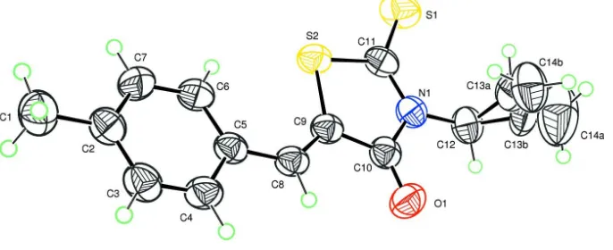
The rhodanine and p-tolyl ring systems (S2—N1—C9—C10—C11 and C2 to C7) are slightly inclined as indicated by
the dihedral angle of 5.51 (12)° between them. The rhodanine moiety is linked to an allyl group at the nitrogen atom and
to a p-tolyl group at C(5) as shown in Fig.1. Moreover, the molecule of the title compound is characterized by a disorder
in the allyl group in which all atoms are split with an occupancy factors of 0.559 (9) : 0.441 (9). No specific
inter-molecular interactions are observed in the crystal packing.
S2. Synthesis and crystallization
To a solution of 3-allylrhodanine (1.15 mmol, 0.2 g) in 10 ml of THF,
(4-methylbenzylidene)-4-methyl-5-oxopyrazolidin-2-ium-1-ide (1.38 mmol) was added. The mixture was refluxed for 8 h, monitored by TLC, the reaction
completed and a yellow spot (TLC Rf = 0.3, using hexane/ethyl acetate 1:9) was generated cleanly. The solvent was
evaporated in vacuo. The crude product was purified on silica gel using hexane: ethyl acetate (1/9) as eluent. The title
compound was recrystallized from ethanol (Yield: 75%; m.p.: 390 K).
S3. Refinement
Crystal data, data collection and structure refinement details are summarized in Table 1. The reflection (0 0 1) affected by
the beamstop was removed during refinement. The refinement of the model requires constraints on the distance C13A—
C14A and C13B—C14B of the disordered allyl group. H atoms were located in a difference map and treated as riding
with C–H = 0.96 Å, C–H = 0.97 Å and C–H = 0.93 Å for methyl, methylene and aromatic hydrogen atoms, respectively.
All hydrogen atoms were refined with a common thermal displacement parameter of Uiso(H) = 1.5 Ueq for methyl and
Figure 1
Plot of the molecule of the title compound with displacement ellipsoids are drawn at the 50% probability level. H atoms
are represented as small circles.
(Z)-3-Allyl-5-(4-methylbenzylidene)-2-sulfanylidene-1,3-thiazolidin- 4-one
Crystal data
C14H13NOS2
Mr = 275.37
Triclinic, P1
a = 7.3606 (4) Å
b = 8.8342 (6) Å
c = 11.3134 (7) Å
α = 109.736 (2)°
β = 95.380 (2)°
γ = 96.502 (2)°
V = 681.10 (7) Å3
Z = 2
F(000) = 288
Dx = 1.343 Mg m−3
Melting point: 390 K
Mo Kα radiation, λ = 0.71073 Å
Cell parameters from 2853 reflections
θ = 2.5–26.6°
µ = 0.38 mm−1
T = 296 K
Block, yellow
0.37 × 0.35 × 0.28 mm
Data collection
Bruker X8 APEX diffractometer
Radiation source: fine-focus sealed tube Graphite monochromator
φ and ω scans
Absorption correction: multi-scan (SADABS; Bruker, 2009)
Tmin = 0.700, Tmax = 0.746
21519 measured reflections 2853 independent reflections 2241 reflections with I > 2σ(I)
Rint = 0.032
θmax = 26.6°, θmin = 2.5°
h = −9→9
k = −11→11
l = −14→14
Refinement
Refinement on F2
Least-squares matrix: full
R[F2 > 2σ(F2)] = 0.043
wR(F2) = 0.133
S = 1.07
2853 reflections 182 parameters 2 restraints
Hydrogen site location: inferred from neighbouring sites
H-atom parameters constrained
w = 1/[σ2(F
o2) + (0.0651P)2 + 0.2196P]
where P = (Fo2 + 2Fc2)/3
(Δ/σ)max < 0.001
Δρmax = 0.34 e Å−3
supporting information
sup-3 Acta Cryst. (2015). E71, o906–o907
Special details
Geometry. All e.s.d.'s (except the e.s.d. in the dihedral angle between two l.s. planes) are estimated using the full covariance matrix. The cell e.s.d.'s are taken into account individually in the estimation of e.s.d.'s in distances, angles and torsion angles; correlations between e.s.d.'s in cell parameters are only used when they are defined by crystal symmetry. An approximate (isotropic) treatment of cell e.s.d.'s is used for estimating e.s.d.'s involving l.s. planes.
Fractional atomic coordinates and isotropic or equivalent isotropic displacement parameters (Å2)
x y z Uiso*/Ueq Occ. (<1)
C1 0.5130 (4) 0.6507 (3) 1.2642 (3) 0.0784 (7)
H1A 0.3857 0.6024 1.2376 0.118*
H1B 0.5440 0.6723 1.3533 0.118*
H1C 0.5329 0.7509 1.2481 0.118*
C2 0.6328 (3) 0.5359 (3) 1.1916 (2) 0.0591 (5)
C3 0.8224 (3) 0.5758 (3) 1.2117 (2) 0.0644 (6)
H3 0.8773 0.6754 1.2720 0.077*
C4 0.9323 (3) 0.4717 (3) 1.1444 (2) 0.0608 (6)
H4 1.0599 0.5021 1.1607 0.073*
C5 0.8563 (3) 0.3212 (2) 1.05205 (18) 0.0507 (5)
C6 0.6656 (3) 0.2794 (3) 1.0338 (2) 0.0604 (5)
H6 0.6103 0.1791 0.9747 0.073*
C7 0.5571 (3) 0.3847 (3) 1.1022 (2) 0.0659 (6)
H7 0.4296 0.3536 1.0882 0.079*
C8 0.9804 (3) 0.2217 (3) 0.98157 (19) 0.0530 (5)
H8 1.1048 0.2647 1.0078 0.064*
C9 0.9485 (3) 0.0782 (3) 0.88491 (19) 0.0518 (5)
C10 1.1041 (3) 0.0018 (3) 0.8283 (2) 0.0552 (5)
C11 0.8509 (3) −0.1882 (3) 0.7007 (2) 0.0594 (5)
C12 1.1670 (4) −0.2379 (3) 0.6539 (2) 0.0695 (6)
H12A 1.1258 −0.3537 0.6306 0.083*
H12B 1.2900 −0.2102 0.7018 0.083*
C13A 1.1640 (13) −0.1898 (15) 0.5393 (11) 0.090 (3) 0.441 (10)
H13A 1.0526 −0.2181 0.4852 0.108* 0.441 (10)
C14A 1.3042 (16) −0.1105 (14) 0.5064 (12) 0.147 (5) 0.441 (10)
H14A 1.4185 −0.0794 0.5573 0.177* 0.441 (10)
H14B 1.2876 −0.0863 0.4326 0.177* 0.441 (10)
C13B 1.2460 (12) −0.1897 (10) 0.5554 (8) 0.077 (2) 0.559 (10)
H13B 1.3415 −0.2408 0.5196 0.092* 0.559 (10)
C14B 1.1877 (12) −0.0760 (8) 0.5147 (6) 0.101 (3) 0.559 (10)
H14C 1.0923 −0.0231 0.5491 0.121* 0.559 (10)
H14D 1.2423 −0.0497 0.4519 0.121* 0.559 (10)
N1 1.0378 (3) −0.1426 (2) 0.72653 (17) 0.0560 (4)
O1 1.2661 (2) 0.0540 (2) 0.86169 (17) 0.0734 (5)
S1 0.73489 (11) −0.35069 (9) 0.59005 (7) 0.0874 (3)
Atomic displacement parameters (Å2)
U11 U22 U33 U12 U13 U23
C1 0.0840 (18) 0.0719 (16) 0.0752 (17) 0.0164 (14) 0.0191 (14) 0.0170 (14)
C2 0.0674 (13) 0.0592 (13) 0.0517 (12) 0.0074 (10) 0.0086 (10) 0.0217 (10)
C3 0.0730 (15) 0.0523 (12) 0.0566 (12) −0.0057 (11) 0.0068 (11) 0.0102 (10)
C4 0.0523 (11) 0.0603 (13) 0.0602 (13) −0.0082 (10) 0.0000 (10) 0.0165 (11)
C5 0.0541 (11) 0.0526 (11) 0.0445 (10) −0.0004 (9) 0.0024 (8) 0.0200 (9)
C6 0.0567 (12) 0.0552 (12) 0.0564 (12) −0.0027 (10) −0.0022 (10) 0.0093 (10)
C7 0.0497 (12) 0.0697 (14) 0.0679 (14) 0.0011 (10) 0.0030 (10) 0.0149 (12)
C8 0.0503 (11) 0.0537 (11) 0.0523 (11) −0.0027 (9) 0.0008 (9) 0.0201 (9)
C9 0.0527 (11) 0.0529 (11) 0.0502 (11) 0.0019 (9) 0.0022 (9) 0.0218 (9)
C10 0.0592 (12) 0.0566 (12) 0.0521 (11) 0.0056 (10) 0.0077 (9) 0.0231 (10)
C11 0.0710 (14) 0.0527 (12) 0.0531 (12) 0.0057 (10) −0.0007 (10) 0.0204 (10)
C12 0.0810 (16) 0.0628 (14) 0.0660 (14) 0.0176 (12) 0.0197 (12) 0.0197 (12)
C13A 0.078 (6) 0.123 (8) 0.077 (5) 0.049 (7) 0.023 (5) 0.030 (5)
C14A 0.171 (11) 0.176 (11) 0.163 (10) 0.100 (10) 0.091 (9) 0.106 (9)
C13B 0.064 (4) 0.102 (4) 0.081 (4) 0.033 (4) 0.036 (4) 0.040 (3)
C14B 0.128 (7) 0.105 (5) 0.092 (4) 0.026 (4) 0.043 (4) 0.054 (4)
N1 0.0635 (11) 0.0541 (10) 0.0526 (10) 0.0100 (8) 0.0082 (8) 0.0211 (8)
O1 0.0546 (9) 0.0745 (11) 0.0810 (11) 0.0037 (8) 0.0101 (8) 0.0164 (9)
S1 0.0934 (5) 0.0627 (4) 0.0795 (5) 0.0007 (3) −0.0076 (4) −0.0002 (3)
S2 0.0540 (3) 0.0597 (4) 0.0580 (3) 0.0004 (2) −0.0020 (2) 0.0126 (3)
Geometric parameters (Å, º)
C1—C2 1.502 (3) C10—O1 1.204 (3)
C1—H1A 0.9600 C10—N1 1.397 (3)
C1—H1B 0.9600 C11—N1 1.364 (3)
C1—H1C 0.9600 C11—S1 1.633 (2)
C2—C3 1.378 (3) C11—S2 1.751 (2)
C2—C7 1.390 (3) C12—C13B 1.465 (8)
C3—C4 1.374 (3) C12—N1 1.466 (3)
C3—H3 0.9300 C12—C13A 1.493 (12)
C4—C5 1.399 (3) C12—H12A 0.9700
C4—H4 0.9300 C12—H12B 0.9700
C5—C6 1.388 (3) C13A—C14A 1.3337 (10)
C5—C8 1.445 (3) C13A—H13A 0.9300
C6—C7 1.378 (3) C14A—H14A 0.9300
C6—H6 0.9300 C14A—H14B 0.9300
C7—H7 0.9300 C13B—C14B 1.3331 (10)
C8—C9 1.344 (3) C13B—H13B 0.9300
C8—H8 0.9300 C14B—H14C 0.9300
C9—C10 1.482 (3) C14B—H14D 0.9300
supporting information
sup-5 Acta Cryst. (2015). E71, o906–o907
H1A—C1—H1B 109.5 N1—C10—C9 110.46 (18)
C2—C1—H1C 109.5 N1—C11—S1 127.64 (18)
H1A—C1—H1C 109.5 N1—C11—S2 110.78 (16)
H1B—C1—H1C 109.5 S1—C11—S2 121.58 (15)
C3—C2—C7 117.3 (2) C13B—C12—N1 119.7 (3)
C3—C2—C1 121.3 (2) N1—C12—C13A 103.2 (5)
C7—C2—C1 121.4 (2) N1—C12—H12A 111.1
C4—C3—C2 121.4 (2) C13A—C12—H12A 111.1
C4—C3—H3 119.3 N1—C12—H12B 111.1
C2—C3—H3 119.3 C13A—C12—H12B 111.1
C3—C4—C5 121.4 (2) H12A—C12—H12B 109.1
C3—C4—H4 119.3 C14A—C13A—C12 126.8 (11)
C5—C4—H4 119.3 C14A—C13A—H13A 116.6
C6—C5—C4 117.1 (2) C12—C13A—H13A 116.6
C6—C5—C8 124.73 (19) C13A—C14A—H14A 120.0
C4—C5—C8 118.14 (19) C13A—C14A—H14B 120.0
C7—C6—C5 120.8 (2) H14A—C14A—H14B 120.0
C7—C6—H6 119.6 C14B—C13B—C12 123.2 (7)
C5—C6—H6 119.6 C14B—C13B—H13B 118.4
C6—C7—C2 121.8 (2) C12—C13B—H13B 118.4
C6—C7—H7 119.1 C13B—C14B—H14C 120.0
C2—C7—H7 119.1 C13B—C14B—H14D 120.0
C9—C8—C5 131.6 (2) H14C—C14B—H14D 120.0
C9—C8—H8 114.2 C11—N1—C10 116.69 (19)
C5—C8—H8 114.2 C11—N1—C12 123.1 (2)
C8—C9—C10 120.57 (19) C10—N1—C12 120.2 (2)
C8—C9—S2 130.12 (17) C9—S2—C11 92.73 (10)