Bilberry (Vaccinium myrtillus L.) fruit is well-known as a source of antioxidants, including polyphenolic compounds (Abby et al. 2013). Antiviral (Sekizawa et al. 2012), antibacterial (Raudsepp et al. 2013), anticholinesterase (Szwajgier & Borowiec 2012; Borowiec et al. 2014), and antidiabetic (Törrönen et al. 2013) effects of bilberry extracts have also been well documented. A phenolic extract from bilberry has been shown to inhibit proliferation of the colon cancer cells (Aaby et al. 2013). Anthocyanidins isolated from bilberry have inhibited the growth of two aggressive non-small-cell lung cancer cell lines (Kausar et al. 2012). Regular consumption of bilberry reportedly reduces low-grade inflammation (Kolehmainen et al. 2012) and decreases lipoprotein levels (Madihi et al. 2013).
The majority of the available studies are concen-trated on bilberry extracts. In this study, a bilberry (BB) preparation obtained by juice processing and ultrafiltration (molecular weight cutoff < 5 kDa) was used. Specific cell lines were selected to examine the influence of BB on the digestive system, as these are the cells that are exposed to high concentrations of compounds derived from food. The IEC-6 (normal rat small intestine epithelial) cells were chosen as a model of the small intestine. Considering the func-tionality of the Caco-2 (human epithelial colorectal adenocarcinoma) cells in the evaluation of passive transport of drugs and food compounds (Balimane & Chong 2005), this cell line was also used as these cells are morphologically and physiologically similar to normal enterocytes. The HepG2 (human
hepa-Effects of a Bilberry Preparation on Selected Cell Lines
of the Digestive System
Kamila Borowiec 1, Dominik Szwajgier 1, anna olejniK 2, Katarzyna KowalSKa2 and zdzisław TargońSKi 1
1Department of Biotechnology, Human nutrition and Science of Food commodities, Faculty
of Food Science and Biotechnology, University of life Sciences, lublin, Poland; 2Department of Biotechnology and Food Microbiology, Faculty of Food Science and nutrition Poznań
University of life Sciences, Poznań, Poland
Abstract
Borowiec K., Szwajgier D., Olejnik A., Kowalska K., Targoński Z. (2016): Effects of a bilberry prepara-tion on selected cell lines of the digestive system. Czech J. Food Sci., 34: 300–305.
Bilberry is a valuable wild forest fruit harvested in many countries in Europe. The biological activities of bilberry include antioxidant, anticancer, antiviral, antibacterial, and anticholinesterase activities. This study examines the protective effects of a bilberry (BB) preparation on IEC-6, Caco-2, and HepG2 cell lines. The 3-(4,5-dimethylthiazol-2-yl)-2,5-diphenyltetrazolium bromide assay was used to study the cytotoxicity of BB. The genotoxicity was determined using single-cell microgel electrophoresis. The Ames test was employed to assay bilberry mutagenicity. No significant effects of BB (12.5–100 µg dry mass/ml) were observed on the viability of IEC-6, Caco-2, and HepG2 cells. There were no differences in the percentage of DNA in the comet tail between the cells treated with BB (100 µg dry mass/ml) and the control cells. However, a significant reduction of oxidative DNA damage in the HepG2 cells was found. BB exhibited neither mutagenic nor promutagenic effects. Our results suggest that bilberry can be a potential tool in the prevention of chronic diseases, without any undesired effects on the cells of the gastrointestinal tract.
Keywords: Vaccinium myrtillus L.; cytotoxicity; functional food; genotoxicity; mutagenicity
tocellular liver carcinoma) cells represented the metabolic function of the liver as a common tool in the study of the metabolism of many compounds (Wilkening & Bader 2003).
MATERIAL AND METHODS
Bilberry preparation. Test samples of fresh bil-berry fruit in harvest maturity were purchased from a local market and frozen (–20°C) until use. Fruits were harvested in forests in the Lublin province in June 2011. The identity of the fruit samples was confirmed by Prof Kazimierz Głowniak (Medical University, Department of Pharmacognosy with Medicinal Plant Laboratory, Lublin, Poland).
Bilberry juice was obtained using a commercial juice extractor (500 g for 15 min, Thermomix TM31; Vorwerk, Wuppertal, Germany) followed by cen-trifugation (4°C, 13 131 g, 30 min). The supernatant solution was ultrafiltered (4°C, Vivaflow 50, 5 kDa PES membrane, Masterflex L/S Economy Drive pump; Cole-Parmer Instrument Co., Vernon Hills, USA). The ultrafiltrate solution was freeze dried (–50°C, Labconco FreeZone 2.5; Labconco, Fort Scott, USA) and frozen (–20°C) until use.
Cell cultures. The IEC-6, Caco-2, and HepG2 cell lines were purchased from the European Collection of Animal Cell Cultures (Sigma-Aldrich, Steinheim, Germany). The cells were cultured in Dulbecco’s modification of Eagle’s medium supplemented with nonessential amino acid solution (1 : 100, v/v), fetal bovine serum (1 : 10, v/v), and 50 mg/ml gentamicin solution (1 : 1000, v/v). In the case of IEC-6 cells, 10 mg/ml insulin (1 : 2000, v/v) was also added. Cells were maintained at 37°C, at 98% relative humidity, in the presence of 5% CO2 and 95% air.
BB was dissolved in the complete media prepared as described above. The concentration of BB was 5 mg dry mass – DM/ml (pH 7.2). The bilberry medium solution was centrifuged (18°C, 3000 g, 10 min) and filtered (0.22 µm low-protein-binding syringe filters; Merck Millipore, Darmstadt, Germany).
Cytotoxicity assay. The cells were grown in 96-well plates at an initial density of 2.5 × 10⁴ cells/cm2. The 24-h cultures were treated with BB at concentrations ranging from 12.5 to 400 µg DM/ml and incubated for 48 h under standard culture conditions.
Cytotoxicity was determined using the 3-(4,5-di-methylthiazol-2-yl)-2,5-diphenyltetrazolium bromide (MTT) reduction assay (Mosmann 1983) according
to the previously described procedure (Olejnik et al. 2016). Briefly, after treatment of the cells with BB, the MTT solution (0.5 mg/ml) was added to each well and the microplate was incubated at 37°C for 3 hours. Acidic isopropanol was added to each well at the end of the incubation period. The absorbance was measured at 570 nm using a Tecan M200 Infinite microplate reader (Tecan Group Ltd., Männedorf, Switzerland). Viability of the cells treated with BB was expressed as % absorbance relative to untreated cells. The test was performed in triplicate.
Genotoxicity assay. The potential DNA damage in the IEC-6, Caco-2, and the HepG2 cells induced following the 48-h treatment with BB (100 µg DM/ml) was determined using the method described by Hart-mann et al. (2003) with modifications proposed by Olejnik et al. (2010). Moreover, the effect of BB on the oxidative DNA damage induced by 100µM H2O2 (37°C, 30 min) was also investigated. The single-cell gel electrophoresis method was used to detect DNA damage. The slides with single cells were placed in the lysis mixture (100 mM EDTA, 5.5 M NaCl, 10 mM Tris, 1% Triton X-100, pH 10.0, 4°C, 1 h), washed and incubated in electrophoresis buffer. Electrophoresis was conducted for 40 min at 300 mA. After staining with SYBRGold (Molecular Probes, Inc., Eugene, USA), 80 comets per slide were analysed using an inverted fluorescence microscope (Axiovert 200 Zeiss; Carl Zeiss AG, Göttingen, Germany). Data were expressed as the mean percentage of DNA in the comet tail using CometScoreTM software (TriTek Corp., Sumerduck, USA). At least three slides per experimental condition were analysed.
the reaction mixtures containing the reference pro-mutagen or BB. PBS was added to a total volume of 1 ml. After 48-h incubation at 37°C, bacterial colonies were counted. Based on the obtained re-sults, the mutagenic activity of BB and mutagens was calculated according to the following equation: AM = (Ri − Rs)/Rs, where Ri stands for the number of revertants induced by standard mutagen or by the analysed extract and Rs is the number of spontaneous revertants in the negative control. A doubling of the mutagenic activity was considered to be significant. Based on the value of AM, 2-AA, 2-AF, AZS, and 9-AA have been recognised as reference mutagens similar to other mutagenic compounds with AM ≥ 2. The experiments were performed in triplicate.
Statistical analysis. All data are expressed as mean ± SD. Data were assessed by analysis of variance and mean comparisons were performed according to Tukey’s test using STATISTICA 6.0 (StatSoft, Kraków, Poland). A significance value of P < 0.05 was used to distinguish significant differences.
RESULTS AND DISCUSSION
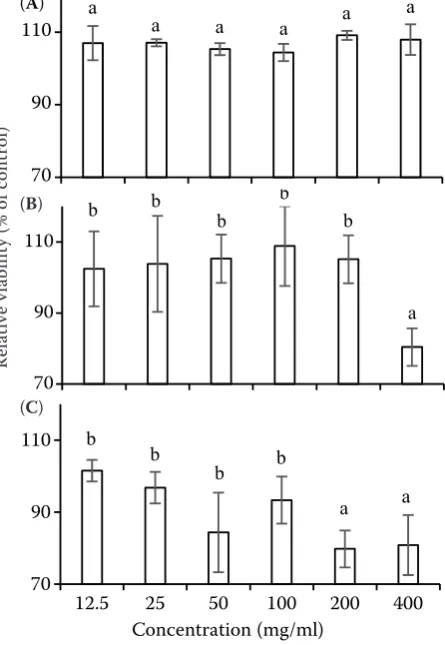
To see any potential cytotoxic effect of BB on the IEC-6, Caco-2, and HepG2 cells, the tetrazolium reduction rate was measured in the MTT assay. No significant influence (P < 0.05) of BB at 12.5–400 µg DM/ml was observed in terms of the cell proliferation and viability of the IEC-6 cells (Figure 1A). The viability of the Caco-2 and HepG2 cells was only decreased (by approximately 20%) by BB at 200 and 400 µg DM/ml (Figures 1B and 1C). However, such high concentra-tions (higher than 100 µg/ml) of BB are unlikely to be observed in vivo. Our results broaden our knowledge of the bilberry cytotoxicity. Admittedly, Schantz et al. (2010) reported that the bilberry extract was not cytotoxic towards the Caco-2 cells in the alamar-Blue assay (10–500 µg DM/ml, 24 h). In contrast, Aaby et al. (2013) proved that the bilberry extract (75, 125, and 250 mg of gallic acid equivalents/ml, 24 h) inhibited the proliferation of the Caco-2 cells in the MTT test. Then, a daily intake of bilberry extract (100 and 400 ppm extract in drinking water) was also recommended to suppress the hepatocar-cinogenic process in piperonyl butoxide-promoted hepatocarcinogenesis in rats (Hara et al. 2014). Last but not least, Valentová et al. (2007) showed no cytotoxic effect of the bilberry concentrate in the MTT assay (100 and 500 µg DM/ml; 4, 24, or 48 h)
on primary cultures of rat hepatocytes. However, the present work has been the first attempt to examine the potential cytotoxicity of the whole bilberry fruit, not just an anthocyanin-rich bilberry extract. At the present moment, no data are available concerning the effect of either bilberry (or its preparation) or of any plant from the genus Vaccinium on the viability of the IEC-6 cells.
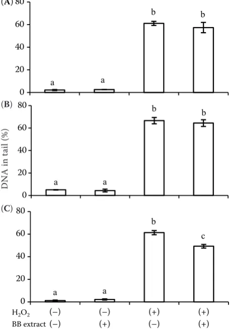
A genotoxic effect of BB on the IEC-6, Caco-2, and HepG2 cells is presented in Figure 2. There were no differences (P < 0.05) in the percentage of DNA in the comet tail between cells treated with BB (100 µg DM/ml, 48 h) and control cells. No effect of BB on oxidative DNA damage caused by 100 µM H2O2 on the Caco-2 and IEC-6 cells was observed. In contrast, a protective effect of BB on the HepG2 cells exposed to oxidative stress was observed. In this case, a significant reduction (P < 0.05) of about 12% in oxidative DNA damage was observed (Figure 2C). Therefore, it is proposed that BB has a protective
ef-70 90 110
12.5 25 50 100 200 400
Re la tiv e ce ll vi ab ili ty (% o f c on tr ol ) Concentration (mg/ml) a
a a a a a
70 90 110
12.5 25 50 100 200 400
Re la tiv e ce ll vi ab ili ty (% of c on tr ol ) Concentration (mg/ml) a a b b b b 70 90 110
12.5 25 50 100 200 400
[image:3.595.308.531.110.433.2]Re la tiv e ce ll vi ab ili ty (% o f c ont ro l) Concentration (mg/ml) a b b b b b
Figure 1. Effect of bilberry on the IEC-6 (A), Caco-2 (B), and the HepG2 (C) cell viability
Mean values ± SD (n = 3); bars with the same letter are not significantly different (P < 0.05)
Re la tiv e vi ab ili ty (% o f c on tr ol )
(A)
(B)
bilberry fruit on the selected cells. Valentová et al. (2007) also reported that the bilberry extract showed a significant (P < 0.01) dose-dependent (100 and 500 µg DM/ml) protective activity against oxidative damage (induced by tert-butyl hydroperoxide and allyl alcohol) in primary cultures of rat hepatocytes. Additionally, Schantz et al. (2010) observed a sig-nificant (P < 0.05) reduction of menadione-induced DNA damage (6 µM, 1 h) in the Caco-2 cells after pretreatment with a bilberry extract (5 and 50 µg/ml, 24 h). Likewise, Juadjur et al. (2015) demonstrated that the oxidative DNA damage in the Caco-2 cells was significantly (P < 0.05) reduced in the presence of a commercially available bilberry extract (5 and 10 µg/ml, 1 or 24 h).
The potential mutagenic properties of BB were determined by the Ames test. The effect of reverse mutation in altered Salmonella typhimurium strains (unable to synthesise histidine) was tested. In the present study, BB (100–300 µg DM/ml) did not ex-ert any mutagenic or promutagenic effects on all strains used (Table 1). There were no differences in the number of revertants induced by BB com-pared to spontaneous revertants. Additionally, the mutagenic activity was estimated to be lower than or equal to 0.5, when the mutagenic activity of the reference mutagens and 2-AA was higher than 3. The only study which focused on mutagenicity of bilberry was the test that examined Myrtocyan®, a
commercial bilberry extract (Seifried et al. 2006). In the cited work, the authors showed that Myrtocyan®
[image:4.595.63.292.110.438.2](100–10000 µg/plate) caused no mutagenic lesions in Salmonella typhimurium TA98, TA100, TA1535, TA1537, and TA1538.
Table 1. Effect of bilberry on mutagenesis in TA98, TA100, TA1535, and TA1537 strains, with (+S9) or without (−S9) metabolic activation
Bacteria
strain Metabolic activation (+S9)/(−S9) Spontaneously induced revertants (CFU/plate) Induced revertants (CFU/plate)mutagen BB mutagenMutagenic activityBB
TA98 −+ 8 ± 216 ± 5 107 ± 1175 ± 6 12 ± 510 ± 2 12.43.7 −0.40.5
TA100 −+ 37 ± 555 ± 7 327 ± 25280 ± 37 58 ± 1029 ± 7 7.84.1 −0.20.1
TA1535 −+ 13 ± 210 ± 4 76 ± 947 ± 3 11 ± 312 ± 4 4.83.7 −0.20.2
TA1537 −+ 27 ± 412 ± 3 216 ± 17 54 ± 12 30 ± 6 8 ± 4 7.03.5 0.10.3
Data were expressed as the mean number of revertants (± SD); mutations were induced by the mutagens: 2-AF (TA98), AZS (TA100, TA1535), 9-AA (TA1537), and the promutagen 2-AA (TA98, TA100, TA1535, TA1537)
0 20 40 60 80
(–) (-)
(–) (+)
(+) (-)
(+) (+)
D
N
A
in
ta
il
(%)
H2O2 BB extract
a a
b b
0 20 40 60 80
(-) (-)
(-) (+)
(+) (-)
(+) (+)
D
N
A
in t
ai
l (%)
H2O2…
a a
b b
0 20 40 60 80
(–)
(–) (–) (+) (+) (–) (+)(+)
D
N
A
in
ta
il
(%)
H2O2
BB extract
c
a a
[image:4.595.64.532.590.732.2]b
Figure 2. Effect of bilberry on DNA damage in the IEC-6 (A), Caco-2 (B), and HepG2 (C) cells untreated (−) or treated (+) with H2O2
Mean values ± SD (n = 3); bars with same letter are not sig-nificantly different (P < 0.05) when compared to the control
D
N
A
in
ta
il
(%
)
fect against oxidative damage in hepatocytes. There are no reports on potential genotoxicity of the whole (A)
(B)
CONCLUSION
In summary, our work suggests that a diet sup-plemented with bilberry fruit can be considered a potential tool in the combined therapy of many diseases, without any undesired effects on the cells present in the gastrointestinal tract. However, more extensive in vitro or in vivo assays should be carried out. We demonstrated that BB exerted no cytotoxic, genotoxic, or mutagenic effects on the Caco-2, IEC-6, and HepG2 cells. At the same time, the reduction of oxidative damage in the HepG2 cells should be stressed. Our results confirmed findings of other authors, however, unlike most of the authors, we deliberately examined potential protective effects of the whole bilberry fruit. This work has evaluated the influence of the whole bilberry fruit on the IEC-6 cells for the first time, which is important because the IEC-6 cells come into direct physical contact with a very complex mixture of food components in the intestine.
References
Aaby K., Grimmer S., Holtung L. (2013): Extraction of phenolic compounds from bilberry (Vaccinium myrtillus L.) press residue: Effects on phenolic composition and cell proliferation. LWT-Food Science and Technology, 54: 257–264.
Balimane P.V., Chong S. (2005): Cell culture-based models for intestinal permeability: a critique. Drug Discovery Today, 10: 335–343.
Borowiec K., Szwajgier D., Targoński Z., Demchuk O.M., Cybulska J., Czernecki T., Malik A. (2014): Cholinesterase inhibitors isolated from bilberry fruit. Journal of Func-tional Foods, 11: 313–321.
Cyplik P., Marecik R., Piotrowska-Cyplik A., Olejnik A., Drożdżyńska A., Chrzanowski Ł. (2012): Biological deni-trification of high nitrate processing wastewaters from explosives production plant. Water, Air and Soil Pollu-tion, 223: 1791–1800.
Hara S., Morita R., Ogawa T., Segawa R., Takimoto N., Su-zuki K., Hamadate N., Hayashi S.M., Odachi A., Ogiwara I., Shibusawa S., Yoshida T., Shibutani M. (2014): Tumor suppression effects of bilberry extracts and enzymatically modified isoquercitrin in early preneoplastic liver cell lesions induced by piperonyl butoxide promotion in a two-stage rat hepatocarcinogenesis model. Experimental and Toxicologic Pathology, 66: 225–234.
Hartmann A., Agurell E., Beevers C., Brendler-Schwaab S., Burlinson B., Clay P., Collins A., Smith A., Speit G.,
Thybaud V., Tice R.R. (2003): Recommendations for con-ducting the in vivo alkaline Comet assay. 4th International
Comet Assay Workshop. Mutagenesis, 18: 45–51. Juadjur A., Mohn C., Schantz M., Baum M., Winterhalter P.,
Richling E. (2015): Fractionation of an anthocyanin-rich bilberry extract and in vitro antioxidative activity testing. Food Chemistry, 167: 418–424.
Kausar H., Jeyabalan J., Aqil F., Chabba D., Sidana J., Singh I.P., Gupta R.C. (2012): Berry anthocyanidins synergistical-ly suppress growth and invasive potential of human non-small-cell lung cancer cells. Cancer Letters, 325: 54–62. Kolehmainen M., Mykkänen O., Kirjavainen P.V., Leppänen
T., Moilanen E., Adriaens M., Laaksonen D.E., Hallikain-en M., PuupponHallikain-en-Pimiä R., PulkkinHallikain-en L., MykkänHallikain-en H., Gylling H., Poutanen K., Törrönen R. (2012): Bilber-ries reduce low-grade inflammation in individuals with features of metabolic syndrome. Molecular Nutrition & Food Research, 56: 1501–1510.
Madihi Y., Merrikhi A., Baradaran A., Ghobadi S., Shahin-fard N., Ansari R., Karimi A., Mesripour A., Rafieian-Kopaei M. (2013): Bioactive components and the effect of hydroalcoholic extract of Vacciniummyrtillus on post-prandial atherosclerosis risk factors in rabbits. Pakistan Journal of Medical Sciences, 29: 384–389.
Maron D.M., Ames B.N. (1983): Revised methods for the Salmonella mutagenicity test. Mutation Research, 113: 173–215.
Mosmann T. (1983): Rapid colorimetric assay for estimation for cellular growth and survival: application to prolifera-tion and cytotoxicity assays. Journal of Immunological Methods, 65: 55–63.
Olejnik A.M., Marecik R., Białas W., Cyplik P., Grajek W. (2010): in vitro studies on atrazine effects on human in-testinal cells. Water, Air and Soil Pollution, 213: 401–411. Olejnik A., Rychlik J., Kidoń M., Czapski J., Kowalska K.,
Juzwa W., Olkowicz M., Dembczyński R., Moyer M.P. (2016): Antioxidant effects of gastrointestinal digested purple carrot extract on the human cells of colonic mu-cosa. Food Chemistry, 190: 1069–1077.
Raudsepp P., Anton D., Roasto M., Meremäe K., Pedastsaar P., Mäesaar M., Raal A., Laikoja K., Püssa T. (2013): The antioxi-dative and antimicrobial properties of the blue honeysuckle (lonicera caerulea L.), Siberian rhubarb (rheum rhaponti-cum L.) and some other plants, compared to ascorbic acid and sodium nitrite. Food Control, 31: 129–135.
Schantz M., Mohn C., Baum M., Richling E. (2010): Antioxi-dative efficiency of an anthocyanin rich bilberry extract in the human colon tumor cell lines Caco-2 and HT-29. Journal of Berry Research, 1: 25–33.
muta-genicity test results with the Ames Salmonella typhimu-rium and L5178Y mouse lymphoma cell mutation assays. Chemical Research in Toxicology, 19: 627–644.
Sekizawa H., Ikuta K., Mizuta K., Takechia S., Suzutani T. (2013): Relationship between polyphenol content and anti-influenza viral effects of berries. Journal of the Sci-ence of Food and Agriculture,93: 2239–2241.
Szwajgier D., Borowiec K. (2012): Screening for cholinester-ase inhibitors in selected fruits and vegetables. Electronic Journal of Polish Agricultural Universities, 15, #06. Törrönen R., Kolehmainen M., Sarkkinen E., Poutanen
K., Mykkänen H., Niskanen L. (2013): Berries reduce postprandial insulin responses to wheat and rye breads in healthy women. Journal of Nutrition, 143: 430–436.
Valentová K., Ulrichová J., Cvak L., Šimánek V. (2007): Cytoprotective effect of a bilberry extract against oxida-tive damage of rat hepatocytes. Food Chemistry, 101: 912–917.
Wilkening S., Bader A. (2003): Influence of culture time on the expression of drug-metabolising enzymes in pri-mary human hepatocytes and hepatoma cell line HepG2. Journal of Biochemical and Molecular Toxicology, 17: 207–213.
Received: 2015–07–28 Accepted after corrections: 2016–07–13
corresponding author: