Received 18 September 2019 Accepted 30 September 2019
Edited by M. Weil, Vienna University of Technology, Austria
Keywords:crystal structure; imidazole-pyridine derivative;–interactions; DFT calculation; Hirshfeld surface analysis; energy framework; frontier molecular orbitals.
CCDC reference:1914069
Supporting information:this article has supporting information at journals.iucr.org/e
Crystal structure, DFT calculation, Hirshfeld
surface analysis and energy framework study of
6-bromo-2-(4-bromophenyl)imidazo[1,2-
a
]pyridine
Hussien Ahmed Khamees,aKumara Chaluvaiah,bNasseem Ahmed El-khatatneh,a Ananda Swamynayaka,aKwong Huey Chong,cJagadeesh Prasad Dasappaband Mahendra Madegowdaa*
a
Department of Studies in Physics, Manasagangotri, University of Mysore, Mysuru 570 006, Karnataka, India, bDepartment of Chemistry, Mangalore University, Mangalagangothri, Mangaluru 574 199, Karnataka, India, and cDepartment of Chemistry, Faculty of Science, Universiti Putra Malaysia 43400, UPM Serdang, Selangor Darul Ehsan,
Malaysia. *Correspondence e-mail: [email protected]
The title imidazo[1,2-a] pyridine derivative, C13H8Br2N2, was synthesizedviaa
single-step reaction method. The title molecule is planar, showing a dihedral angle of 0.62 (17)between the phenyl and the imidazo[1,2-a] pyridine rings. An intramolecular C—H N hydrogen bond with anS(5) ring motif is present. In the crystal, a short H H contact links adjacent molecules into inversion-related dimers. The dimers are linked in turn by weak C—H and slipped–
stacking interactions, forming layers parallel to (110). The layers are connected into a three-dimensional network by short Br H contacts. Two-dimensional fingerprint plots and three-Two-dimensional Hirshfeld surface analysis of the intermolecular contacts reveal that the most important contributions for the crystal packing are from H Br/Br H (26.1%), H H (21.7%), H C/ C H (21.3%) and C C (6.5%) interactions. Energy framework calculations suggest that the contacts formed between molecules are largely dispersive in nature. Analysis of HOMO–LUMO energies from a DFT calculation reveals the purecharacter of the aromatic rings with the highest electron density on the phenyl ring, and character of the electron density on the Br atoms. The HOMO–LUMO gap was found to be 4.343 eV.
1. Chemical context
Five-membered heterocyclic compounds comprising a nitro-gen atom and at least one other non-carbon atom (i.e. nitro-gen, sulfur, or oxygen) as part of the ring are known as azoles. To date, numerous azoles have found a wide range of appli-cations in various fields, including agriculture (Berger et al., 2017), and because of their biological activities (Pozharskiiet al., 2011; Kumbaret al., 2018). Among the various classes of azoles, the imidazole moiety with two nitrogen atoms is extremely common in nature and forms the core of many biomolecules (Chopra & Sahu, 2019) and synthetic drugs (Pozharskii et al., 2011). Furthermore, pyridine and its deri-vatives are present in many important compounds, including pharmaceuticals, vitamins (Al-Ghorbani et al., 2016) and drugs, acting as antimicrobial, antiviral, antioxidants, anti-diabetic, anti-malarial, anti-inflammatory or antiamoebic agents, as well as psychopharmacological antagonists (Altafet al., 2015). Hence, the combination of pyridine and imidazole derivatives has been proven to result in highly active agents in
diverse biological fields that include anticancer (Kamalet al., 2014; Mantu et al., 2016), anti-HIV (Bodeet al., 2011), anti-bacterial (Rivalet al., 1992) and anti-inflammatory (Rupertet al., 2003) properties. In addition, such a combination showed significant activity against the humancytomegalovirus and the
varicella-zostervirus (Gueiffieret al., 1998; Mavelet al., 2002).
In this context, we synthesized a new imidazo[1,2-a] pyri-dine derivative, C13H8Br2N2, and report herein its molecular
and crystal structure, as well as the quantification of supra-molecular interactions by Hirshfeld surface analysis. This study is supplemented by DFT calculations and a comparison of structural details with related compounds.
2. Structural commentary
The molecular structure of the title compound is depicted in Fig. 1. The molecular system is planar, showing a dihedral angle of 0.62 (17)between the phenyl ring (C1–C6) and the imidazo[1,2-a] pyridine ring system (C7–C13,N1,N2). The torsion angles about the terminal bromine atoms, Br1 and Br2, are 177.3 (3) (Br1—C1—C6—C5) and 178.9 (4) (Br2—
C11—C12—C13), respectively. The planar arrangement between the two rings enables an intramolecular C—H N interaction (Fig. 1, Table 1) forming anS(5) ring motif (Tan & Tiekink, 2019). The Br1—C1 and Br2—C11 bond lengths are 1.886 (4) A˚ and 1.880 (4) A˚, respectively, in good agreement with structures comprising bromophenyl moieties (Zhang & Hu, 2005; Arif Tawfeeq et al., 2019). The N1 C9 bond is slightly longer than similar bonds of reported imidazo[1,2-a] pyridine structures (see x7 for a listing of these structures), which may be attributed to the presence of the intramolecular bond (H5 N1). Overall, the bond lengths and angles of the phenyl ring and the imidazo[1,2-a]pyridine ring system are in normal ranges and compare well with those of other imidazo[1,2-a]pyridine derivatives (Zhang et al., 2005; Dhanalakshmiet al., 2018).
3. Supramolecular features
The crystal packing is mainly based on short contacts and weak–interactions, similar to reported structures with the same kind of terminal bromine atoms (Arif Tawfeeq et al., 2019). In the title compound, two inversion-related molecules are linked by a short H5 H5(1x, 2y,z) contact (Fig. 2). These dimers are connected to each other through C—H
interactions (Table 1), forming sheets propagating parallel to (110). Slipped–stacking interactions [Cg3 Cg1(x, 1y,
z) = 3.655 (2) A˚ , slippage of 0.885 A˚;Cg3 Cg2(x, 1y,
z) = 3.819 (2) A˚ , slippage of 1.473 A˚], whereCg1, Cg2 and
Cg3 are the centroids of the imidazole, pyridine and phenyl rings, respectively, are also present within these sheets (Fig. 2). Adjacent sheets are linked along [001] into a three-dimen-sional network through short contacts of 3.01 A˚ between Br1 and H12(x,3
2y, 1
2+z), formingS(11) chain motifs (Fig. 3).
Table 1
Hydrogen-bond geometry (A˚ ,).
Cg3 is the centroid of C1–C6 ring
D—H A D—H H A D A D—H A
C5—H5 N1 0.93 2.47 2.827 (5) 103
C3—H3 Cg3i 0.93 2.91 3.5670 (1) 129
Symmetry code: (i)x;y3 2;z
[image:2.610.68.273.160.216.2] [image:2.610.315.564.428.677.2]1 2.
Figure 2
[image:2.610.46.298.612.699.2]The crystal packing of the title compound in a view along [001], showing interactions in the sheets. H5 H5 short contacts are represented as blue dashed lines, C3—H3 Cg3 interactions as red dashed lines (slippage 1.676 A˚ ) and Cg3 Cg1 and Cg3 Cg2 interactions as light-green dashed lines.
Figure 1
4. DFT study and FMOs
Density functional theory (DFT) calculations were carried out by using the B3LYP basis set (Becke, 1993) at the highest basis set level of 6-311 ++G(d,p) in the GAUSSIAN09 program (Frischet al., 2009). The DFT-optimized structure of the title compound is generally found to be in good agreement with the experimental data for all bond lengths and angles.
Frontier molecular orbitals (FMOs) are useful to specify the distribution of electronic densities and other quantum chemical parameters including hardness (), softness (), chemical potential (), electrophilicity ( ) and electro-negativity () by foreseeing the highest occupied molecular orbitals (HOMO) and the lowest-unoccupied molecular
orbitals (LUMO), as well as the energy gap (Eg =EH -EL)
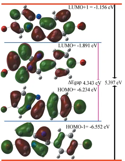
(Khamees et al., 2018). The results of these calculations are compiled in Table 2, and orbital energy plots of (EH,EH-1) and
(EL,EL+1) are depicted in Fig. 4. The HOMO (ground state)
manifests the highestcharacterization for phenyl ring (C1– C6) that displays bifurcated–stacking interactions as well as C—H interactions in the supramolecular network, as discussed in Section 3. Pronouncedcharacter of the electron density is located on the two Br atoms, with the higher amount located on Br1. The other FMOs orbitals, i.e. HOMO-1, LUMO and LUMO+1, exhibit a mix ofandcharacter on the rings with variations of the electron density distribution (Fig. 4). The HOMO–LUMO gap is 4.343 eV for the title compound.
5. Hirshfeld surface analysis
The nature of intermolecular interactions in the title compound has been computed byCrystalExplorer17.5(Turner
et al., 2017), using Hirshfeld surface analysis (Spackman & Jayatilaka, 2009) and two-dimensional fingerprint plots (McKinnon et al., 2007). The dnorm plot was estimated via
calculations of the external (de) and internal (di) distances to
[image:3.610.45.294.71.212.2]the nearest nucleus and built over the volume of 363.34 A˚3and an area of 339.81 A˚2, with scaled colour of0.1544 (red) a.u. to 1.0479 (blue) a.u. (Fig. 5a). The plots of shape-index and curvedness were generated in the range of4.0 to 4.0 a.u. and
Table 2
HUMO–LUMO energies and values of quantum chemical parameters (eV).
Property Symbol and formula Value
HOMO energy EH(eV) 6.234
LUMO energy EL(eV) 1.891
HOMO-1 energy EH-1(eV) 6.552
LUMO+1 energy EL+1(eV) 1.156
Energy gap 1 Eg1= (EH-EL) (eV) 4.343
Energy gap 2 Eg2= (EH-1-EL+1) (eV) 5.397
Global hardness = (EL-EH)/2 2.172
Softness = 1/ 2 0.230
Chemical potential = (EL+EH)/2 4.062
Electrophilicity =2
/2 3.799
[image:3.610.315.566.102.223.2]Electronegativity = - 4.062
Figure 4
Electron distribution and molecular orbital energies of HOMO-1, HOMO, LUMO and LUMO+1 of the title compound.
Figure 3
[image:3.610.68.274.443.708.2]The three-dimensional supramolecular network of the title compound viewed approximately along [010].
Figure 5
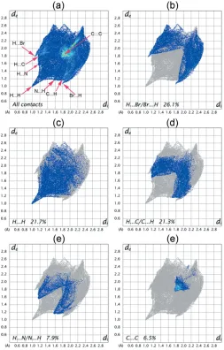
[image:3.610.318.566.581.706.2]1.00 to 1.00 a.u., respectively, (Fig. 5b,c). The medium dark and side pale-red spots on the Hirshfeld surface (Fig. 5a) symbolize the H5 H5 and Br1 H12 short contacts, respectively. The two-dimensional fingerprint plot for all contacts is depicted in Fig. 6a. The H Br/Br H contacts make the largest contribution (26.1%) to the Hirshfeld surface (Fig. 6b). These contacts also make a significant contribution to the crystal packing as the distance between the atoms involved is slightly less than their van der Waals radii (di+de
’ 3.01 A˚ ). The interatomic contacts of H H interactions generated 22.7% of the Hirshfeld surface (Fig. 6c), showing a short spike at diagonal axesdi+de’2.24 A˚ < 2.4 A˚, denoting
H H short contacts with another significant effect on the molecular packing. The two symmetrical broad wings in Fig. 6d
belong to H C/C H contacts that represent 21.3% of total surface and indicate the presence of C—H interactions in the crystal packing, where di + de ’ 2.77 A˚ < 2.90 A˚. The
proportion of H N/N H contacts is 7.9% of the Hirshfeld surface (Fig. 6e) and they appear as two close wings pointing at
a distance greater than the van der Waals radii of N and H atoms (di + de > 2.75A˚ ), with no significant contribution
towards the crystal packing of the title molecule. The small contribution of the C C contacts (6.5%) to the Hirshfeld surface appears as an intense triangle (Fig. 6f) atdi+ de ’
3.6 A˚ , indicating – stacking interactions in the crystal packing. This type of stacking interaction appears as a flat region on the curvedness (Fig. 5c) and also on the shape-index as red and blue triangles on the rings (Fig. 5b), in particular on the phenyl ring (C1–C6). The contributions from other contacts have negligible effects on the packing.
6. Energy framework
[image:4.610.45.298.316.712.2]Quantification of energy framework energies is considered a powerful method for understanding the topology of the overall interactions of molecules in the crystal. This method allowed us to calculate and compare different energy components,i.e.repulsion (E_rep), electric (E_ele), dispersion (E_dis), polarization (E_pol) and total (E_tot) energy based on the anisotropy of the topology of pairwise intermolecular interaction energies.CrystalExplorer17.5(Turneret al., 2017) was used to calculate the energy framework of the title compound by generating new wave functions using the DFT method under 3-21G basis set with exchange and potential functions (B3LYP) for a molecular cluster environment for a 111 unit cell. The thickness of the cylinder radius indicates the grade of interactions and is directly related to the energy magnitude and offers information about the stabilization of the crystal packing. In order to avoid the crowdedness of less significant interaction energies, we set the cylindrical radii with a cut-off value of 5 kJ mol1 and a scale factor of 50 to all
Figure 7
Energy framework of the title molecules viewed along [001], showing: (a) electrostatic, (b) dispersion, (c) total energy force diagrams and (d) the details of interaction with colour-coded, symmetry operation (Symop) and distances between molecular centroids (R) in A˚ .
Figure 6
[image:4.610.315.564.476.700.2]energy components. The benchmarked energies were scaled according to Mackenzieet al.(2017) whileE_rep,E_ele,E_dis and E_pol were scaled as 0.618, 1.057, 0.740, 0.871, respec-tively (Edwards et al., 2017). The results of the calculations revealed that dispersion interactions exhibit approximately chair-shaped energy topologies through the rings, having a maximum energy value of 180.558 kJ mol1 (Fig. 7). The other energy components have values of 62.232 kJ mol1,
29.38 kJ mol1 and9.176 kJ mol1for repulsion, electro-static and polarization energies, respectively. The small value of electrostatic energy is attributed to the absence of classical hydrogen bonds. The total interaction energy that resulted from all four main components is156.886 kJ mol1(Fig. 7d).
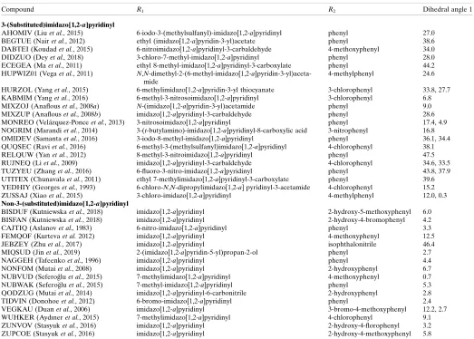
7. Database survey
36 structures containing the 2-phenylimidazo[1,2-a]pyridine moiety with different substituents were found in a search of the Cambridge Structural Database (CSD, version 5.40, last update May 2019; Groomet al., 2016). The different
substit-uentsR1(on the imidazo[1,2-a]pyridinyl ring) andR2(on the
phenyl ring) together with the dihedral angles between the mean planes of the corresponding imidazo[1,2-a]pyridinyl and phenyl rings (dihedral angle 1) are compiled in Table 3. By comparing the substitution positions, the structures can be divided into ‘3-(substituted)imidazo[1,2-a]pyridinyl’ compounds and ‘non-3-(substituted)imidazo[1,2-a]pyridinyl’ compounds. In general, the 3-(substituted)imidazo[1,2-a ]-pyridinyl compounds have a greater dihedral angle 1 values (12.0–47.5). This may arise from steric repulsion between the
3-(substituted) group and the phenyl ring. However, there are four outliers (KABMIM, MIXZOJ, MONREO and ZUSSAJ) whose dihedral angle 1 values are lower than 10. Most of the non-3-(substituted)imidazo[1,2-a]pyridinyl compounds have dihedral angle 1 values between 0.7 and 12.5, which indicates
that the imidazo[1,2-a]pyridinyl rings are close to coplanar to their attached phenyl rings. Here, the outlier is JEBZEY where the imidazo[1,2-a]pyridinyl ring is attached to a
[image:5.610.44.567.114.488.2]di-ortho-substituted isophthalonitrile ring. The dihedral angle 1 is 46.4in this structure.
Table 3
Comparison of structural details in related imidazo[1,2-a]pyridinyl derivatives containing phenyl rings.
Dihedral angle 1 is the angle between the mean planes of imidazo[1,2-a]pyridinyl and phenyl rings. Two sets of dihedral angles 1 are stated for compounds HURZOL, MONREO, OMIDEV, RUJNEQ, TUZYEU, ZUSSAJ and VEGKAU because there are two molecules in their asymmetric units.
Compound R1 R2 Dihedral angle 1
3-(Substituted)imidazo[1,2-a]pyridinyl
AHOMIV (Liuet al., 2015) 6-iodo-3-(methylsulfanyl)-imidazo[1,2-a]pyridinyl phenyl 27.0
BEGTUE (Nairet al., 2012) ethyl (imidazo[1,2-a]pyridin-3-yl)acetate phenyl 38.6
DABTEI (Koudadet al., 2015) 6-nitroimidazo[1,2-a]pyridinyl-3-carbaldehyde 4-methoxyphenyl 34.0
DIDZUO (Deyet al., 2018) 3-chloro-7-methyl-imidazo[1,2-a]pyridinyl phenyl 28.0
ECEGEA (Maet al., 2011) ethyl 8-methyl-imidazo[1,2-a]pyridinyl-3-carboxylate phenyl 44.2
HUPWIZ01 (Vegaet al., 2011) N,N-dimethyl-2-(6-methyl-imidazo[1,2-a ]pyridin-3-yl)aceta-mide
4-methylphenyl 24.6
HURZOL (Yanget al., 2015) 6-methylimidazo[1,2-a]pyridin-3-yl thiocyanate 3-chlorophenyl 33.8, 27.7
KABMIM (Yanget al., 2016) 6-methyl-3-nitrosoimidazo[1,2-a]pyridinyl 3-chlorophenyl 6.8
MIXZOJ (Anaflouset al., 2008a) N-(imidazo[1,2-a]pyridin-3-yl)acetamide phenyl 9.0
MIXZUP (Anaflouset al., 2008b) imidazo[1,2-a]pyridinyl-3-carbaldehyde phenyl 28.6
MONREO (Vela´zquez-Ponceet al., 2013) 3-nitrosoimidazo[1,2-a]pyridinyl phenyl 17.4, 4.9
NOGRIM (Marandiet al., 2014) 3-(t-butylamino)-imidazo[1,2-a]pyridinyl-8-carboxylic acid 3-nitrophenyl 16.8
OMIDEV (Samantaet al., 2016) 3-iodo-8-methyl-imidazo[1,2-a]pyridinyl phenyl 36.1, 34.4
QUQSEC (Raviet al., 2016) 6-methyl-3-(methylsulfanyl)imidazo[1,2-a]pyridinyl 4-chlorophenyl 38.1
RELQUW (Yanet al., 2012) 8-methyl-3-nitroimidazo[1,2-a]pyridinyl phenyl 47.5
RUJNEQ (Liet al., 2009) imidazo[1,2-a]pyridinyl-3-carbaldehyde 4-chlorophenyl 34.6, 33.5
TUZYEU (Zhanget al., 2016) 6-fluoro-3-nitro-imidazo[1,2-a]pyridinyl phenyl 43.8, 37.9
UTITEX (Chunavalaet al., 2011) ethyl 7-methylimidazo[1,2-a]pyridinyl-3-carboxylate phenyl 39.6
YEDHIY (Georgeset al., 1993) 6-chloro-N,N-dipropylimidazo[1,2-a] pyridinyl-3-acetamide 4-chlorophenyl 15.2
ZUSSAJ (Xiaoet al., 2015) 3-chloro-imidazo[1,2-a]pyridinyl 4-methylphenyl 12.0, 0.3
Non-3-(substituted)imidazo[1,2-a]pyridinyl
BISDUF (Kutniewskaet al., 2018) imidazo[1,2-a]pyridinyl 2-hydroxy-5-methoxyphenyl 6.0
BISFAN (Kutniewskaet al., 2018) imidazo[1,2-a]pyridinyl 2-hydroxy-4-bromophenyl 4.2
CAJTIQ (Aslanovet al., 1983) 6-nitro-imidazo[1,2-a]pyridinyl phenyl 3.3
FEMQOF (Kurtevaet al.2012) imidazo[1,2-a]pyridinyl 4-methoxyphenyl 12.5
JEBZEY (Zhuet al., 2017) imidazo[1,2-a]pyridinyl isophthalonitrile 46.4
MIQSUD (Jinet al., 2019) 2-(imidazo[1,2-a]pyridin-5-yl)propan-2-ol phenyl 2.7
NAGGEH (Tafeenkoet al., 1996) imidazo[1,2-a]pyridinyl phenyl 4.4
NONFOM (Mutaiet al., 2008) imidazo[1,2-a]pyridinyl 2-hydroxyphenyl 6.7
NUBVUD (Seferog˘luet al., 2015) 7-methylimidazo[1,2-a]pyridinyl 4-methoxyphenyl 0.7
NUBWAK (Seferog˘luet al., 2015) 7-methyl-imidazo[1,2-a]pyridinyl phenyl 5.3
QODZUG (Mutaiet al., 2014) imidazo[1,2-a]pyridinyl-6-carbonitrile 2-hydroxyphenyl 2.8
TIDVIN (Donohoeet al., 2012) 6-bromo-imidazo[1,2-a]pyridinyl phenyl 2.4
VEGKAU (Duanet al., 2006) imidazo[1,2-a]pyridinyl 3-bromo-4-methoxyphenyl 12.2, 2.7
WUHKER (Aydıneret al., 2015) 7-methylimidazo[1,2-a]pyridinyl 4-chlorophenyl 9.1
ZUNVOV (Stasyuket al., 2016) imidazo[1,2-a]pyridinyl 2-hydroxy-4-florophenyl 3.2
8. Synthesis and crystallization
5-Bromopyridin-2-amine (1.211 g, 0.007 mol) and phenacyl bromide (0.007 mol) were refluxed for 14 h in 50 ml of abso-lute ethanol. The progress of the reaction was monitored by thin layer chromatography using Merck alumina backed silica gel 60 F254. After completion of the reaction, the resulting product was poured into crushed ice to obtain a fine grained solid product that was filtered off, separated and dried. The crude product was then recrystallized from hot ethanol with a yield of70%. The melting point of 345 K was determined in an open capillary and is uncorrected. IR (KBr, cm1): 3080 (Ar C—H stretch), 2918 (aliphatic C—H stretch, 4-bromo-phenyl moiety), 1587 (C N stretch), 1332 (C—N), 792 and 595 (C—Br).1H NMR (400 MHz, DMSO,ppm): 7.37 (d, 1H,
5-bromopyridine moiety), 7.55 (d, 2H, 4-bromophenyl moiety), 7.56 (d, 1H, 5-bromopyridine moiety), 7.78 (d, 2H, 4-bromophenyl moiety), 8.37 (s, 1H, imidazole ring), 8.87 (s, 1H, 5-bromopyridine moiety).13C NMR (400 MHz, ppm): 145.13 (imidazopyridine carbon atom), 110.24, 119.82, 125.12 and 132.14 (four carbon atoms of 5-bromopyridine moiety), 123.12, 128.30, 132.11, and 132.32 (six carbon atoms of 4-bromophenyl moiety), 113.13 and 130.10 (two carbon atoms of imidazole ring). LC–Mass m/z 350 [M+], 352 [M+2], 354 [M+4]. Analysis calculated for C13H8Br2N2(350): C, 44.36; H,
2.29; N, 7.96. Found: C, 44.31; H, 2.23; N, 7.92%.
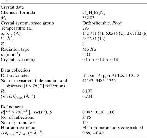
9. Refinement
Crystal data, data collection and structure refinement details are summarized in Table 4. Hydrogen atoms were placed in
calculated positions (C—H = 0.93 A˚ ) and were included in the refinement in the riding-model approximation, with Uiso(H)
set to 1.2Ueq(C). The reflection (002) was affected by the
beam-stop and was removed from the refinement
Acknowledgements
The authors thank the Sophisticated Analytical Instruments Facility (SAIF), IIT Madras, for providing single-crystal and spectroscopic data.
References
Al-Ghorbani, M., Thirusangu, P., Gurupadaswamy, H. D., Girish, V., Shamanth Neralagundi, H. G., Prabhakar, B. T. & Khanum, S. A. (2016).Bioorg. Chem.65, 73–81.
Altaf, A. A., Shahzad, A., Gul, Z., Rasool, N., Badshah, A., Lal, B. & Khan, E. (2015).J. Drug Des. Med. Chem.1, 1–11.
Anaflous, A., Albay, H., Benchat, N., El Bali, B., Dusˇek, M. & Fejfarova´, K. (2008a).Acta Cryst.E64, o926.
Anaflous, A., Albay, H., Benchat, N., El Bali, B., Dusˇek, M. & Fejfarova´, K. (2008b).Acta Cryst.E64, o927.
Arif Tawfeeq, N., Kwong, H. C., Mohamed Tahir, M. I. & Ravoof, T. B. S. A. (2019).Acta Cryst.E75, 774–779.
Aslanov, L. A., Tafeenko, V. A., Paseshnichenko, K. A., Bundel, Y. G., Gromov, S. P. & Gerasimov, B. G. (1983).Zh. Strukt. Khim.24, 115–123.
Aydıner, B., Yalc¸ın, E., Ihmels, H., Arslan, L., Ac¸ık, L. & Seferog˘lu, Z. (2015).J. Photochem. Photobiol. Chem.310, 113–121.
Becke, A. D. (1993).J. Chem. Phys.98, 5648–5652.
Berger, S., El Chazli, Y., Babu, A. F. & Coste, A. T. (2017).Front. Microbiol.8, 1–6.
Bode, M. L., Gravestock, D., Moleele, S. S., van der Westhuyzen, C. W., Pelly, S. C., Steenkamp, P. A., Hoppe, H. C., Khan, T. & Nkabinde, L. A. (2011).Bioorg. Med. Chem.19, 4227–4237. Bruker (2016). APEX2 and SAINT. Bruker AXS Inc., Madison,
Wisconsin, USA.
Chopra, P. N. & Sahu, J. K. (2019).Curr. Drug Discov. Technol.1, 1570–1638.
Chunavala, K. C., Joshi, G., Suresh, E. & Adimurthy, S. (2011). Synthesis,2011, 635–641.
Dey, A., Singsardar, M., Sarkar, R. & Hajra, A. (2018).ACS Omega3, 3513–3521.
Dhanalakshmi, G., Ramanjaneyulu, M., Thennarasu, S. & Aravindhan, S. (2018).Acta Cryst.E74, 1913–1918.
Donohoe, T. J., Kabeshov, M. A., Rathi, A. H. & Smith, I. E. D. (2012).Org. Biomol. Chem.10, 1093–1101.
Duan, G.-Y., Tu, C.-B., Sun, Y.-W., Zhang, D.-T. & Wang, J.-W. (2006). Acta Cryst.E62, o1141–o1142.
Edwards, A. J., Mackenzie, C. F., Spackman, P. R., Jayatilaka, D. & Spackman, M. A. (2017).Faraday Discuss.203, 93–112.
Frisch, M. J., Trucks, G. W., Schlegel, H. B., Scuseria, G. E., Robb, M. A., Cheeseman, J. R.,et al. (2009).GAUSSIAN09. Gaussian Inc., Wallingford, CT, USA.
Georges, G. J., Vercauteren, D. P., Evrard, G. H., Durant, F. V., George, P. G. & Wick, A. E. (1993).Eur. J. Med. Chem.28, 323–335. Groom, C. R., Bruno, I. J., Lightfoot, M. P. & Ward, S. C. (2016).Acta
Cryst.B72, 171–179.
Gueiffier, A., Mavel, S., Lhassani, M., Elhakmaoui, A., Snoeck, R., Andrei, G., Chavignon, O., Teulade, J. C., Witvrouw, M., Balzarini, J., De Clercq, E. & Chapat, J. (1998).J. Med. Chem.41, 5108–5112. Jin, S., Xie, B., Lin, S., Min, C., Deng, R. & Yan, Z. (2019).Org. Lett.
21, 3436–3440.
[image:6.610.44.295.88.324.2]Kamal, A., Reddy, V. S., Santosh, K., Bharath Kumar, G., Shaik, A. B., Mahesh, R., Chourasiya, S. S., Sayeed, I. B. & Kotamraju, S. (2014). Med. Chem. Commun.5, 1718–1723.
Table 4
Experimental details.
Crystal data
Chemical formula C13H8Br2N2
Mr 352.03
Crystal system, space group Orthorhombic,Pbca
Temperature (K) 293
a,b,c(A˚ ) 14.1711 (4), 6.0546 (2), 27.7102 (8)
V(A˚3) 2377.54 (12)
Z 8
Radiation type MoK
(mm1) 6.80
Crystal size (mm) 0.150.140.14
Data collection
Diffractometer Bruker Kappa APEXII CCD
No. of measured, independent and observed [I> 2(I)] reflections
41143, 3485, 1726
Rint 0.100
(sin/)max(A˚
1) 0.704
Refinement
R[F2> 2(F2)],wR(F2),S 0.047, 0.118, 1.00
No. of reflections 3485
No. of parameters 154
H-atom treatment H-atom parameters constrained
max,min(e A˚
3
) 0.88,0.49
Khamees, H. A., Jyothi, M., Khanum, S. A. & Madegowda, M. (2018). J. Mol. Struct.1161, 199–217.
Koudad, M., Elaatiaoui, A., Benchat, N., Saadi, M. & El Ammari, L. (2015).Acta Cryst.E71, o979–o980.
Kumbar, M. N., Kamble, R. R., Dasappa, J. P., Bayannavar, P. K., Khamees, H. A., Mahendra, M., Joshi, S. D., Dodamani, S., Rasal, V. P. & Jalalpure, S. (2018).J. Mol. Struct.1160, 63–72.
Kurteva, V. B., Lubenov, L. A., Nedeltcheva, D. V., Nikolova, R. P. & Shivachev, B. L. (2012).Arkivoc,13, 282–294.
Kutniewska, S. E., Jarzembska, K. N., Kamin´ski, R., Stasyuk, A. J., Gryko, D. T. & Cyran´ski, M. K. (2018).Acta Cryst.B74, 725–737. Li, Y.-H., Liu, W.-Y., Gao, Y. & Wang, Y.-P. (2009).Acta Cryst.E65,
o3192.
Liu, S., Xi, H., Zhang, J., Wu, X., Gao, Q. & Wu, A. (2015).Org. Biomol. Chem.13, 8807–8811.
Ma, L., Wang, X., Yu, W. & Han, B. (2011). Chem. Commun. 47, 11333–11335.
Mackenzie, C. F., Spackman, P. R., Jayatilaka, D. & Spackman, M. A. (2017).IUCrJ,4, 575–587.
Macrae, C. F., Bruno, I. J., Chisholm, J. A., Edgington, P. R., McCabe, P., Pidcock, E., Rodriguez-Monge, L., Taylor, R., van de Streek, J. & Wood, P. A. (2008).J. Appl. Cryst.41, 466–470.
Mantu, D., Antoci, V., Moldoveanu, C., Zbancioc, G. & Mangalagiu, I. I. (2016).J. Enzyme Inhib. Med. Chem.31, 96–103.
Marandi, G., Saghatforoush, L., Mendoza-Meron˜o, R. & Garcı´a-Granda, S. (2014).Tetrahedron Lett.55, 3052–3054.
Mavel, S., Renou, J. L., Galtier, C., Allouchi, H., Snoeck, R., Andrei, G., De Clercq, E., Balzarini, J. & Gueiffier, A. (2002).Bioorg. Med. Chem.10, 941–946.
McKinnon, J. J., Jayatilaka, D. & Spackman, M. A. (2007). Chem. Commun.pp. 3814–3816.
Mutai, T., Shono, H., Shigemitsu, Y. & Araki, K. (2014). CrystEng-Comm,16, 3890–3895.
Mutai, T., Tomoda, H., Ohkawa, T., Yabe, Y. & Araki, K. (2008). Angew. Chem. Int. Ed.47, 9522–9524.
Nair, D. K., Mobin, S. M. & Namboothiri, I. N. N. (2012).Org. Lett.14, 4580–4583.
Pozharskii, A. F., Soldatenkov, A. T. & Katritzky, A. R. (2011). Heterocycles in Life and Society. John Wiley & Sons.
Ravi, C., Chandra Mohan, D. & Adimurthy, S. (2016).Org. Biomol. Chem.14, 2282–2290.
Rival, Y., Grassy, G. & Michel, G. (1992).Chem. Pharm. Bull. 40, 1170–1176.
Rupert, K. C., Henry, J. R., Dodd, J. H., Wadsworth, S. A., Cavender, D. E., Olini, G. C., Fahmy, B. & Siekierka, J. J. (2003).Bioorg. Med. Chem. Lett.13, 347–350.
Samanta, S., Jana, S., Mondal, S., Monir, K., Chandra, S. K. & Hajra, A. (2016).Org. Biomol. Chem.14, 5073–5078.
Seferog˘lu, Z., Ihmels, H. & S¸ahin, E. (2015).Dyes Pigments,113, 465– 473.
Sheldrick, G. M. (2015a).Acta Cryst.A71, 3–8. Sheldrick, G. M. (2015b).Acta Cryst.C71, 3–8.
Spackman, M. A. & Jayatilaka, D. (2009).CrystEngComm,11, 19–32. Spek, A. L. (2009).Acta Cryst.D65, 148–155.
Stasyuk, A. J., Bultinck, P., Gryko, D. T. & Cyran´ski, M. K. (2016).J. Photochem. Photobiol. Chem.314, 198–213.
Tafeenko, V., Paseshnichenko, K. & Schenk, H. (1996).Z. Kristallogr. 211, 457–263.
Tan, S. L. & Tiekink, E. R. T. (2019).Acta Cryst.E75, 1–7.
Turner, M. J., McKinnon, J. J., Wolff, S. K., Grimwood, D. J., Spackman, P. R., Jayatilaka, D. & Spackman, M. A. (2017). CrystalExplorer17. University of Western Australia. http://hirsh-feldsurface. net.
Vega, D. R., Baggio, R., Roca, M. & Tombari, D. (2011).J. Pharm. Sci. 100, 1377–1386.
Vela´zquez-Ponce, M., Salgado-Zamora, H., Jime´nez-Va´zquez, H. A., Campos-Aldrete, M. E., Jime´nez, R., Cervantes, H. & Hadda, T. B. (2013).Chem. Cent. J.7: 20.
Westrip, S. P. (2010).J. Appl. Cryst.43, 920–925.
Xiao, X., Xie, Y., Bai, S., Deng, Y., Jiang, H. & Zeng, W. (2015).Org. Lett.17, 3998–4001.
Yan, R.-L., Yan, H., Ma, C., Ren, Z.-Y., Gao, X.-A., Huang, G.-S. & Liang, Y.-M. (2012).J. Org. Chem.77, 2024–2028.
Yang, D., Yan, K., Wei, W., Li, G., Lu, S., Zhao, C., Tian, L. & Wang, H. (2015).J. Org. Chem.80, 11073–11079.
Yang, D., Yan, K., Wei, W., Liu, Y., Zhang, M., Zhao, C., Tian, L. & Wang, H. (2016).Synthesis,48, 122–130.
Zhang, M., Lu, J., Zhang, J.-N. & Zhang, Z.-H. (2016). Catal. Commun.78, 26–32.
Zhang, R. & Hu, Y. (2005).Acta Cryst.E61, o4037–o4038.
sup-1
Acta Cryst. (2019). E75, 1620-1626
supporting information
Acta Cryst. (2019). E75, 1620-1626 [https://doi.org/10.1107/S2056989019013410]
Crystal structure, DFT calculation, Hirshfeld surface analysis and energy
framework study of 6-bromo-2-(4-bromophenyl)imidazo[1,2-
a
]pyridine
Hussien Ahmed Khamees, Kumara Chaluvaiah, Nasseem Ahmed El-khatatneh, Ananda
Swamynayaka, Kwong Huey Chong, Jagadeesh Prasad Dasappa and Mahendra Madegowda
Computing details
Data collection: APEX2 (Bruker, 2016); cell refinement: SAINT (Bruker, 2016); data reduction: SAINT (Bruker, 2016);
program(s) used to solve structure: SHELXT (Sheldrick, 2015a); program(s) used to refine structure: SHELXL
(Sheldrick, 2015b); molecular graphics: Mercury (Macrae et al., 2008) and PLATON (Spek, 2009); software used to
prepare material for publication: publCIF (Westrip, 2010).
6-Bromo-2-(4-bromophenyl)imidazo[1,2-a]pyridine
Crystal data
C13H8Br2N2 Mr = 352.03
Orthorhombic, Pbca a = 14.1711 (4) Å b = 6.0546 (2) Å c = 27.7102 (8) Å V = 2377.54 (12) Å3 Z = 8
F(000) = 1360
Dx = 1.967 Mg m−3
Mo Kα radiation, λ = 0.71073 Å Cell parameters from 3485 reflections θ = 2.1–30.1°
µ = 6.80 mm−1 T = 293 K Block, colourless 0.15 × 0.14 × 0.14 mm
Data collection
Bruker Kappa APEXII CCD diffractometer
ω and φ scan
41143 measured reflections 3485 independent reflections 1726 reflections with I > 2σ(I)
Rint = 0.100
θmax = 30.1°, θmin = 2.1° h = −19→19
k = −8→8 l = −39→38
Refinement
Refinement on F2
Least-squares matrix: full R[F2 > 2σ(F2)] = 0.047 wR(F2) = 0.118 S = 1.00 3485 reflections 154 parameters 0 restraints
Hydrogen site location: inferred from neighbouring sites
H-atom parameters constrained w = 1/[σ2(F
o2) + (0.0442P)2 + 2.6166P]
where P = (Fo2 + 2Fc2)/3
(Δ/σ)max = 0.002
Δρmax = 0.88 e Å−3
sup-2
Acta Cryst. (2019). E75, 1620-1626
Special details
Geometry. All esds (except the esd in the dihedral angle between two l.s. planes) are estimated using the full covariance matrix. The cell esds are taken into account individually in the estimation of esds in distances, angles and torsion angles; correlations between esds in cell parameters are only used when they are defined by crystal symmetry. An approximate (isotropic) treatment of cell esds is used for estimating esds involving l.s. planes.
Fractional atomic coordinates and isotropic or equivalent isotropic displacement parameters (Å2)
x y z Uiso*/Ueq
Br2 0.15325 (4) 0.05914 (9) 0.27691 (2) 0.0700 (2) Br1 0.12673 (4) 0.88905 (10) 0.68410 (2) 0.0740 (2) N2 0.1377 (2) 0.3517 (6) 0.40975 (12) 0.0449 (8) N1 0.0885 (2) 0.6735 (6) 0.44091 (12) 0.0484 (9) C7 0.1238 (3) 0.5400 (6) 0.47646 (14) 0.0401 (9) C1 0.1234 (3) 0.7762 (8) 0.62067 (15) 0.0482 (11) C11 0.1297 (3) 0.2554 (7) 0.32808 (15) 0.0498 (11) C4 0.1241 (3) 0.6173 (7) 0.52687 (15) 0.0424 (9) C12 0.0872 (3) 0.4597 (8) 0.31737 (16) 0.0571 (12)
H12 0.070603 0.493692 0.285760 0.069*
C8 0.1549 (3) 0.3438 (8) 0.45819 (15) 0.0479 (10)
H8 0.182324 0.228190 0.475266 0.058*
C3 0.1597 (3) 0.4933 (7) 0.56424 (16) 0.0495 (10)
H3 0.185335 0.355055 0.557750 0.059*
C6 0.0866 (3) 0.9064 (7) 0.58414 (16) 0.0539 (11)
H6 0.061410 1.044944 0.590752 0.065*
C13 0.0707 (3) 0.6058 (8) 0.35300 (15) 0.0561 (12)
H13 0.041977 0.740195 0.346048 0.067*
C2 0.1582 (3) 0.5687 (8) 0.61068 (16) 0.0532 (11)
H2 0.180652 0.480001 0.635558 0.064*
C5 0.0885 (3) 0.8244 (7) 0.53745 (15) 0.0479 (10)
H5 0.065063 0.911265 0.512485 0.057*
C9 0.0968 (3) 0.5559 (7) 0.40055 (15) 0.0442 (10) C10 0.1555 (3) 0.2005 (7) 0.37352 (15) 0.0492 (11)
H10 0.184202 0.065712 0.380142 0.059*
Atomic displacement parameters (Å2)
U11 U22 U33 U12 U13 U23
sup-3
Acta Cryst. (2019). E75, 1620-1626
C3 0.046 (2) 0.045 (2) 0.058 (3) 0.008 (2) −0.001 (2) 0.008 (2) C6 0.051 (3) 0.049 (3) 0.062 (3) 0.003 (2) 0.004 (2) −0.001 (2) C13 0.067 (3) 0.057 (3) 0.044 (2) 0.009 (2) 0.005 (2) 0.008 (2) C2 0.047 (3) 0.066 (3) 0.046 (2) 0.007 (2) −0.0042 (19) 0.008 (2) C5 0.044 (2) 0.051 (3) 0.048 (2) 0.006 (2) −0.0076 (19) 0.013 (2) C9 0.044 (2) 0.041 (2) 0.048 (2) 0.0035 (19) 0.0013 (18) 0.0050 (19) C10 0.047 (3) 0.042 (2) 0.059 (3) 0.002 (2) 0.002 (2) 0.000 (2)
Geometric parameters (Å, º)
Br2—C11 1.880 (4) C4—C5 1.383 (6)
Br1—C1 1.886 (4) C12—C13 1.346 (6)
N2—C8 1.365 (5) C12—H12 0.9300
N2—C10 1.382 (5) C8—H8 0.9300
N2—C9 1.389 (5) C3—C2 1.366 (6)
N1—C9 1.331 (5) C3—H3 0.9300
N1—C7 1.369 (5) C6—C5 1.386 (6)
C7—C8 1.364 (6) C6—H6 0.9300
C7—C4 1.473 (6) C13—C9 1.401 (6)
C1—C2 1.378 (6) C13—H13 0.9300
C1—C6 1.385 (6) C2—H2 0.9300
C11—C10 1.353 (6) C5—H5 0.9300
C11—C12 1.407 (6) C10—H10 0.9300
C4—C3 1.375 (6)
C8—N2—C10 131.2 (4) C2—C3—C4 121.4 (4)
C8—N2—C9 106.6 (3) C2—C3—H3 119.3
C10—N2—C9 122.2 (4) C4—C3—H3 119.3
C9—N1—C7 104.8 (3) C1—C6—C5 118.1 (4)
C8—C7—N1 111.4 (4) C1—C6—H6 120.9
C8—C7—C4 128.9 (4) C5—C6—H6 120.9
N1—C7—C4 119.7 (4) C12—C13—C9 120.1 (4)
C2—C1—C6 120.5 (4) C12—C13—H13 119.9
C2—C1—Br1 120.6 (3) C9—C13—H13 119.9
C6—C1—Br1 119.0 (4) C3—C2—C1 120.0 (4)
C10—C11—C12 121.9 (4) C3—C2—H2 120.0
C10—C11—Br2 119.9 (3) C1—C2—H2 120.0
C12—C11—Br2 118.2 (3) C4—C5—C6 122.0 (4)
C3—C4—C5 118.0 (4) C4—C5—H5 119.0
C3—C4—C7 122.8 (4) C6—C5—H5 119.0
C5—C4—C7 119.2 (4) N1—C9—N2 111.0 (3)
C13—C12—C11 119.8 (4) N1—C9—C13 130.6 (4)
C13—C12—H12 120.1 N2—C9—C13 118.3 (4)
C11—C12—H12 120.1 C11—C10—N2 117.6 (4)
C7—C8—N2 106.1 (4) C11—C10—H10 121.2
C7—C8—H8 127.0 N2—C10—H10 121.2
sup-4
Acta Cryst. (2019). E75, 1620-1626
C9—N1—C7—C8 0.9 (5) C6—C1—C2—C3 2.4 (7)
C9—N1—C7—C4 −178.9 (4) Br1—C1—C2—C3 −176.7 (3)
C8—C7—C4—C3 1.1 (6) C3—C4—C5—C6 −1.0 (6)
N1—C7—C4—C3 −179.1 (4) C7—C4—C5—C6 179.9 (4)
C8—C7—C4—C5 −179.8 (4) C1—C6—C5—C4 1.2 (6)
N1—C7—C4—C5 0.0 (6) C7—N1—C9—N2 −0.7 (4)
C10—C11—C12—C13 −0.5 (7) C7—N1—C9—C13 178.7 (5)
Br2—C11—C12—C13 −178.9 (4) C8—N2—C9—N1 0.3 (5)
N1—C7—C8—N2 −0.8 (5) C10—N2—C9—N1 −178.8 (4)
C4—C7—C8—N2 179.0 (4) C8—N2—C9—C13 −179.2 (4)
C10—N2—C8—C7 179.3 (4) C10—N2—C9—C13 1.7 (6)
C9—N2—C8—C7 0.3 (4) C12—C13—C9—N1 179.3 (5)
C5—C4—C3—C2 1.5 (6) C12—C13—C9—N2 −1.4 (7)
C7—C4—C3—C2 −179.4 (4) C12—C11—C10—N2 0.8 (6)
C2—C1—C6—C5 −1.8 (6) Br2—C11—C10—N2 179.2 (3)
Br1—C1—C6—C5 177.3 (3) C8—N2—C10—C11 179.8 (4)
C11—C12—C13—C9 0.8 (7) C9—N2—C10—C11 −1.4 (6)
C4—C3—C2—C1 −2.2 (7)
Hydrogen-bond geometry (Å, º)
Cg3 is the centroid of C1–C6 ring
D—H···A D—H H···A D···A D—H···A
C5—H5···N1 0.93 2.47 2.827 (5) 103
C3—H3···Cg3i 0.93 2.91 3.5670 (1) 129
Symmetry code: (i) x, −y−3/2, z−1/2.
Summary of short interatomic contacts (Å)
Contact Distance symmetry
H5···H5 2.24 1-x,2-y,-z
![Figure 1The crystal packing of the title compound in a view along [001], showing](https://thumb-us.123doks.com/thumbv2/123dok_us/378212.535250/2.610.46.298.612.699/figure-the-crystal-packing-title-compound-view-showing.webp)
![Crystal structure and Hirshfeld surface analysis of 2 [(2 oxo 2H chromen 4 yl)oxy]acetic acid dimethyl sulfoxide monosolvate](data:image/gif;base64,R0lGODlhAQABAIAAAP///wAAACH5BAEAAAAALAAAAAABAAEAAAICRAEAOw==)