Received 24 April 2019 Accepted 10 May 2019
Edited by A. V. Yatsenko, Moscow State University, Russia
Keywords:crystal structure; Schiff base; hydrogen bonding; Hirshfeld surface analysis.
CCDC reference:1915380
Supporting information:this article has supporting information at journals.iucr.org/e
Crystal structure and Hirshfeld surface analysis of
(
Z
)-6-[(2-hydroxy-5-nitroanilino)methylidene]-4-methylcyclohexa-2,4-dien-1-one
Sevgi Kansiz,a* Necmi Dege,bAlev Sema Aydin,bErbil Ag˘arcand Igor P. Matushkod*
aOndokuz Mayıs University, Faculty of Arts and Sciences, Department of Physics, 55139, Kurupelit, Samsun, Turkey, bOndokuz Mayıs University, Faculty of Arts and Sciences, Department of Physics, 55139, Samsun, Turkey,cOndokuz Mayıs University, Faculty of Arts and Sciences, Department of Chemistry, 55139, Samsun, Turkey, anddTaras Shevchenko National University of Kyiv, Department of Chemistry, 64, Vladimirska Str., Kiev 01601, Ukraine. *Correspondence e-mail: [email protected], [email protected]
The title compound, C14H12N2O4, is a Schiff base that exists in the keto–enamine
tautomeric form and adopts aZconfiguration. The molecule is almost planar, the rings making a dihedral angle of 4.99 (7). The molecular structure is stabilized by an intramolecular N—H O hydrogen bond forming anS(6) ring motif. In the crystal, inversion-related molecules are linked by pairs of O— H O hydrogen bonds, forming dimers with anR22(18) ring motif. The dimers
are linked by pairs of C—H O contacts with an R22(10) ring motif, forming
ribbons extended along the [210] direction. Hirshfeld surface analysis, two-dimensional fingerprint plots and the molecular electrostatic potential surfaces were used to analyse the intermolecular interactions present in the crystal, indicating that the most important contributions for the crystal packing are from H H (33.9%), O H/H O (29.8%) and C H/H C (17.3%) interactions.
1. Chemical context
Compounds containing theRHC NRfragment, obtained by the condensation reaction of primary amines with aldehydes or ketones under proper conditions, are named Schiff bases after Hugo Schiff (Schiff, 1864). Schiff bases have a wide variety of applications in many areas such as analytical, biological, and inorganic chemistry (Jainet al., 2008; Lozieret al., 1975; Calligaris & Randaccio, 1987). Many Schiff bases are biologically active and some bases show phototochromism which can be used for radiation intensity measurements, display systems or optical devices (Hadjoudis et al., 1987). In the present study, a new Schiff base, (Z )-6-[(2-hydroxy-5-nitroanilino)methylidene]-4-methylcyclohexa-2,4-dien-1-one, was obtained in crystalline form from the reaction of 2-amino-4-nitrophenol with 2-hydroxy-5-methylbenzaldehyde. We report here the synthesis and the crystal and molecular structures of the title compound along with the results of a Hirshfeld surface analysis.
2. Structural commentary
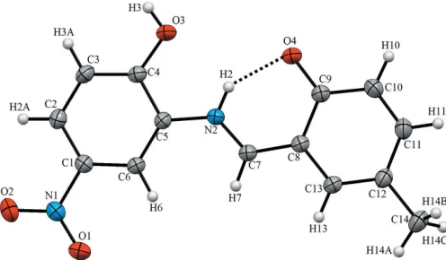
Fig. 1 illustrates the molecular structure of the title compound. Its asymmetric unit contains one independent molecule, which adopts the keto–enamine tautomeric form. The molecule is almost planar, the C1–C6 and C8–C13 rings making a dihedral angle of 4.99 (7). The O4 C9, C9—C8, C8 C7, C7—N2 and N2—C5 bond lengths are typical of double and single bonds, respectively (Table 1), thus indicating that the title molecule exists as a keto–enamine tautomer (Kansiz et al., 2018). The bond lengths at the N1 atom are typical of aromatic nitro groups. The molecular structure is stabilized by the intramolecular N—H O hydrogen bond involving the keto O4 and amine N2 atoms (Fig. 1, Table 2).
3. Supramolecular features
The most important intermolecular interactions in the title structure are the medium–strong O3—H3 O4i hydrogen bonds, which link inversion-related molecules into dimers with an R22(18) ring motif (Table 2). These dimers are further
connected by pairs of weak C—H O hydrogen bonds with anR2
2(10) ring motif to form ribbons extended along the [210]
direction (Fig. 2).
4. Database survey
A search of the Cambridge Structural Database (CSD, version 5.40, update November 2018; Groomet al., 2016) for the 2-[(2-hydroxyphenyliminio)methyl]phenolate fragment revealed 25 hits where this fragment adopts the keto–enamine tautomeric
form. The enamine (N2—C7) and keto (C9—O4) bond lengths in the title compound are the same within standard uncertainties as the corresponding bond lengths in the struc-tures of 2-{[(2-hydroxyphenyl)iminio]methyl}-4-methoxy-phenolate (BALGUR02; Makalet al., 2011), 4-bromo-2-{[(2-hydroxy-5-methylphenyl)iminio]methyl}phenolate (EYUSIC; Takjooet al., 2017), 2-hydroxy-6-{[(2-hydroxyphenyl)iminio]-methyl}phenolate methanol solvate (HEKSIC; Ezeorahet al., 2018) and 2-{(E )-[(5-bromo-2-hydroxyphenyl)methylidene]-amino}-4-chlorophenol (SEFKUL; Ebrahimipouret al., 2012). In the structures of these typical keto–enamine tautomers, the bonds corresponding to C7—C8 in the title structure are distinctly longer, being in the range of 1.416–1.423 A˚ . As for the C9—O4 bond, its length compares well with 1.286 (2) A˚ for HEKSIC and 1.291 (2) A˚ for SEFKUL, but this bond is shorter than 1.298 (2) A˚ for EYUSIC and 1.310 (2) A˚ for BALGUR02. It is likely that the intermolecular O—H O hydrogen bond, where the keto O atom acts as a hydrogen-bond acceptor, is an important prerequisite for the tautomeric shift toward the keto–enamine form. In fact, in all 25 struc-tures of the keto–enamine tautomers, hydrogen bonds of this type are observed.
5. Hirshfeld surface analysis
The Hirshfeld surface analysis (Spackman & Jayatilaka, 2009) and the associated two-dimensional fingerprint plots (McKinnon et al., 2007) were performed with Crystal-Explorer17(Turneret al., 2017). The Hirshfeld surfaces were mapped with different properties namely,dnorm, electrostatic
potential,diandde (Fig. 3). The Hirshfeld surfaces mapped
overdnormutilize the function of normalized distancesdeand di, wheredeanddiare the distances from a given point on the
research communications
Acta Cryst.(2019). E75, 812–815 Kansizet al. C
14H12N2O4
813
Table 1Selected bond lengths (A˚ ).
O3—C4 1.3329 (14) O2—N1 1.2213 (15)
O4—C9 1.2887 (15) N1—C1 1.4530 (15)
N2—C7 1.3070 (15) C7—C8 1.4054 (16)
N2—C5 1.4035 (15) C8—C9 1.4340 (15)
O1—N1 1.2202 (15)
Table 2
Hydrogen-bond geometry (A˚ ,).
D—H A D—H H A D A D—H A
N2—H2 O4 0.86 1.86 2.5661 (13) 139
O3—H3 O4i 0.82 1.77 2.5465 (12) 156
C6—H6 O1ii 0.93 2.51 3.4383 (16) 172
C7—H7 O1ii 0.93 2.40 3.3182 (14) 172
[image:2.610.46.294.553.703.2]Symmetry codes: (i)xþ2;y;zþ1; (ii)x;yþ1;zþ1.
Figure 2
A view of the crystal packing of the title compound. Dashed lines denote the intermolecular C—H O and O—H O hydrogen bonds forming dimers withR2
2(10) andR22(18) ring motifs (Table 1). Figure 1
[image:2.610.312.565.596.706.2]surface to the nearest atom outside and inside, respectively. The blue, white and red colour conventions used for thednorm
-mapped Hirshfeld surfaces recognize long intermolecular contacts, the contacts at the van der Waals separations, and short intermolecular contacts, respectively. The red region is apparent around the keto oxygen atom (O4) participating in
the O—H O and N—H O contacts mentioned above
(Fig. 3, Table 2). Fig. 4 illustrates the Hirshfeld surface of the molecule in the crystal, with the evident hydrogen-bonding interactions indicated as intense red spots.
The two-dimensional fingerprint plot derived from a Hirshfeld surface provides a convenient visual summary of the frequency of each combination ofdeanddiacross the surface
of a molecule. A fingerprint plot delineated into specific interatomic contacts contains information related to specific intermolecular interactions (Tanet al., 2019). The blue colour refers to the frequency of occurrence of the (di,de) pairs with
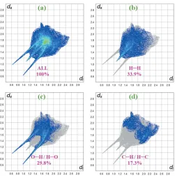
the full fingerprint outlined in grey. Fig. 5a shows the two-dimensional fingerprint of the sum of the contacts contributing to the Hirshfeld surface represented in normal mode. Indivi-dual fingerprint plots (Fig. 5b) reveal that the H H contacts clearly make the most significant contribution to the Hirshfeld surface (33.9%). It is usually the case that the main contri-bution to the overall surface arises from H H contacts. In
addition, O H/H O and C H/H C contacts contribute 29.8% and 17.3%, respectively, to the Hirshfeld surface. In particular, the O H/H O and C H/H C contacts indi-cate the presence of intermolecular O—H O and C—H O interactions, respectively. Much weaker C O/O C (6.8%) and C C (4.8%) contacts also occur.
The view of the electrostatic potential obtained using
CrystalExplorer17enables the visualization of the donors and acceptors of intermolecular interactions through blue and red regions around the participating atoms corresponding to positive and negative electrostatic potential, respectively, on
Figure 4
[image:3.610.315.566.67.319.2]dnormmapped on the Hirshfeld surfaces to visualize the intermolecular interactions for the title compound.
Figure 5
[image:3.610.45.294.70.232.2] [image:3.610.316.563.538.716.2]Two-dimensional fingerprint plots for the title compound, with adnorm view and the relative contributions of the atom pairs to the Hirshfeld surface.
Figure 6
A view of the molecular electrostatic potential of the title compound in the range0.0500 to 0.0500 a.u. calculated at the HF/STO-3 G level. Figure 3
[image:3.610.45.300.602.719.2]the surface. The view of the electrostatic potential in the range
0.0500 to 0.0500 a.u., calculated for the title compound at the HF/STO-3G level, is shown in Fig. 6. The acceptors for N— H O and O—H O hydrogen bonds are shown as red areas around the O4 atom related with negative electrostatic potentials (Fig. 6).
6. Synthesis and crystallization
The title compound was prepared by mixing the solutions of 2-hydroxy-5-methylbenzaldehyde (34.0 mg, 0.25 mmol) in ethanol (15 ml) and 2-amino-4-nitrophenol (38.5 mg, 0.25 mmol) in ethanol (15 ml) with subsequent stirring for 5 h under reflux. Single crystals of the title compound suitable for X-ray analysis were obtained by slow evaporation of an ethanol solution (yield 65%, m.p. 523–525 K).
7. Refinement
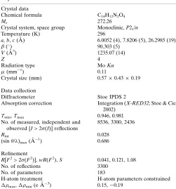
Crystal data, data collection and structure refinement details are summarized in Table 3. The hydroxy H atom was located
in a difference-Fourier map, and the OH group was allowed to rotate during the refinement procedure (AFIX 147). The C-bound H atoms were positioned geometrically and refined using a riding model: C—H = 0.93 A˚ withUiso(H) = 1.2Ueq(C)
for aromatic H atoms and C—H = 0.96 A˚ with Uiso(H) =
1.5Ueq(C) for methyl H atoms. The amine H atom was also
refined using a riding model: N—H = 0.86 A˚ with Uiso(H) =
1.2Ueq(N).
Funding information
This study was supported by Ondokuz Mayıs University under project No. PYOFEN.1906.19.001.
References
Calligaris, M. & Randaccio, L. (1987).Comprehensive Coordination Chemistry, Vol. 2, edited by G. Wilkinson, pp. 715–738. London: Pergamon.
Ebrahimipour, S. Y., Mague, J. T., Akbari, A. & Takjoo, R. (2012).J. Mol. Struct.1028, 148–155.
Ezeorah, J. C., Ossai, V., Obasi, L. N., Elzagheid, M. I., Rhyman, L., Lutter, M., Jurkschat, K., Dege, N. & Ramasami, P. (2018).J. Mol. Struct.1152, 21–28.
Farrugia, L. J. (2012).J. Appl. Cryst.45, 849–854.
Groom, C. R., Bruno, I. J., Lightfoot, M. P. & Ward, S. C. (2016).Acta Cryst.B72, 171–179.
Hadjoudis, E., Vittorakis, M. & Moustakali-Mavridis, I. (1987).
Tetrahedron,43, 1345–1360.
Jain, J., Masand, N., Sinha, R., Garg, V. & Patil, V. (2008).J. Cell Tissue Res.8, 1431–1431.
Kansiz, S., C¸ akmak, S¸., Dege, N., Meral, G. & Ku¨tu¨k, H. (2018).X-ray Struct. Anal. Online,34, 17–18.
Lozier, R. H., Bogomolni, R. A. & Stoeckenius, W. (1975).Biophys. J. 15, 955–962.
Makal, A., Schilf, W., Kamien´ski, B., Szady-Chelmieniecka, A., Grech, E. & Woz´niak, K. (2011).Dalton Trans.40, 421–430. McKinnon, J. J., Jayatilaka, D. & Spackman, M. A. (2007).Chem.
Commun.pp. 3814–3816.
Schiff, H. (1864).Ann. Chem. Pharm.131, 118–119. Sheldrick, G. M. (2015a).Acta Cryst.A71, 3–8. Sheldrick, G. M. (2015b).Acta Cryst.C71, 3–8.
Spackman, M. A. & Jayatilaka, D. (2009).CrystEngComm,11, 19–32. Spek, A. L. (2009).Acta Cryst.D65, 148–155.
Stoe & Cie (2002). X-AREAand X-RED32. Stoe & Cie GmbH, Darmstadt, Germany.
Takjoo, R., Akbari, A., Ebrahimipour, S. Y., Kubicki, M., Mohamadi, M. & Mollania, N. (2017).Inorg. Chim. Acta,455, 173–182. Tan, S. L., Jotani, M. M. & Tiekink, E. R. T. (2019).Acta Cryst.E75,
308–318.
Turner, M. J., MacKinnon, J. J., Wolff, S. K., Grimwood, D. J., Spackman, P. R., Jayatilaka, D. & Spackman, M. A. (2017).
CrystalExplorer17.5. University of Western Australia. http:// hirshfeldsurface.net.
Westrip, S. P. (2010).J. Appl. Cryst.43, 920–925.
research communications
Acta Cryst.(2019). E75, 812–815 Kansizet al. C
[image:4.610.44.295.88.357.2]14H12N2O4
815
Table 3Experimental details.
Crystal data
Chemical formula C14H12N2O4
Mr 272.26
Crystal system, space group Monoclinic,P21/n
Temperature (K) 296
a,b,c(A˚ ) 6.0052 (4), 7.8206 (5), 26.2985 (19)
() 90.303 (5)
V(A˚3) 1235.07 (14)
Z 4
Radiation type MoK
(mm1) 0.11
Crystal size (mm) 0.570.430.19
Data collection
Diffractometer Stoe IPDS 2
Absorption correction Integration (X-RED32; Stoe & Cie, 2002)
Tmin,Tmax 0.946, 0.981
No. of measured, independent and observed [I> 2(I)] reflections
8536, 3300, 2436
Rint 0.028
(sin/)max(A˚
1) 0.686
Refinement
R[F2> 2(F2)],wR(F2),S 0.041, 0.121, 1.08
No. of reflections 3300
No. of parameters 183
H-atom treatment H-atom parameters constrained
max,min(e A˚
3) 0.15,0.19
Computer programs: X-AREA and X-RED (Stoe & Cie, 2002), SHELXT2017
(Sheldrick, 2015a), WinGX (Farrugia, 2012), SHELXL2018 (Sheldrick, 2015b),
sup-1 Acta Cryst. (2019). E75, 812-815
supporting information
Acta Cryst. (2019). E75, 812-815 [https://doi.org/10.1107/S205698901900673X]
Crystal structure and Hirshfeld surface analysis of (
Z
)-6-[(2-hydroxy-5-nitro-anilino)methylidene]-4-methylcyclohexa-2,4-dien-1-one
Sevgi Kansiz, Necmi Dege, Alev Sema Aydin, Erbil A
ğ
ar and Igor P. Matushko
Computing details
Data collection: X-AREA (Stoe & Cie, 2002); cell refinement: X-AREA (Stoe & Cie, 2002); data reduction: X-RED (Stoe
& Cie, 2002); program(s) used to solve structure: SHELXT2017 (Sheldrick, 2015a); program(s) used to refine structure:
SHELXL2018 (Sheldrick, 2015b); molecular graphics: PLATON (Spek, 2009); software used to prepare material for
publication: WinGX (Farrugia, 2012), SHELXL2018 (Sheldrick, 2015b), PLATON (Spek, 2009) and publCIF (Westrip,
2010).
(Z)-6-[(2-Hydroxy-5-nitroanilino)methylidene]-4-methylcyclohexa-2,4-dien-1-one
Crystal data
C14H12N2O4
Mr = 272.26
Monoclinic, P21/n
a = 6.0052 (4) Å
b = 7.8206 (5) Å
c = 26.2985 (19) Å
β = 90.303 (5)°
V = 1235.07 (14) Å3
Z = 4
F(000) = 568
Dx = 1.464 Mg m−3
Mo Kα radiation, λ = 0.71073 Å
Cell parameters from 9358 reflections
θ = 2.6–29.6°
µ = 0.11 mm−1
T = 296 K
Plate, orange
0.57 × 0.43 × 0.19 mm
Data collection
STOE IPDS 2 diffractometer
Radiation source: sealed X-ray tube, 12 x 0.4 mm long-fine focus
Detector resolution: 6.67 pixels mm-1
rotation method scans
Absorption correction: integration (X-RED32; Stoe & Cie, 2002)
Tmin = 0.946, Tmax = 0.981
8536 measured reflections 3300 independent reflections 2436 reflections with I > 2σ(I)
Rint = 0.028
θmax = 29.2°, θmin = 2.7°
h = −7→8
k = −10→10
l = −27→36
Refinement
Refinement on F2
Least-squares matrix: full R[F2 > 2σ(F2)] = 0.041
wR(F2) = 0.121
S = 1.08
3300 reflections
183 parameters 0 restraints
Hydrogen site location: inferred from neighbouring sites
supporting information
sup-2 Acta Cryst. (2019). E75, 812-815
w = 1/[σ2(F
o2) + (0.0685P)2 + 0.0139P]
where P = (Fo2 + 2Fc2)/3
(Δ/σ)max < 0.001
Δρmax = 0.15 e Å−3
Δρmin = −0.19 e Å−3
Special details
Geometry. All esds (except the esd in the dihedral angle between two l.s. planes) are estimated using the full covariance matrix. The cell esds are taken into account individually in the estimation of esds in distances, angles and torsion angles; correlations between esds in cell parameters are only used when they are defined by crystal symmetry. An approximate (isotropic) treatment of cell esds is used for estimating esds involving l.s. planes.
Fractional atomic coordinates and isotropic or equivalent isotropic displacement parameters (Å2)
x y z Uiso*/Ueq
O3 0.85290 (16) 0.10060 (14) 0.53689 (4) 0.0593 (3)
H3 0.926262 0.063519 0.560850 0.089*
O4 0.85793 (15) 0.05497 (13) 0.40914 (4) 0.0573 (3)
N2 0.57371 (16) 0.20069 (12) 0.46738 (4) 0.0413 (2)
H2 0.697995 0.148587 0.462636 0.050*
O1 0.00525 (18) 0.53706 (15) 0.56524 (4) 0.0687 (3)
O2 0.0910 (2) 0.50110 (16) 0.64372 (4) 0.0716 (3)
N1 0.12631 (18) 0.47944 (14) 0.59848 (4) 0.0482 (3)
C5 0.52750 (19) 0.24767 (14) 0.51773 (4) 0.0377 (2)
C1 0.32008 (19) 0.38097 (14) 0.58331 (5) 0.0400 (3)
C6 0.34505 (19) 0.34263 (14) 0.53221 (4) 0.0394 (2)
H6 0.241371 0.379997 0.508283 0.047*
C7 0.45170 (19) 0.22613 (14) 0.42678 (4) 0.0405 (3)
H7 0.315808 0.281942 0.430109 0.049*
C4 0.68303 (19) 0.19257 (15) 0.55447 (5) 0.0423 (3)
C8 0.51880 (18) 0.17203 (15) 0.37826 (4) 0.0397 (2)
C13 0.3761 (2) 0.20126 (16) 0.33610 (5) 0.0440 (3)
H13 0.242313 0.258453 0.341190 0.053*
C9 0.72607 (19) 0.08425 (16) 0.37131 (5) 0.0432 (3)
C12 0.4290 (2) 0.14798 (17) 0.28810 (5) 0.0476 (3)
C2 0.4709 (2) 0.32843 (16) 0.61998 (5) 0.0457 (3)
H2A 0.450177 0.356630 0.653984 0.055*
C3 0.6520 (2) 0.23381 (16) 0.60524 (5) 0.0480 (3)
H3A 0.754588 0.197052 0.629487 0.058*
C11 0.6332 (2) 0.06220 (19) 0.28190 (5) 0.0541 (3)
H11 0.672078 0.024806 0.249571 0.065*
C10 0.7763 (2) 0.03164 (19) 0.32125 (5) 0.0540 (3)
H10 0.909625 −0.024955 0.315049 0.065*
C14 0.2767 (3) 0.1771 (2) 0.24318 (5) 0.0638 (4)
H14A 0.141128 0.229334 0.254508 0.096*
H14B 0.243485 0.069530 0.227267 0.096*
sup-3 Acta Cryst. (2019). E75, 812-815
Atomic displacement parameters (Å2)
U11 U22 U33 U12 U13 U23
O3 0.0503 (5) 0.0818 (6) 0.0458 (5) 0.0315 (5) −0.0095 (4) −0.0046 (5)
O4 0.0477 (5) 0.0811 (6) 0.0430 (5) 0.0271 (5) −0.0075 (4) −0.0024 (4)
N2 0.0384 (5) 0.0503 (5) 0.0352 (5) 0.0132 (4) −0.0015 (4) −0.0015 (4)
O1 0.0591 (6) 0.0891 (7) 0.0578 (6) 0.0363 (5) −0.0010 (5) −0.0009 (5)
O2 0.0699 (7) 0.0984 (8) 0.0467 (6) 0.0202 (6) 0.0138 (5) −0.0104 (5)
N1 0.0454 (6) 0.0538 (6) 0.0455 (6) 0.0084 (5) 0.0058 (5) −0.0033 (5)
C5 0.0383 (5) 0.0422 (5) 0.0327 (5) 0.0055 (4) −0.0029 (4) −0.0001 (4)
C1 0.0389 (6) 0.0429 (6) 0.0382 (6) 0.0042 (4) 0.0009 (4) −0.0006 (4)
C6 0.0375 (5) 0.0456 (6) 0.0351 (6) 0.0068 (4) −0.0033 (4) 0.0022 (4)
C7 0.0386 (5) 0.0459 (6) 0.0369 (6) 0.0103 (4) −0.0031 (4) −0.0002 (4)
C4 0.0379 (5) 0.0479 (6) 0.0409 (6) 0.0088 (5) −0.0056 (5) −0.0004 (5)
C8 0.0392 (6) 0.0443 (6) 0.0356 (5) 0.0068 (4) −0.0014 (4) 0.0011 (4)
C13 0.0416 (6) 0.0527 (6) 0.0378 (6) 0.0091 (5) −0.0042 (5) 0.0025 (5)
C9 0.0390 (6) 0.0521 (6) 0.0384 (6) 0.0080 (5) −0.0023 (5) 0.0011 (5)
C12 0.0492 (7) 0.0590 (7) 0.0344 (6) 0.0014 (5) −0.0037 (5) 0.0033 (5)
C2 0.0513 (7) 0.0517 (6) 0.0339 (6) 0.0043 (5) −0.0029 (5) −0.0021 (5)
C3 0.0477 (6) 0.0579 (7) 0.0382 (6) 0.0091 (5) −0.0125 (5) 0.0007 (5)
C11 0.0537 (7) 0.0733 (9) 0.0354 (6) 0.0060 (6) 0.0043 (5) −0.0040 (6)
C10 0.0453 (7) 0.0722 (8) 0.0445 (7) 0.0148 (6) 0.0044 (5) −0.0040 (6)
C14 0.0669 (9) 0.0863 (10) 0.0381 (7) 0.0072 (8) −0.0102 (6) 0.0039 (7)
Geometric parameters (Å, º)
O3—C4 1.3329 (14) C8—C13 1.4162 (16)
O3—H3 0.8200 C8—C9 1.4340 (15)
O4—C9 1.2887 (15) C13—C12 1.3683 (18)
N2—C7 1.3070 (15) C13—H13 0.9300
N2—C5 1.4035 (15) C9—C10 1.4134 (18)
N2—H2 0.8600 C12—C11 1.4077 (19)
O1—N1 1.2202 (15) C12—C14 1.5074 (19)
O2—N1 1.2213 (15) C2—C3 1.3726 (17)
N1—C1 1.4530 (15) C2—H2A 0.9300
C5—C6 1.3789 (15) C3—H3A 0.9300
C5—C4 1.4077 (15) C11—C10 1.3629 (19)
C1—C2 1.3823 (17) C11—H11 0.9300
C1—C6 1.3857 (16) C10—H10 0.9300
C6—H6 0.9300 C14—H14A 0.9600
C7—C8 1.4054 (16) C14—H14B 0.9600
C7—H7 0.9300 C14—H14C 0.9600
C4—C3 1.3873 (18)
C4—O3—H3 109.5 C12—C13—H13 119.0
C7—N2—C5 128.19 (10) C8—C13—H13 119.0
C7—N2—H2 115.9 O4—C9—C10 122.28 (11)
supporting information
sup-4 Acta Cryst. (2019). E75, 812-815
O1—N1—O2 122.72 (11) C10—C9—C8 116.59 (11)
O1—N1—C1 118.29 (10) C13—C12—C11 117.30 (12)
O2—N1—C1 118.99 (11) C13—C12—C14 122.28 (12)
C6—C5—N2 124.25 (10) C11—C12—C14 120.42 (12)
C6—C5—C4 120.05 (10) C3—C2—C1 118.70 (11)
N2—C5—C4 115.70 (10) C3—C2—H2A 120.6
C2—C1—C6 122.58 (11) C1—C2—H2A 120.6
C2—C1—N1 119.25 (11) C2—C3—C4 120.53 (11)
C6—C1—N1 118.17 (10) C2—C3—H3A 119.7
C5—C6—C1 118.33 (10) C4—C3—H3A 119.7
C5—C6—H6 120.8 C10—C11—C12 122.80 (12)
C1—C6—H6 120.8 C10—C11—H11 118.6
N2—C7—C8 122.26 (10) C12—C11—H11 118.6
N2—C7—H7 118.9 C11—C10—C9 121.30 (12)
C8—C7—H7 118.9 C11—C10—H10 119.4
O3—C4—C3 124.50 (10) C9—C10—H10 119.4
O3—C4—C5 115.68 (10) C12—C14—H14A 109.5
C3—C4—C5 119.81 (10) C12—C14—H14B 109.5
C7—C8—C13 119.15 (10) H14A—C14—H14B 109.5
C7—C8—C9 120.86 (10) C12—C14—H14C 109.5
C13—C8—C9 119.97 (10) H14A—C14—H14C 109.5
C12—C13—C8 122.04 (11) H14B—C14—H14C 109.5
C7—N2—C5—C6 −4.9 (2) C9—C8—C13—C12 0.05 (19)
C7—N2—C5—C4 175.61 (12) C7—C8—C9—O4 −1.11 (19)
O1—N1—C1—C2 173.86 (12) C13—C8—C9—O4 −179.47 (12)
O2—N1—C1—C2 −6.20 (19) C7—C8—C9—C10 178.60 (12)
O1—N1—C1—C6 −6.51 (18) C13—C8—C9—C10 0.25 (18)
O2—N1—C1—C6 173.42 (12) C8—C13—C12—C11 −0.1 (2)
N2—C5—C6—C1 −179.62 (11) C8—C13—C12—C14 179.34 (13)
C4—C5—C6—C1 −0.16 (17) C6—C1—C2—C3 −0.3 (2)
C2—C1—C6—C5 0.28 (18) N1—C1—C2—C3 179.26 (11)
N1—C1—C6—C5 −179.33 (10) C1—C2—C3—C4 0.3 (2)
C5—N2—C7—C8 179.95 (11) O3—C4—C3—C2 −179.58 (13)
C6—C5—C4—O3 179.56 (11) C5—C4—C3—C2 −0.2 (2)
N2—C5—C4—O3 −0.93 (17) C13—C12—C11—C10 −0.1 (2)
C6—C5—C4—C3 0.12 (18) C14—C12—C11—C10 −179.58 (15)
N2—C5—C4—C3 179.62 (11) C12—C11—C10—C9 0.4 (2)
N2—C7—C8—C13 178.88 (11) O4—C9—C10—C11 179.23 (13)
N2—C7—C8—C9 0.51 (19) C8—C9—C10—C11 −0.5 (2)
C7—C8—C13—C12 −178.33 (12)
Hydrogen-bond geometry (Å, º)
D—H···A D—H H···A D···A D—H···A
N2—H2···O4 0.86 1.86 2.5661 (13) 139
sup-5 Acta Cryst. (2019). E75, 812-815
C6—H6···O1ii 0.93 2.51 3.4383 (16) 172
C7—H7···O1ii 0.93 2.40 3.3182 (14) 172