Anti free radical & anti inflammatory effect of rebamipide
in chronic gastritis
Marcellus Simadibrata1, Ari Fahrial Syam1, Aziz Rani1, Septelia Inawati Wanandi2, Achmad Fauzi1, Murdani Abdullah1
1Gastroenterology Division, Department of Internal Medicine, Faculty of Medicine, University of Indonesia, Depok, Indonesia 2Department of Biochemistry, Faculty of Medicine, University of Indonesia,Jakarta, Indonesia
Email: cgono@indosat.net.id
Received 11 October 2012; revised 12 November 2012; accepted 19 November 2012
ABSTRACT
Background/Aim: Free radicals have a role in the development of chronic gastritis. The aim of this study to know the effect and efficacy of rebamipide on free radicals in chronic gastritis. Method: Forty five patients in the division gastroenterology Cipto Mangunkusumo Hospital Jakarta 2009-2010 with moderate and severe gastritis endoscopically were in-cluded in this study. Before and after rebamipide treatment the patient were performed endoscopical examination and were taken 5 biopsies for histopa-thological examination and free radicals (MDA & Carbonyl Compound) examination. All patients were given rebamipide 100 mg three times a day for 28 days. Data were analyzed with t test or wilcoxon signed rank test. Exclusion: GERD, Peptic ulcer, PPI treatment, NSAID consumption etc. The symptoms were recorded on day-0 and day-28. The severity symptoms were measured by VAS. Result: The mu-cosal damage on day-0 was 2.268 ± 0.45 vs day-28 was 1.707 ± 0.78 (P < 0.001). The antrum neutrophil: day-0: 0.12 ± 0.46 vs day-28: 0.10 ± 0.37 (P = 0.710) and corpus neutrophil: day-0: 0.12 ± 0.40 vs day-28: 0.07 ± 0.26 (P = 0.421). The mean endoscopical mu-cosal severity score was decreased significantly on day- 28 compared to day-0 (1.707 ± 0.78 vs 2.268 ± 0.45;
P < 0.05). The other histopathological appearances
between day-0 and day-28 were not different. Re-bamipide can reduce the mean of MDA from 5.28 ± 3.54 on day-0 to 4.15 ± 2.71 on day-28 (P = 0.047). The mean of carbonyl compound on day-0 was 4.14 ± 3.01 and on day-28 was 5.12 ± 2.71 (P = 0.642). Con-clusion: Rebamipide significantly reduced the extend of symptoms associated with chronic gastritis. The improvement in symptoms was associated with the decreased of endoscopic severity score and the mean gastric mucosal malondialdehyde (MDA) significantly but not the histopathologic appearance and carbonyl compound.
Keywords:Rebamipide; Chronic Gastritis;
Histopathologic Severity; MDA; Carbonyl Compound
1. INTRODUCTION
Chronic gastritis is an inflammatory condition of the gas-tric mucosa characterized by elementary lesions, whose extent and distribution are related to their etiology and host response.
Chronic gastritis has a multietiopathogenic factors with dyspeptic syndrome. It is widely accepted that a major underlying factor of this disorder is the generation of free radicals. There is substantial evidence that oxygen de-rived free radicals play an important role in the patho-genesis of the injury of various tissues, including the digestive system [1]. Furthermore, Helicobacter pylori and non steroidal anti-inflammatory drug (NSAID) are two major causes related to gastric injury. Helicobacter pylori plays an important role in the induction of chronic gastritis and has been accepted that there is a strong as-sociation between Helicobacter pylori-associated gastri-tis and gastric diseases including peptic ulcer and gastric cancer.
Rebamipide is a gastro-protective drug used by pa-tients suffering from gastric ailments and has been widely used as an anti-gastritis and anti-ulcer agent in patients with dyspepsia and gastroesophageal reflux diseases (GERD) [2,3]. Rebamipide is classified as anti-inflam- matory agent which will suppress inflammation process, and as anti free-radical agent by inhibiting the release of
the free radical, superoxide ( 2) and also scavenge
hy-droxyl radical (OH−). Rebamipide is one of the most
commonly used gastro protective agents in East Asia and had been widely proven that the drug exhibits preventive effects in gastric mucosa by increasing endogenous pros-taglandin or by suppressing oxygen free-radicals as well as increasing blood flow. Unlike in Western countries, gastric atrophy is more prominent in Asia, which indi-cates that mucosal protection is more important than
mono antiacid therapy.
Helicobacter pylori eradication therapy in patients with functional dyspepsia, gastritis, and gastro esophag-eal reflux disease remains controversial. Therefore, the therapeutic strategy without H. pylori eradication therapy is needed for these symptomatic patients [4,5]. Pre-clini- cal studies have indicated that rebamipide contributes to the enhancement of the defense mechanism in gastric mucosa, which results from increasing gastric mucus and the stimulation of the production of endogenous pros-taglandins without gastric acid suppression. Rebamipide is known to suppress gastric mucosal inflammation, which is thought to be related to its activity in the inhibition of superoxide anion production from neutrophils and scav-enging hydroxyl radicals, and in inhibiting interleukin-8 production. Rebamipide appears to be a suitable drug for the treatment of chronic gastritis patients whose symp-toms and inflammation in gastric mucosa are not im-proved by acid-suppressive agents [6,7].
The aim of this study was to evaluate the efficacy and safety of rebamipide for the relief of dyspeptic symptoms due to free radicals and inflammation, and also the im-provement in endoscopic and histological appearances in patients suffering from chronic gastritis in Indonesia.
2. MATERIALS AND METHODS
2.1. Patients
Patients with chronic gastritis by evaluating endoscopic and histological biopsy and with dyspeptic symptoms were enrolled in this study.
Written informed consent was obtained from all par-ticipants.
The protocol was approved by the Ethics Committee of the Faculty of Medicine, University of Indonesia.
At screening, upper gastrointestinal endoscopy was performed in all patients so that the presence of endo-scopic and histological features of chronic mucosal in-flammation could be confirmed. Helicobacter pylori was evaluated by histology and/or rapid urease test.
Patients might also have other symptoms, including stomach heaviness, abdominal fullness, poor appetite, and diarrhea.
Exclusion criteria were: 1) patients who are treated with drugs that may induce gastritis or ulcer or may af-fect the evaluation before enrolled (NSAIDs, teprenone,
sucralfate, PPIs, H2RAs, antibiotics, mesalazine); 2)
chronic alcoholism; 3) drug abuser; 4) contraindicated for endoscopic examination; 5) erosive or ulcerative esophagitis; 6) peptic ulcer; 7) pyloric stenosis; 8) active gastrointestinal bleeding; 9) major absorption disorder; 10) history of gastric surgery; 11) renal disorder (creatinine > 2 mg/dL); 12) liver diseases; 13) hematologic disorder; 14) suffering from congestive gastropathy due to liver
cirrhosis; 15) congestive heart disease; 16) pregnancy or breast feeding; 17) hypersensitive to rebamipide.
Patients who are under treatment of PPIs or H2RAs
should undergo wash-out period at least 1 week before enrolled to the study. Only antacid is allowed during the wash-out period.
2.2. Study Design
The study was carried out as an open, non-randomized and single group treatment clinical trial performed in Dr. Cipto Mangunkusumo General Hospital, Jakarta, Indo-nesia between October 2007 and February 2010.
Patients orally received an open-labeled treatment of rebamipide 100 mg three times a day for 4 weeks. Re-bamipide used in the current study was an original drug (Mucosta).
3. EVALUATIONS
3.1. Endpoints
At the end of study, the patients were examined on the basis of their dyspepsia symptoms. Endoscopy was per-formed to compare endoscopic score and grading of chronic gastritis on day-0 and day-28.
The primary endpoint of this study was to assess the efficacy of rebamipide in reducing gastric mucosal damage due to free radicals and inflammation. The free radicals were measured accordingly to the malondial- dehyde and dicarbonyl compound levels in biochemis- try assessment while the latter were measured by the level of mononuclear inflammatory cells and polymer- phonuclear neutrophil activity in the laboratory evalua-tion. The secondary endpoint was to confirm the im-provement of dyspeptic symptoms which were measured using the visual analogue scale of the Updated Sydney System [8].
3.2. Clinical Assessments
A visual analogue scale-based standard questionnaire was used to investigate and to access the dyspepsia symptoms, such as epigastric pain, nausea, vomiting, ano-rexia, bloating, belching and early satiety. All of these symptoms were rated by patients using a visual analogue scale from 0, none, to 10, worst symptom in day-0, day-7 and day-28 observation.
3.3. Endoscopic Evaluation
Univer-sity of Indonesia). Endoscopic was performed before and after treatment (day-0 and day-28) by 4 endoscopists.
The endoscopic score was expressed from score 0 to score 3: score 0 = no hyperaemia or erosion (normal), score 1 = mild hyperaemia or mild erosion (mild gastri-tis), score 2 = mild hyperaemia with moderate erosion or moderate hyperaemia with mild erosion or moderate hy-peraemia with moderate erosion (moderate gastritis), score 3 = severe hyperaemia or severe hyperaemia with mild erosion or severe hyperaemia with moderate erosion or severe hyperaemia with severe erosion or moderate hyperaemia with severe erosion or mild hyperaemia with severe erosion (severe gastritis).
3.4. Laboratory and Histological Evaluation
During endoscopy, two mucosal biopsy specimens were taken from each of two sites, the corpus and antrum. Giemsa stains for identification of Helicobacter pylori. All specimens were graded for gastritis according to the Updated Sydney System for chronic mononuclear in-flammatory cells, polymorphonuclear neutrophil activity,
and Helicobacter pylori density. Inflammation and
neu-trophil activity were classified and graded into four cate-gories: absent, mild, moderate and severe. All the biopsy specimens were reviewed by a pathologist. Hematologic screening such as haemoglobin, erythrocyte, leukocyte, neutrophil, eosinophil, monocytes, lymphocytes, serum creatinine, blood urea nitrogen, AST, ALT, bilirubin were done at day-0 and day-28. Biochemistry assessment (malondialdehyde and dicarbonyl compound) were done at day-0 and day-28.
3.5. Safety Assessment
Patients who are under treatment of PPIs or H2RAs
should undergo wash-out period at least 1 week before enrolled to the study. Only antacid is allowed during the wash-out period.
Any complaint, adverse drug reaction or adverse events during the study will be recorded in the safety report form in the case study report form.
3.6. Statistical Analysis
The data was analyzed with STATA version 9.0. Shapiro wilk normality test was used to describe the data. Then the data was analyzed with dependent t-test or Wilcoxon match paired signed rank test. Statistical significance was defined at the P < 0.05 level.
4. RESULTS
Forty five patients diagnosed with chronic gastritis were enrolled and completed the study. All patients were proven to have dyspeptic symptoms and features of chronic
gas-tritis by endoscopy and histology.
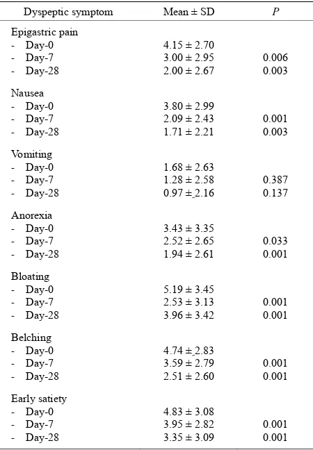
There was a significantly improvement of epigastric pain score on day 28 and day 7 after rebamipide com-pared to day-0 (P < 0.05). The score of nausea, anorexia, bloating, belching and early satiety score on day 28 and day 7 after rebamipide was also improved significantly compared to day 0 (P < 0.05). There was no significant improvement of the vomiting score on day 28 or day 7
after rebamipide compared to day 0 (see Table 1).
The mean gastric mucosal malondialdehyde (MDA) was decreased significantly on day-28 rebamipide com-pared to day-0 and the mean gastric mucosal dicarbonyl compound (CC) was slightly increased on day-28
re-bamipide compared to day-0 but not significant (see
Ta-ble 2 and Figure 1). The endoscopic mucosal severity
score on day-28 was improved significantly compared to
day-0 (see Table 2).
Histological features of chronic gastritis according to
the updated Sydney system are summarized in Table 2,
Figures 2 and 3.
[image:3.595.312.537.412.736.2]The degree of activity of chronic mononuclear inflam- matory cells in both corpus and antrum was higher than polymorphonuclear neutrophil activity in the same area (corpus 1.39 ± 0.54 vs 0.12 ± 0.40; antrum 1.41 ± 0.59 vs 0.12 ± 0.46). There were no differences in mononuclear
Table 1. The score of dyspeptic symptom.
Dyspeptic symptom Mean ± SD P
Epigastric pain
‐ Day-0
‐ Day-7
‐ Day-28
4.15 ± 2.70 3.00 ± 2.95
2.00 ± 2.67 0.006 0.003
Nausea
‐ Day-0
‐ Day-7
‐ Day-28
3.80 ± 2.99 2.09 ± 2.43 1.71 ± 2.21
0.001 0.003
Vomiting
‐ Day-0
‐ Day-7
‐ Day-28
1.68 ± 2.63 1.28 ± 2.58
0.97 ± 2.16 0.387 0.137
Anorexia
‐ Day-0
‐ Day-7
‐ Day-28
3.43 ± 3.35 2.52 ± 2.65
1.94 ± 2.61 0.033 0.001
Bloating
‐ Day-0
‐ Day-7
‐ Day-28
5.19 ± 3.45 2.53 ± 3.13 3.96 ± 3.42
0.001 0.001
Belching
‐ Day-0
‐ Day-7
‐ Day-28
4.74 ± 2.83 3.59 ± 2.79
2.51 ± 2.60 0.001 0.001
Early satiety
‐ Day-0
‐ Day-7
‐ Day-28
4.83 ± 3.08 3.95 ± 2.82 3.35 ± 3.09
Table 2. The result of free radical and endoscopic examination.
Variable Per protocol (n = 45)
Univariate: Day-0 Day-28 P
Mean malondialdehyde
(MDA) 5.28 ± 3.54 4.15 ± 2.71 0.047
Mean dicarbonyl
compound (CC) 4.14 ± 3.01 5.12 ± 2.71 0.642
Endoscopic mucosal
severity score 2.268 ± 0.45 1.707 ± 0.78 0.001
P<0.05 P>0.05 5.28
4.15 4.14
5.12
Figure 1. Biochemistry assessment.
1.41
0.12 0.15 0.15 0.10
1.49
0.10 0.10
0.24
0.15
0 0.2 0.4 0.6 0.8 1 1.2 1.4 1.6
Mononuclear
cells
Neutrophyl H pylori Atrophy Metaplasia
intestinal Day‐0
Day‐28
[image:4.595.312.535.102.443.2]P>0.05
Figure 2. Histopathology assessment of the gastric antrum.
1.38
0.12 0.07 0.10 0.15
1.41
0.07 0.15 0.15 0.10
0 0.2 0.4 0.6 0.8 1 1.2 1.4 1.6
Mononuclear
cells
Neutrophyl H pylori Atrophy Metaplasia
intestinal Day‐0 Day‐28
P>0.05 P>0.05
Figure 3. Histopathology assessment of the gastric corpus.
cell infiltration and neutrophil activity between corpus and antrum on day-0 and day-28 (P > 0.05). The density
of H. pylori colonization in the corpus at day-0 was
lower than in the antrum, with no significant differences before and after treatment (P > 0.05). There were no sig-nificant changes in atrophy and intestinal metaplasia between corpus and antrum of the stomach before and
after treatment (P > 0.05) (See Table 3).
Table 3. Histopathological score.
Histopathology Mean score SD P
Antrum mononuclear cell
‐ Day-0
‐ Day-28
1.41
1.49 0.59 0.55 0.372
Corpus mononuclear cell
‐ Day-0
‐ Day-28
1.38 1.41
0.54
0.50 0.800
Antrum neutrophil
‐ Day-0
‐ Day-28
0.12 0.10
0.46
0.37 0.710
Corpus neutrophil
‐ Day-0
‐ Day-28
0.12 0.07
0.40
0.26 0.421
Antrum H. pylori
‐ Day-0
‐ Day-28
0.15 0.10
0.48
0.37 0.323
Corpus H. pylori
‐ Day-0
‐ Day-28
0.07
0.15 0.26 0.48 0.262
Antrum atrophy
‐ Day-0
‐ Day-28
0.15 0.24
0.36
0.49 0.253
Corpus atrophy
‐ Day-0
‐ Day-28
0.10 0.15
0.30
0.42 0.486
Antrum intestinal metaplasia
‐ Day-0
‐ Day-28
0.10 0.15
0.30
0.36 0.323
Corpus intestinal metaplasia
‐ Day-0
‐ Day-28
0.15 0.10
0.48
0.30 0.534
5. DISCUSSION
Rebamipide, a gastro-protective agent, has been used clinically for the treatment of acute gastritis and peptic ulcer [9,10].
One of the major roles of rebamipide
[2-(4chlorobenzoylamino)-3[2(1H)-quinolonin-4-yl] pro- pionic acid], is to stimulate the generation of endogenous prostaglandins in the gastric mucosa and it has been re-ported to facilitate and accelerate ulcer healing.
Depending on the extensive published studies in vitro or in vivo, this clinical trial represents a reliable result of rebamipide on chronic gastritis. Open-label design had been chosen in this study, because this was a pilot study only and further trials involving larger sample size and longer observation/treatment period with well-controlled and randomized trials are needed.
[image:4.595.55.289.103.326.2] [image:4.595.55.280.491.605.2]compared with day-0 (P < 0.05). While symptom of vo- mitus was not improved at day-7 and day-28 compared to day 0 (P > 0.05). Chitapanarux et al. and Miwa et al. showed that rebamipide can improve the score of dys-pepsia symptoms [5,11]. Rebamipide may have an effect on Nitric oxide (NO), and improve these functional symptoms including bloating, belching etc. It has been proven that Nitric oxide (NO) plays multiple roles in inflammation and was found to be an essential mediator of the maintenance of resting blood flow of the gastric mucosa.
The most frequent characteristic of the patients with gastritis was men 51.1%. The mean age of patients in this study was 42.73 ± 11.52 years. The mean Body Mass Index (BMI) of the patients in this study was 22.69 ± 4.45. Jeffery et al. [12] found that chronic gastritis, gastric ulceration and atrophy and Helicobacter pylori infection is associated with low body mass index (BMI).
There was a significant improvement of endoscopic mucosal severity score on day-28 compared to day-0 (1.707 ± 0.78 vs 2.268 ± 0.45; P < 0.05).
The mean gastric mucosal malondialdehyde (MDA) was decreased significantly on day-28 compared to day-0 (P < 0.05). So, in this study, malondialdehyde (MDA) was measured and this result may show relationship be-tween these values and improvement of gastric mucosa.
Du et al. [13] also showed the improvement of
endo-scopy scores and gastric mucosa appearances and a sig-nificant reduction of malondialdehyde (MDA) after ad-ministration of rebamipide in chronic erosive gastritis.
Everett et al. [14] also described the levels of malon-
dialdehyde in the gastric mucosa in relationship with lipid peroxidation and Helicobacter pylori infection.
The mean gastric mucosal dicarbonyl compound was slightly increased on day-28 compared to day-0 but not significant (P > 0.05).
Although there was a non-significant small increase in dicarbonyl compound levels from day-0 to day-28 after rebamipide therapy, the level on day-28 was not signifi-cantly affecting the effectiveness of rebamipide. This may be explained by other effects of infection that com-pensate for increased dicarbonyl compound levels, such as carbonyl reactions with proteins including not only lipid peroxidation reactions but also other physiological oxidative stress reactions [15].
In histopathologic assessment, our study demonstrated that the degree of histological gastritis was not different between the corpus and the antrum. There were no sig-nificant changes in the histopathologic examination on
day-28 compared to day-0 (P > 0.05). There were no
significant changes in atrophy and intestinal metaplasia between corpus and antrum of the stomach as well (P > 0.05). Chitapanarux et al. [5] has demonstrated that treat-ment with rebamipide for 8 weeks not only significantly
improved the dyspepsia symptoms (epigastralgia, stom-ach heaviness, abdominal fullness, poor appetite and diarrhea) but also promoted the healing of both endo-scopic and histological features of chronic gastritis. The differences of these results to our study may be related to the short term observation period of the treatment dura-tion.
The anti-free radicals and anti-inflammatory properties of rebamipide in chronic gastritis had been proven in some studies and this is only a pilot study with a rela-tively small sample size and short term observation pe-riod to the treatment duration.
Importantly, no serious adverse event was reported during the study period. The data in the current study indicates the safety of rebamipide.
6. CONCLUSIONS
In conclusion, rebamipide was effective in healing gas-tritis and significantly reduced the extend of symptoms associated with chronic gastritis. The improvement in symptoms was associated with the decreased of endo-scopic severity score and the mean gastric mucosal malondialdehyde (MDA) significantly but not the histo-pathologic appearance and carbonyl compound.
The main shortcoming of this pilot study is the rela-tively small sample size and short term observation pe-riod to the treatment duration.
7. SUGGESTION
Regarding the relatively small sample size and short term of observation period, further longer duration, well-con- trolled and randomized trials are warranted in the future.
8. ACKNOWLEDGEMENTS
The authors would like to greatly thank Aan Santi and Indri Rizkiyani for their help and assistance in this study. The authors also thank the endoscopic nurses in Cipto Mangun kusumo Hospital for their assis-tance as well. Credit should also go to P. T. Otsuka Indonesia for sup-porting and providing the drugs for the study.
REFERENCES
[1] Santra, A., Chowdhury, A., Chaudhury, S., et al. (2000) Oxidative stress in gastric mucosa in Helicobacterpylori infection. Indian Journal of Gastroenterology, 19, 21-23.
[2] Hayashi, S., Sugiyama, T., Amano, K., Isogai, H., Isogai, E., Aihara, M., et al. (1998) Effect of rebamipide, a novel antiulcer agent, on Helicobacter pylori adhesion to gas-tric epithelial cells. Antimicrobial Agents and Chemo-therapy, 42, 1895-1889.
[4] Chey, W.D. and Wong, B.C. (2007) Practice parameters committee of the American College of Gastroenterology: American College of Gastroenterology guideline on the management of Helicobacter pylori infection. American Journal of Gastroenterology, 102, 1808-1825.
doi:10.1111/j.1572-0241.2007.01393.x
[5] Chitapanarux, T., Praisontarangkul, O.A. and Lertpraser- tsuke, N. (2008) An open-labeled study of rebamipide treatment in chronic gastritis patients with dyspeptic symptoms refractory to proton pump inhibitors.Digestive Diseases and Sciences, 53, 2896-2903.
doi:10.1007/s10620-008-0255-5
[6] Naito, Y., Yoshikawa, T., Tanigawa, T., et al. (1995) Hydroxyl radical scavenging by rebamipide and related compounds: Electron paramagnetic resonance study. Free Radical Biology & Medicine, 18, 117-123.
doi:10.1016/0891-5849(94)00110-6
[7] Arakawa, T., Kobayashi, K., Yoshikawa, T., Tarnawski, A. (1998) Rebamipide: Overview of its mechanisms of action and efficacy in mucosal protection and ulcer heal-ing. Digestive Diseases and Sciences, 43, 5S-13S. [8] Rugge, M. and Genta, R.M. (2005) Staging and grading
of chronic gastritis. Human Pathology, 36, 228-233. doi:10.1016/j.humpath.2004.12.008
[9] Naito, Y., Yoshikawa, T., Iinuma, S., Yagi, N., Matsu- yama, K., Boku, Y., Fujii, T., Yoshida, N., Kondo, M. and Sasaki, E. (1998) Rebamipide protects against indo-methacin-induced gastric mucosal injury in healthy vol-unteers in double-blind, placebo-controlled study. Diges-tive Diseases and Sciences, 43, 83S-89S.
[10] Higuchi, K., Arakawa, T., Nebiki, H., Uchida, T.,
Fuji-wara, Y., et al. (1998) Rebamipide prevents recurrence of gastric ulcers without affecting Helicobacter pylori status.
Digestive Diseases and Sciences, 43, S99-S106.
[11] Miwa, T., Osada, T., Nagahara, A., Ohsuka, T., Hojo, M., Tomita, T., et al. (2006) Effect of a gastro-protective agent, rebamipide, on symptom improvement in patients with functional dyspepsia: A double-blind placebo-con- trolled study in Japan. Journal of Gastroenterology and Hepatology, 21, 1826-1831.
doi:10.1111/j.1440-1746.2006.04446.x
[12] Jeffery, P.L., McGuckin, M.A. and Linde, S.K. (2011)
Endocrine impact of Helicobacter pylori: Focus on
ghre-lin and ghreghre-lin o-acyltransferase. World Journal of Gas-troenterology, 17, 1249-1260.
doi:10.3748/wjg.v17.i10.1249
[13] Du, Y., Li, Z., Zhan, X., Chen, J. and Gao, J. (2008) Anti- inflammatory effects of rebamipide according to
Helico-bacterpylori status in patients with chronic erosive
gas-tritis: A randomized sucralfate-controlled multicenter trial in China-STARS study. Digestive Diseases and Sciences, 53, 2886-2895.doi:10.1007/s10620-007-0180-z
[14] Everett, S.M., Singh, R., Leuratti, C. et al. (2001) Levels of malondialdehyde-deoxyguanosine in the gastric mu-cosa: Relationship with lipid peroxidation, ascorbic acid,
and Helicobacterpylori. Cancer Epidemiology, Biomark-
ers & Prevention, 10, 369-376.