inorganic papers
Acta Cryst.(2006). E62, i135–i137 doi:10.1107/S1600536806017697 Xuet al. GeO
3Pb
i135
Acta Crystallographica Section EStructure Reports Online
ISSN 1600-5368
Lead germanium oxide
Yan Xu,a* Li-Ying Cheng,a
Guang-Peng Zhouband Yan-Li
Wanga
aCollege of Chemistry and Chemical
Engineering, Liaoning Normal University, 116029, Dalian, People’s Republic of China, andbInstitute of Chemistry for Functionalized Materials, College of Chemistry and Chemical Engineering, Liaoning Normal University, Dalian, 116029, People’s Republic of China
Correspondence e-mail: [email protected]
Key indicators
Single-crystal X-ray study
T= 293 K
Mean(Ge–O) = 0.019 A˚
Rfactor = 0.050
wRfactor = 0.164
Data-to-parameter ratio = 13.4
For details of how these key indicators were automatically derived from the article, see http://journals.iucr.org/e.
Received 26 April 2006 Accepted 12 May 2006
#2006 International Union of Crystallography All rights reserved
The crystal structure of the title compound, PbGeO3, shows a
three-dimensional framework assembled by Pb6 and Ge6
structural building units. The Pb atom is coordinated by five O atoms in square-pyramidal coordination, while the Ge atom is tetrahedrally coordinated by four O atoms.
Comment
Over the past decades, the synthesis of new open-framework materials with either pure tetrahedral or mixed polyhedral microporous arrangements have received great attention due to their functional applications in catalysis, adsorption, ion-exchange and radioactive waste remediation. Not only aluminium and silicon, but also boron, gallium, phosphorus, germanium and transition metals have been choosen as open-framework building elements to synthesize new three-dimensional materials (Liet al., 1998; Linet al., 2003; Ple´vertet al., 2001; Xu, Fan, Chinoet al., 2004; Xu, Fan, Elangovanet al., 2004). Compared with silicon, germanium not only adopts
longer metal–oxygen distances (about 1.76 A˚ for Ge—O,
1.61 A˚ for Si—O), but also exhibits three types of coord-ination polyhedra,viz. GeO4, GeO5and GeO6. Therefore, the flexibility of the polyhedral model for germanium allows the formation of various open frameworks. In particular, germa-nium shows a great ability to form rings containing three metal atoms. Some germanates with such rings have already been reported (Buet al., 1998; Liet al., 2000; Xu, Ogura & Okubo, 2004).
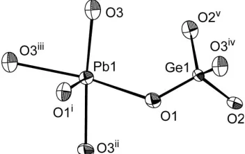
[image:1.610.244.420.604.715.2]In this work, we have designed and synthesized the title compound, which features a three-dimensional framework derived from Pb6 and Ge6 structural building units. Both building units are connected by a new type of Ge2Pb ring. The molecular structure of the title compound is shown in Fig. 1. The asymmetric unit of PbGeO3 contains one formula unit. The O atoms in the open framework can be divided into three groups,viz.O2b, O3band O4b, where O2brepresents an O atom
Figure 1
connected to two Ge atoms, O3bis connected to one Ge and
two Pb atoms, and O4bto one Ge and three Pb atoms. The Pb
atom is five-coordinated by two O3band three O4batoms.
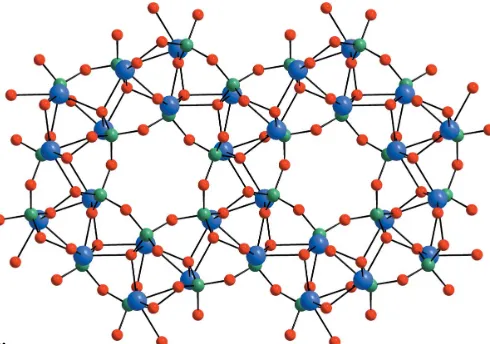
As shown in Fig.2, six PbO5square pyramids form a Pb6ring structure by sharing corners, and six corner-sharing GeO4 tetrahedra form a Ge6ring (Fig. 3). These two building units are connected by Ge2Pb rings (two GeO4 and one PbO5), yielding a three-dimensional open framework with channels along the [111] direction (the rhombohedral symmetry axis) (Fig. 4).
The Pb—O and Ge—O bond lengths (Table 1) are in agreement with those found in previously reported lead and germanium compounds (Buet al., 1998; Shiet al., 2002). The
shortest Pb Pb distance in the Pb6 building unit is
3.6910 (8) A˚ , while Pb Pb Pb is 118.066 (12), which is very close to 120.
Experimental
The title compound was hydrothermally synthesized from a mixture of GeO2, H3BO3, Pb(OAc)23H2O, diethylenetriamine, HF, pyridine
and H2O in the molar ratio 2:1:2:4:4:74:180. In a typical synthesis,
GeO2(0.01 g) and H3BO3(0.03 g) were dissolved in a mixed solvent
of pyridine (2.84 g) and water (1.60 g), followed by the addition of
Pb(OAc)23H2O (0.36 g) and diethylenetriamine (0.23 g) with
constant stirring. Finally, hydrofluoric acid (0.1 ml, 40 wt %) was added to the above mixture. The mixture was kept in a 25 ml Teflon-lined steel autoclave at 443 K for 7 d. The autoclave was slowly cooled to room temperature, and then the product was filtered off, washed with distilled water and acetone, and dried at room temperature. Colourless block-shaped crystals were obtained. We have attempted to find crystals of better quality and to optimize the synthesis, but no better single crystals could be obtained.
Crystal data
GeO3Pb Mr= 327.78 Rhombohedral,R3
a= 9.3282 (2) A˚
= 113.4560 (2)
V= 512.43 (2) A˚3 Z= 6
Dx= 6.373 Mg m3 MoKradiation
= 57.82 mm1
T= 293 (2) K Block, colourless 0.040.030.03 mm
Data collection
Bruker APEX2 CCD diffractometer
!scans
Absorption correction: multi-scan (SADABS; Sheldrick, 2003)
Tmin= 0.206,Tmax= 0.276
(expected range = 0.132–0.176)
2654 measured reflections 628 independent reflections 553 reflections withI> 2(I)
Rint= 0.047
max= 25.5
Refinement
Refinement onF2 R[F2> 2(F2)] = 0.050 wR(F2) = 0.164 S= 1.21 628 reflections 47 parameters
w= 1/[2(F
o2) + (0.1096P)2]
whereP= (Fo2+ 2Fc2)/3
(/)max= 0.001 max= 4.40 e A˚
3 min=3.49 e A˚
3
Extinction correction:SHELXL97
Extinction coefficient: 0.0022 (8)
Table 1
Selected bond lengths (A˚ ).
Pb1—O1i
2.270 (14) Pb1—O3ii
2.416 (15) Pb1—O1 2.458 (14) Pb1—O3 2.551 (16) Pb1—O3iii
2.590 (15)
Ge1—O1 1.723 (14) Ge1—O3iv
1.725 (15) Ge1—O2 1.779 (15) Ge1—O2v
1.781 (15)
Symmetry codes: (i) z;x1;y; (ii) yþ1;z;xþ1; (iii) xþ1;y;z; (iv) xþ2;yþ1;zþ1; (v)zþ2;xþ2;yþ1.
inorganic papers
i136
Xuet al. GeO [image:2.610.318.563.71.243.2]3Pb Acta Cryst.(2006). E62, i135–i137
Figure 4
The three-dimensional open framework with channels, viewed along [111].
Figure 3
[image:2.610.95.244.71.236.2]The structure of the Ge6building unit.
Figure 2
The highest peak in the difference map is located 1.08 (2) A˚ from Pb1, while the deepest hole is 1.13 (3) A˚ from Pb1.
Data collection:APEX2(Bruker, 2005); cell refinement:SAINT
(Bruker, 2002); data reduction: SAINT; program(s) used to solve structure:SHELXS97(Sheldrick, 1997a); program(s) used to refine structure: SHELXL97 (Sheldrick, 1997a); molecular graphics:
SHELXTL(Sheldrick, 1997b); software used to prepare material for publication:SHELXTL.
This work was supported by the Education Office Found-ation of Liaoning Province (grant No. 05L220).
References
Bruker (2002).SAINT. Version 6.36a. Bruker AXS Inc., Madison, Wisconsin, USA.
Bruker (2005).APEX2. Version 1.27. Bruker AXS Inc., Madison, Wisconson, USA.
Bu, X., Feng, P. & Stucky, G. D. (1998). J. Am. Chem. Soc. 120, 11204– 11205.
Li, H., Eddaoudi, M., Ple´vert, J., O’Keeffe, M. & Yaghi, O. M. (2000).J. Am. Chem. Soc.122, 12409–12410.
Li, H., Eddaoudi, M., Richardson, D. A. & Yaghi, O. M. (1998).J. Am. Chem. Soc.120, 8567–8568.
Lin, Z., Zhang, J. & Yang, G. (2003).Inorg. Chem.42, 1797–1799.
Ple´vert, J., Gentz, T. M., Laine, A., Li, H., Young, V. G., Yaghi, O. M. & O’Keeffe, M. (2001).J. Am. Chem. Soc.123, 12706–12707.
Sheldrick, G. M. (1997a). SHELXS97 and SHELXL97. University of Go¨ttingen, Germany.
Sheldrick, G. M. (1997b).SHELXTLVersion 5.10. Bruker AXS Inc., Madison, Wisconsin, USA.
Sheldrick, G. M. (2003).SADABS. University of Go¨ttingen, Germany. Shi, Y., Li, L., Li, Y., Xu, Y., Chen, X., Xue, Z. & You, X. (2002).Inorg. Chem.
Commun.5, 1090–1094.
Xu, Y., Fan, W., Chino, N., Uehara, K., Hikichi, S., Mizuno, N., Ogura, M. & Okubo, T. (2004).Chem. Lett.33, 74–75.
Xu, Y., Fan, W., Elangovan, S. P., Ogura, M. & Okubo, T. (2004).Eur. J. Inorg. Chem.pp. 4547–4549.
Xu, Y., Ogura, M. & Okubo, T. (2004).Microporous Mesoporous Mater.70, 1– 6.
inorganic papers
Acta Cryst.(2006). E62, i135–i137 Xuet al. GeO
supporting information
sup-1 Acta Cryst. (2006). E62, i135–i137
supporting information
Acta Cryst. (2006). E62, i135–i137 [https://doi.org/10.1107/S1600536806017697]
Lead germanium oxide
Yan Xu, Li-Ying Cheng, Guang-Peng Zhou and Yan-Li Wang
(I)
Crystal data
GeO3Pb
Mr = 327.78 Rhombohedral, R3 Hall symbol: -P 3*
a = 9.3282 (2) Å
α = 113.4560 (2)°
V = 512.43 (2) Å3
Z = 6
F(000) = 828
Dx = 6.373 Mg m−3
Mo Kα radiation, λ = 0.71073 Å Cell parameters from 2654 reflections
θ = 2.6–25.5°
µ = 57.82 mm−1
T = 293 K Block, colourless 0.04 × 0.03 × 0.03 mm
Data collection
Bruker Apex2 CCD diffractometer
Radiation source: fine-focus sealed tube Graphite monochromator
ω scans
Absorption correction: multi-scan (SADABS; Sheldrick, 2003)
Tmin = 0.206, Tmax = 0.276
2654 measured reflections 628 independent reflections 553 reflections with I > 2σ(I)
Rint = 0.047
θmax = 25.5°, θmin = 2.6°
h = −10→10
k = −8→7
l = −11→11
Refinement
Refinement on F2 Least-squares matrix: full
R[F2 > 2σ(F2)] = 0.050
wR(F2) = 0.164
S = 1.21 628 reflections 47 parameters 18 restraints
Primary atom site location: structure-invariant direct methods
Secondary atom site location: difference Fourier map
w = 1/[σ2(F
o2) + (0.1096P)2] where P = (Fo2 + 2Fc2)/3 (Δ/σ)max = 0.001
Δρmax = 4.40 e Å−3 Δρmin = −3.49 e Å−3
Extinction correction: SHELXL, Fc*=kFc[1+0.001xFc2λ3/sin(2θ)]-1/4 Extinction coefficient: 0.0022 (8)
Special details
supporting information
sup-2 Acta Cryst. (2006). E62, i135–i137
Refinement. Refinement of F2 against ALL reflections. The weighted R-factor wR and goodness of fit S are based on F2, conventional R-factors R are based on F, with F set to zero for negative F2. The threshold expression of F2 > σ(F2) is used only for calculating R-factors(gt) etc. and is not relevant to the choice of reflections for refinement. R-factors based on F2 are statistically about twice as large as those based on F, and R- factors based on ALL data will be even larger.
Fractional atomic coordinates and isotropic or equivalent isotropic displacement parameters (Å2)
x y z Uiso*/Ueq
Pb1 0.71054 (12) 0.01082 (12) 0.17220 (12) 0.0139 (5) Ge1 1.2560 (3) 0.5203 (3) 0.6560 (3) 0.0114 (7) O1 1.033 (2) 0.287 (2) 0.540 (2) 0.018 (3) O2 1.478 (2) 0.545 (2) 0.777 (2) 0.019 (3) O3 0.684 (2) 0.265 (2) 0.152 (2) 0.020 (3)
Atomic displacement parameters (Å2)
U11 U22 U33 U12 U13 U23
Pb1 0.0151 (6) 0.0162 (6) 0.0148 (6) 0.0111 (5) 0.0112 (5) 0.0108 (5) Ge1 0.0109 (12) 0.0089 (11) 0.0113 (12) 0.0056 (10) 0.0067 (10) 0.0065 (10) O1 0.019 (4) 0.020 (5) 0.015 (4) 0.011 (3) 0.013 (3) 0.012 (3) O2 0.019 (5) 0.017 (4) 0.021 (5) 0.010 (3) 0.012 (3) 0.015 (4) O3 0.017 (4) 0.021 (5) 0.021 (5) 0.013 (3) 0.016 (4) 0.011 (3)
Geometric parameters (Å, º)
Pb1—O1i 2.270 (14) Ge1—O3v 1.725 (15)
Pb1—O3ii 2.416 (15) Ge1—O2 1.779 (15)
Pb1—O1 2.458 (14) Ge1—O2vi 1.781 (15)
Pb1—O3 2.551 (16) O1—Pb1vii 2.270 (14)
Pb1—O3iii 2.590 (15) O2—Ge1viii 1.781 (15)
Pb1—Pb1iv 3.6910 (8) O3—Ge1v 1.725 (15)
Pb1—Pb1ii 3.6910 (8) O3—Pb1iv 2.416 (15)
Ge1—O1 1.723 (14) O3—Pb1iii 2.590 (15)
O1i—Pb1—O3ii 78.6 (6) O3iii—Pb1—Pb1ii 118.2 (3) O1i—Pb1—O1 86.5 (5) Pb1iv—Pb1—Pb1ii 118.066 (12) O3ii—Pb1—O1 72.9 (5) O1—Ge1—O3v 113.3 (7)
O1i—Pb1—O3 78.1 (5) O1—Ge1—O2 111.9 (7)
O3ii—Pb1—O3 153.4 (5) O3v—Ge1—O2 106.0 (7)
O1—Pb1—O3 93.1 (5) O1—Ge1—O2vi 107.6 (7)
supporting information
sup-3 Acta Cryst. (2006). E62, i135–i137
O3iii—Pb1—Pb1iv 92.5 (3) Pb1iv—O3—Pb1 96.0 (5) O1i—Pb1—Pb1ii 121.4 (4) Ge1v—O3—Pb1iii 110.9 (7) O3ii—Pb1—Pb1ii 43.4 (4) Pb1iv—O3—Pb1iii 100.2 (5) O1—Pb1—Pb1ii 71.5 (4) Pb1—O3—Pb1iii 96.0 (5) O3—Pb1—Pb1ii 153.0 (3)