General information
Thesis title The effect of cardiopulmonary resuscitation on ventricular fibrillation waveform measures: a new tool to optimize in-field identification of an acute coronary occlusion during cardiac arrest
Author Jeanne van der Waal, BSc. Email jeannevanderwaal@outlook.com
Organization
Address Radboud University Medical Centre Geert Grooteplein-zuid 10
6525 GA Nijmegen Department Department of Cardiology
Institution
Address University of Twente Drienerlolaan 5 Postbus 217 7500 AE Enschede
Faculty Faculty of Science and Technology Program Technical Medicine
Master Medical Sensing and Stimulation
Committee
Chairman Prof. dr. H.J. Zwart Medical supervisor Dr. M.A. Brouwer Technical supervisor Dr. ir. G. Meinsma
Process supervisor Drs. P.A. van Katwijk External member M.C. Hermans, MSc. Additional member J. Nas, MSc.
Summary
The effect of cardiopulmonary resuscitation on ventricular fibrillation waveform measures: a new tool to optimize in-field identification of an acute coronary occlusion
during cardiac arrest
by Jeanne van der Waal
Since the ventricular fibrillation (VF) waveform has been shown to decrease over time, it has been regarded as marker of arrest duration and has been investigated to predict defibrilla-tion success. However, exact predicdefibrilla-tion of arrest duradefibrilla-tion is complicated by other factors also influencing the VF waveform. The VF waveform has been shown to increase with unin-terrupted chest compressions, and decrease with pauses in chest compressions. Animal and human studies have also shown that myocardial infarction (MI) affects VF, and animal stud-ies suggest that the change in VF waveform in response to cardiopulmonary resuscitation (CPR) may be altered in the presence of an acute coronary occlusion (ACO). This study in-vestigated the change in VF waveform characteristics in relation to CPR quality, whether this is altered in the presence of an ACO, and whether this information can help in identifying these patients during out-of-hospital cardiac arrest (OHCA).
For the change in VF characteristics in response to CPR, we compared this change between patients with and without adequate CPR (defined as chest compression fraction (CCF)≥ or < 0.6) between the first and second defibrillation. In a sub analysis we investigated this change in characteristics in a sub population of patients with and without ACO. In patients with CCF≥0.6 (n=90), an increase in all VF amplitude characteristics was detected, while this did not occur in patients with CCF<0.6 (n=48). Furthermore, this numeric increase was significantly higher in patients with CCF≥0.6 compared to patients with CCF<0.6. The sub analysis showed a difference in change in VF amplitude characteristics between CCF≥0.6 and CCF<0.6 in patients without ACO (n=22), whereas this difference was not found in patients with ACO (n=38).
Next the VF waveform parameters were investigated to determine their ability to predict the presence of an ACO. In patients with an underlying ACO (n=62), the VF amplitude characteristics were significantly lower when compared to patients without an underlying ACO (n=40), showing a limited discriminative ability with an AUC of 0.66. Combining the VF waveform parameter with the change in that parameter in response to CPR using binary logistic regression led to an improved discriminative ability, with an AUC of 0.75.
vii
Acknowledgements
In the period of September to November 2016 I did the last of my 10-week internships in the second year of the Master Technical Medicine at the Cardiology department of the Rad-boudumc. This confirmed my interest in cardiology in general, and more specifically for the electrophysiological aspects. Since there was a very good work atmosphere at the research department, I was glad that I could stay there to start my graduation internship in February 2017. Here, I would like to take the opportunity to thank the people that made this possible. I would like to thank Joris Nas for guiding me throughout the year, for helping me with research questions, the explanation of some (for me) complicated statistical concepts and the critical revisions of the manuscript. For critical revisions of the manuscript I would also like to thank Jos Thannhauser, who provided a fresh set of eyes to further improve the research and the writing. I would like to thank Marc Brouwer for the opportunity to do my gradu-ation internship at the cardiology department. Even though our contact during the project was limited, I was amazed by your ability to read people.
I would like to thank Gjerrit Meinsma for investing time and helping me construct the De-trended Fluctuation Analysis chapter. I would like to thank Paul van Katwijk for being my mentor for the past two years and asking difficult questions that made me think about who I am and who I want to be. Furthermore I would like to thank Hans Zwart for being the chairman in the committee, and Mathilde Hermans for also taking place in the committee. Thanks to all my colleagues for the nice collaboration and making me feel welcome, and a special thanks to the PhD candidates for the frequent educational moments. Thanks to all fellow students in the research department during this internship, for always filling my mug of tea without complaining. Also thanks for some great times, I hope the bucket list keeps growing and also keeps getting checked off.
Contents
Acknowledgements vii
List of Abbreviations x
1 Introduction 1
2 Background 2
2.1 Anatomy and physiology of the heart . . . 2
2.2 Electrical activity in the heart . . . 3
2.3 Myocardial infarction . . . 6
2.4 Ventricular arrhythmias . . . 7
2.5 Out-of-hospital cardiac arrest therapy . . . 9
2.6 Ventricular fibrillation waveform analysis . . . 9
3 The effect cardiopulmonary resuscitation on ventricular fibrillation waveform mea-sures: The role of CPR quality and underlying acute coronary occlusions 15 3.1 Introduction . . . 15
3.2 Methods . . . 16
3.3 Results . . . 19
3.4 Discussion . . . 21
3.5 Conclusion . . . 25
4 Differentiating between patients with and without acute coronary occlusion in out-of-hospital cardiac arrests based on ventricular fibrillation waveform measures 26 4.1 Introduction . . . 26
4.2 Methods . . . 27
4.3 Results . . . 29
4.4 Discussion . . . 32
4.5 Conclusion . . . 35
5 Discussion 36
6 Conclusion 37
A Detrended Fluctuation analysis 38
B Acute coronary occlusion categorisation criteria 51
C Flowchart sub-analysis acute coronary occlusion 52
D Change in VF waveform characteristics of OHCA-patients with or without an
un-derlying acute coronary occlusion 53
E Discrimination of inferior coronary occlusion 54
List of Abbreviations
ACO Acute Coronary Occlusion
AED Automated External Defibrillator
AMI Acute Myocardial Infarction
AMSA Amplitude Spectrum Area
AP Action potential
AV Atrioventricular
AUC Area under the curve
BLS Basic Life Support
CABG Coronary artery bypass graft
CAG Coronary angiography
CCF Chest Compression Fraction
COP Cardioversion Outcome Predictor
CPR Cardiopulmonary resuscitation
CWT Continuous Wavelet Transform
DF Dominant Frequency
DFA Detrended Fluctuation Analysis
ECC Extracorporeal Circulation
ECG Electrocardiogram
ED Emergency Department
EMS Emergency Medical Services
FFT Fast Fourier Transform
FR Frequency Ratio
ICD Implantable Cardioverter Defibrillator
LAD Left anterior descending artery
LBBB Left bundle branch block
LCA Left coronary artery
MAA Mean Absolute Amplitude
MdF Median Frequency
MI Myocardial Infarction
OHCA Out-of-hospital cardiac arrest
PCI Percutaneous Coronary Intervention
PDA Posterior descending artery
PEA Pulseless electrical activity
PSA Power Spectrum Area
PSD Power Spectral Density
PVC Premature Ventricular Complex
PVT Pulseless Ventricular Tachycardia
RCA Right coronary artery
RCx Ramus circumflex artery
RMS Root-mean-square
ROC Receiver Operating Characteristic
ROOR Return of Organized Rhythm
ROSC Return of Spontaneous Circulation
SA Sinoatrial
SDW Scale Distribution Width
STEMI ST-elevation myocardial infarction
TTI Transthoracic Impedance
VF Ventricular Fibrillation
VS Variance of Slope
VT Ventricular Tachycardia
1
1 Introduction
Ventricular fibrillation (VF) is the first observed cardiac rhythm in about 20-50% of out-of-hospital cardiac arrests (OHCAs) [1, 2]. Survival after OHCA is poor, although VF as first observed rhythm has a better outcome in terms of survival to hospital discharge (15-40%) when compared to non-shockable rhythms (2-8%) [2–5]. Still, VF will gradually deterio-rate into asystole with passage of time and this will consequently decrease chance of sur-vival [6, 7]. Defibrillation is considered the only therapy to establish return of spontaneous circulation (ROSC) in patients with ventricular fibrillation [8]. Therefore, the guidelines for cardiopulmonary resuscitation (CPR) recommend immediate defibrillation as soon as a de-fibrillator is made available [9]. However, if the myocardial metabolic state is compromised, success rates of defibrillation are poorer [8, 10]. Additionally, defibrillation can also have the adverse effect of producing asystole [11].
In absence of an automated external defibrillator (AED) or in between defibrillations, chest compressions and ventilations can be administered to keep oxygenated blood flowing to the brain and other vital organs. Studies have shown that CPR does not only slow the deteri-oration of myocardial cells, but also increases the chances of survival [9, 12, 13]. Therefore, it has been suggested that for VF of longer duration, withholding defibrillation in order to apply CPR might increase myocardial readiness for defibrillation and increase chances of successful defibrillation [14, 15]. This was shown in two studies where patients with ambu-lance response time of more than four to five minutes showed increased rates of ROSC and survival if 1.5 or 3 minutes of CPR was administered by ambulance personnel before de-fibrillation [16, 17]. Unfortunately, the onset time of VF is (especially in the out-of-hospital setting) rarely known, making it difficult to determine the priority of CPR intervention based on the duration of the untreated cardiac arrest. In this light, the VF waveform on the electro-cardiogram (ECG) has been investigated for its ability to reflect myocardial metabolic state. However, the application of VF waveform to guide initial therapy may be complicated by the largely unknown effect of factors influencing the VF waveform. The VF waveform deterio-rates after an episode without CPR [18, 19], accompanied by a decrease in survival chances. Since VF characteristics are associated with shock success and long-term outcome [20–23], and CPR improves the chance of ROSC [13, 16, 17], VF characteristics are also likely to be affected by CPR. In addition, VF characteristics have shown to be influenced by the presence of myocardial ischemia. Animal studies have indicated that VF characteristics were lower in the presence of an induced myocardial infarction (MI) [24–26]. Similarly, in human stud-ies lower VF characteristics were found in patients with acute MI as underlying aetiology of VF [27], as well as in patients with a previous MI where VF was induced during ICD-testing [28, 29]. One animal study also shows that the reaction of the VF waveform on CPR may be altered in the presence of acute coronary occlusion [30].
2 Background
2.1
Anatomy and physiology of the heart
The heart is a pump, allowing blood to flow through the body. The right side of the heart contributes to the pulmonary circulation, and the left side of the heart contributes to the systemic circulation. The flow through the heart is schematically illustrated in Figure 2.1. In the pulmonary circulation, deoxygenated blood enters the right atrium and continues to flow to the right ventricle. The right ventricle pumps the blood towards the lungs via the pulmonary arteries. In the lungs exchange of oxygen and carbon dioxide occurs, after which oxygen enriched blood is transported from the lungs to the left atrium via the pulmonary veins. In the systemic circulation, the oxygenated blood travels from the left atrium to the left ventricle. Through forceful contraction of the left ventricle, the blood is pumped into the aorta, via which the blood is distributed towards the organs and tissues. After supplying the tissue with oxygen the blood is returned to the right atrium via the superior and inferior vena cava [31, 32].
A cardiac cycle, defined as all cardiac events from the beginning of one heartbeat to the beginning of the next, consists of two phases: the diastole and the systole. In the diastole the ventricles are relaxed, allowing blood to flow from the atria to the ventricles. By the end
FIGURE2.1: Blood flow through the heart. The right atrium receives blood via the superior vena cava and inferior vena cava and the right ventricle pumps blood via the pulmonary artery to the lungs. The left atrium re-ceives blood from the lungs and the left ventricle pumps blood into the aorta, for distribution to the organs. Backflow of blood from the ventricles to the atria is prevented by the atrioventricular valves (i.e. the tricuspid and mi-tral valve), and backflow from the pulmonary artery and aorta is prevented by the semilunar valves (i.e. the pulmonary and aortic valve). Reproduced
2.2. Electrical activity in the heart 3
of the diastole, the atria contract to pump the remaining volume of blood to the ventricles. During relaxation of the ventricles, backflow of blood from the pulmonary artery and aorta is prevented by the pulmonary valve and aortic valve respectively (see Figure 2.1). After the diastole, the systole starts by isovolumetric contraction, i.e. contraction without a change in volume, of both ventricles simultaneously. When the pressure in the ventricle exceeds the pressure in the associated outflow tract, the aortic and pulmonary valve are forced to open and bood starts flowing out. During contraction of the ventricles, backflow of blood from the ventricles to the atria is prevented by the atrioventricular valves, with the tricuspid valve between the right atrium and ventricle and the mitral valve between the left atrium and ventricle (see Figure 2.1). After the blood is ejected in the pulmonary artery and aorta, the diastole starts with isovolumetric relaxation until the pressure in the ventricles drops below the atrial pressure, after which the ventricles are filled with blood again and the cycle repeats [31, 32].
To be able to provide the oxygen-rich blood to the body tissues, the heart itself also needs a steady oxygen supply. This is provided through the coronary arteries, originating from the root of the aorta (Figure 2.2). The left coronary artery (LCA) bifurcates into the left anterior descending artery (LAD) and the ramus circumflex artery (RCx). The LAD descends into the anterior inter-ventricular groove, where its branches mainly supply the anterior wall of the left ventricle, the inter-ventricular septum and parts of the conduction system. The RCx travels in the left atrioventricular groove, where it supplies most of the left atrium and the posterolateral wall of the left ventricle. The right coronary artery (RCA) runs through the right atrioventricular groove, and distributes blood to the right atrium, the right ventricle, and parts of the conduction system of the heart. The inferior wall of the heart is provided with blood by the posterior descending artery (PDA), which originates either from the RCA (in 85% of people) or from the RCx (in 15% of people) [32, 34].
FIGURE2.2: Anterior view of the heart showing the main coronary arter-ies. The left main coronary artery bifurcates into the left anterior descend-ing artery, descenddescend-ing into the anterior interventricular groove, and the ra-mus circumflex artery, running in the left atrioventricular groove. The right coronary artery runs through the right atrioventricular groove towards the posterior region of the heart. SVC = Superior vena cava, Ao = Aorta, PA =
Pulmonary artery, IVC = Inferior vena cava. Adapted from [32].
2.2
Electrical activity in the heart
FIGURE2.3: Electrical conduction system of the heart. The impulse originates from the sinoatrial node, travels through the atria toward the atrioventricular node. After a short delay it is conducted trough the bundle of His to the Purk-inje fibers, resulting in depolarization of the ventricles. Adapted from [39].
heart: pacemaker cells that have the ability to generate electrical impulses and cardiomy-ocytes which can only conduct an impulse. Normally, the electrical impulse originates from a group of pacemaker cells that is located in the high right atrium near the superior vena cava, called the sinoatrial (SA) node (Figure 2.3) [35]. The impulse propagates through neighbouring cells in the atria and stimulates the myocardium of the atria for contraction. When it reaches the atrioventricular (AV) groove, a fibrous structure which is electrically in-ert disables conduction directly from the atria to the ventricles. Therefore, conduction from the atria to the ventricles is only possible through the AV node, located close to the tricus-pid valve in the interatrial septum. The AV node has specific electrophysiologic properties, which slows the conduction velocity. This results in a delay in conduction between the atria and ventricles, allowing sufficient emptying of the atria. When leaving the AV node, the impulse enters the bundle of His, which penetrates the fibrous tissue to allow conduction toward the ventricles. The His bundle branches into the right and left bundle branches, and the rapidly conducting Purkinje fibers reaching to the more distal and lateral parts of the ventricular myocardium ensure an almost simultaneous depolarization of the ventricles [32, 35–38].
2.2. Electrical activity in the heart 5
2.2.1
The electrocardiogram
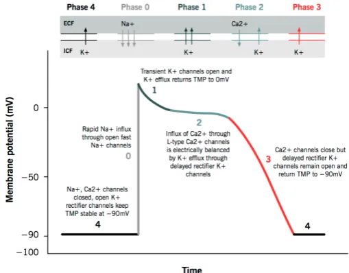
The cardiac action potential represents the electrical activity of a single cardiac cell, which cannot be measured from the outside. To find information about the electrical properties of the heart, a surface electrocardiogram (ECG) can be measured. Tissues surrounding the heart are able to conduct electrical currents, allowing these currents to be detected at the body surface by an array of electrodes. This ECG represents the sum of electrical activity in the heart, which is measured as voltage changes from a baseline voltage. A normal rhythm consists of a P wave, a QRS complex and a T wave, as seen in Figure 2.5. The P wave represents the depolarization wave spreading through the atria. The conduction delay in the AV node as discussed earlier leads to a brief isoelectric (i.e. zero voltage) period. The fast ventricular depolarization results in the QRS complex, only lasting about 0.06 to 0.1 seconds. The isoelectric ST segment is the period at which the entire ventricle is depolarized. It roughly corresponds to the plateau phase of the ventricular action potential as seen in Figure 2.4. The T wave represents repolarization of the ventricles (phase 3 of the action potential) [32, 35].
The height and direction of the different deflections of the ECG is dependent on the record-ing direction. The ECG is measured as a potential difference between a positive and a nega-tive electrode. A wave of depolarization travelling toward a posinega-tive electrode will result in a positive deflection in the ECG trace. A wave of depolarization or repolarization oriented perpendicular to an electrode axis produces no net deflection (i.e. equally positive and neg-ative voltages). Therefore, the ECG is conventionally measured in 12 directions, i.e. leads, using 10 electrodes. Four electrodes are placed on each arm and leg and six electrodes are placed at defined locations on the chest. The electrodes on the left arm, right arm and left leg together compose the triangle of Einthoven, and form the three bipolar limb leads I, II and III (see Figure 2.6). With the same three electrodes, augmented limb leads (aVR, aVL, aVF) are measured by using a single positive electrode referenced against a combination of the other two electrodes. These limb leads record the ECG in the frontal plane. The same three elec-trodes are used as a combined negative electrode to the positive precordial elecelec-trodes. The resulting precordial leads (V1-V6) record electrical activity in the horizontal plane, perpen-dicular to the frontal plane in which the limb leads record. A visualization of the different recording directions is given on the right side of Figure 2.6 [32, 35–37].
An ECG gives diagnostic information about a possible underlying condition of the heart. It can be used to determine the heart rate and rhythm, and therefore also detects if the rhythm does not follow the usual conduction pathway as described above. Furthermore, the shape
[image:17.595.173.428.536.735.2]FIGURE2.5: Electrocardiogram of a normal cardiac cycle. P-wave: atrial de-polarization, QRS-complex: ventricular dede-polarization, T-wave: ventricular repolarization.The PR interval is the time from the onset of atrial depolariza-tion to the time from the onset of ventricular depolarizadepolariza-tion. The ST segment represents the isoelectric period when the entire ventricle is depolarized.
Re-produced from [32].
of the P wave can give information about the size of the atria, whereas the height of the R wave (in the precordial leads) can give an indication for left ventricular wall thickening. Events of ischemia of the heart can be detected on the ECG by looking for elevated or de-pressed ST segments, inverted T waves or deep Q waves [35, 37].
FIGURE2.6: Left: Triangle of Einthoven. Right: Recording directions of the 12 leads of the electrocardiogram.
2.3
Myocardial infarction
Myocardial infarction (MI) is a major cause of death and disability worldwide. It is defined as a clinical event caused by myocardial ischemia in which there is evidence of myocardial injury or necrosis [41]. Cell death is reached when ischemia exceeds a critical threshold, as a result of decreased delivery of oxygen via the coronary arteries, increased myocardial metabolic demand or a combination of both.
2.4. Ventricular arrhythmias 7
disrupted plaque can cause an (almost) complete occlusion of the coronary artery [43–45]. However, occlusion of a coronary artery can also occur due to a blood clot without under-lying plaque (although less frequently). Conditions associated with increased myocardial metabolic demand include physical activity, severe hypertension, hypertrophic cardiomy-opathy and severe aortic valve stenosis [31, 32].
Acute MI can have different manifestations in individual patients. The most characteristic symptoms are chest pain described as a pressure sensation or squeezing of the thorax, radia-tion of chest pain into jaw, shoulder, arm and/or back, shortness of breath, nausea, syncope or near syncope and (excessive) sweating [41, 42].
MIs can be subcategorized on the basis of diagnostic clinical information, meaning symp-toms, myocardial biomarkers, ECG findings and imaging techniques. Two types of acute MIs are commonly distinguished by a classification scheme based on ECG findings: (1) MI with segment elevation in at least two contiguous leads (STEMI) and (2) MI without ST-segment elevation (non-STEMI) [41]. In case of a STEMI, the ECG leads with ST elevation give an indication of the localization of the MI [35].
Treatment of MI consist of three main options: (1) With percutaneous coronary intervention (PCI) a catheter is used to place a stent over the obstructed lumen to reinstate blood flow, (2) coronary artery bypass grafting (CABG) provides an alternative route for the blood via a bypass vein or artery, thereby circumventing the obstructed coronary artery and (3) conser-vative treatment with pharmacological therapy. In STEMI, one of the first two treatments is essential to restore coronary blood flow. Non-STEMI on the other hand can sometimes ini-tially also be treated conservatively, while planning a PCI or CABG in the non-acute setting [42, 46].
Ischemia is a common cause of premature ventricular contractions (PVC’s), which in turn can trigger ventricular arrhythmias. This mechanism and different kinds of arrhythmias are discussed in the next section.
2.4
Ventricular arrhythmias
Abnormal rhythms (arrhythmias) can be caused by abnormal formation of an action po-tential. They are generally divided into two categories based on the origin of the action potential: Supraventricular and ventricular arrhythmia. Supraventricular arrhythmias initi-ate in the area above the ventricles (i.e. atria or AV-node), whereas ventricular arrhythmias initiate in the ventricles. In ventricular arrhythmias, depolarization does not follow the nor-mal conduction pathways, which in combination with an increased heartbeat (tachycardia) results in inefficient filling and contraction of the heart. This in turn causes a decrease in cardiac output, therefore insufficient oxygen supply to the body tissues, eventually leading to death. Two important/well-known ventricular arrhythmias are ventricular tachycardia (VT) and ventricular fibrillation (VF) (Figure 2.7).
Ventricular tachycardia
FIGURE 2.7: ECG examples of a normal sinus rhythm and the abnormal rhythms ventricular tachycardia and ventricular fibrillation. Reproduced
from [32]
is an anatomical re-entry. This is the case when depolarization waves circle around (non-conducting) scar tissue and initiates re-entry. These mechanism are illustrated in Figure 2.8. As a result of the fast ventricular rhythm, the ventricles are continuously in motion and therefore do not pump efficiently. This can cause symptoms such as palpitations, dyspnea and dizziness, but could lead to sudden cardiac death [47]. Therefore it requires immediate attention to convert to sinus rhythm. A VT can eventually evolve into VF [48].
Ventricular fibrillation
VF has been defined as turbulent cardiac electrical activity with varying frequency and am-plitude, indicating a large amount of irregularity in the depolarization waves causing the ventricular excitation [50]. During VF, there is no efficient contraction of the ventricles, re-sulting in an inadequate cardiac output [32].
Despite a lot of investigation, the exact mechanism of VF remains unknown. The two princi-pal proposed mechanisms are mother rotors and multiple wavelets. The mother rotors the-ory hypothesizes that VF is maintained by a single, stable re-entrant circuit, i.e. the mother rotor, which gives rise to variable daughter wavelets that spread through the remainder of the ventricular myocardium [50–52]. The multiple wavelet theory also indicates initiation of VF by a re-entrant circuit, but it hypothesizes that this circuit breaks into other wavelet circuits. These ‘wandering wavelets’ follow constantly changing pathways and are easily terminated. However, these wavelets create new re-entry circuits, allowing the fibrillation
FIGURE2.8: Examples of initiation mechanisms of ventricular tachycardia: (a) Premature ventricular contraction (PVC); (b) functional re-entry due to heterogeneity of the myocardial cells; (c) anatomical re-entry due to scar
2.5. Out-of-hospital cardiac arrest therapy 9
to be sustained [50, 51, 53]. Both theories remain under investigation, and studies have pro-vided evidence for (a combination of) both mechanisms [54–56].
Since VF results in a cardiac output decreasing to zero, it requires immediate attention to convert to a normal (perfusing) rhythm. VF is the most commonly identified rhythm in out-of-hospital cardiac arrest (OHCA) [3], and can be caused by several diseases. Here, we will focus on underlying cardiovascular diseases. The most common cause of VF, and therefore also the most common cardiovascular cause, is coronary artery disease [57]. This can be due to either acute MI (75%) or scarring from a previous MI (25%) [58]. (Acute) MI causes increased extracellular potassium concentration, and this causes a disruption of the normal repolarization. This causes heterogeneity in conductive properties and therefore could give rise to an arrhythmia [59]. Other (less common) cardiovascular causes are cardiomyopathy, ion-channel abnormalities or congenital heart disease.
2.5
Out-of-hospital cardiac arrest therapy
A cardiac arrest is defined as sudden cessation of cardiac mechanical activity, leading to the absence of signs of circulation (pulse) [60]. Several initial cardiac rhythms can occur during this cardiac arrest, which can be divided in shockable and non-shockable rhythms. Shock-able rhythms include pulseless ventricular tachycardia (PVT) and ventricular fibrillation (VF), whereas non-shockable rhythms are pulseless electrical activity (PEA) and asystole. Early recognition and immediate cardiopulmonary resuscitation (CPR) are important deter-minants of survival. Therefore, the first steps of the American Heart Association guidelines for OHCAs are recognition of the arrest, calling for help and initiating chest compressions and ventilations. In the shockable rhythms, defibrillation of the heart can be achieved with application of an electrical shock. The goal of an electrical defibrillation is to depolarize all myocardial cells at the same time. This could result in the extinction of the propagating wavefronts that preserve VF, so that a natural pacemaker cell (e.g. SA or AV node) can take over again [61]. This defibrillation can be performed by lay rescuers when an automated external defibrillator (AED) is available. When a team of professionally trained emergency medical service providers take over responsibility, intravenous access can be acquired to ad-minister drugs, and the patient can be transported to an emergency department and/or car-diac catheterization lab. OHCA with VF as initially observed rhythm has a survival rate of 19-22% [2, 3], but early initiation of CPR combined with defibrillation can double or quadru-ple chances of survival [62].
2.6
Ventricular fibrillation waveform analysis
Over time studies have focused on several methods to find non-invasive markers of myocar-dial metabolic state that allow prediction of whether or not a shock would achieve ROSC [22, 63–65]. Measurement of the VF waveform from the ECG offers a non-invasive, real-time analysis of the myocardial cells, with a deteriorating VF waveform over time indicating a worse prognosis [6, 66, 67]. There is evidence that this decrease is related to a depletion of myocardial energy phosphates during untreated VF [68], therefore the VF waveform gives an indication of the metabolic state of the myocardium. A frequently investigated parameter is amplitude spectrum area (AMSA), which comprises information on both the amplitude and the frequency of the VF waveform. This is considered a promising outcome predictor, with studies showing that a high AMSA correlates with defibrillation success and long-term outcome [21, 23, 64, 69]. Additionally, the change in AMSA throughout the first three shock sequences was associated with the likelihood of survival [70, 71]. Because of these relation-ships, VF waveform analysis has been proposed to guide the priority of interventions and to predict the best timing for defibrillation delivery, thereby reducing the number of failed defibrillation attempts and CPR interruptions.
To investigate this new approach, in 2013 a randomized controlled trial was conducted using a VF waveform analysis based algorithm to guide initial treatment of OHCA patients. Un-fortunately, this study did not find improved survival rates [72]. A possible explanation for this is that recent studies showed that altered VF waveforms are also influenced by the un-derlying aetiology of the arrhythmia, especially by the presence of myocardial ischemia [25, 27, 28, 73]. Therefore, low VF waveform characteristics may not only be caused by longer arrest duration, indicating a smaller chance of defibrillation success, but could also be an expression of myocardial ischemia in a patients with a short arrest duration, with possibly a better chance of successful defibrillation.
Calculation of VF waveform parameters
During resuscitation the two paddles of a defibrillator record the ECG signal as well as the transthoracic impedance (TTI) data. As often with physiological signals, the raw ECG can be noisy, containing low-frequency baseline drift and high-frequency noise. This can be removed by pre-processing the signal using a bandpass filter, which removes frequencies below and above the lower and higher cut-off frequency (i.e. 2 and 48 Hz in VF analysis). To cancel out the phase shift that is introduced by filtering, the filter is applied once forward and once backward. Furthermore, the ECG also contains artefacts induced by chest compres-sions. Due to variations in the rate (i.e. frequency) of chest compressions, complete removal of these artefacts by filtering is difficult. Therefore, only chest compression free segments of the ECG can be used for VF analysis. The presence of chest compressions is identified with the TTI data, which shows peaks for each chest compression. To assure inter- and intra-patient comparability, a VF period of equal length needs to be selected. After selecting and pre-processing the VF segments, several parameters can be calculated. The parameters investigated in this study are described below.
First parameters are calculated from the ECG segment in the time domain. The mean abso-lute amplitude (MAA) represents the mean absoabso-lute deviation (i.e. amplitude) of the mean of the waveform, and the median slope (MdS) is the median steepness of the waveform [25]. The variance of the slope (VS) gives an indication about the diversity of the slope in the seg-ment. Then, the VF segment is transformed to the frequency domain using the Fast Fourier Transform (FFT). From this signal the amplitude spectrum area (AMSA) is computed as the summed product of individual frequencies and their corresponding amplitudes [19, 22, 64, 74]. From the Fourier transform of the original VF signal, the power spectral density (PSD) is estimated asP SDk = N2·f
s · |F F Tk|
2, in whichN is the number of samples,kis the
2.6. Ventricular fibrillation waveform analysis 11
TABLE2.1: Mathematical description of VF waveform characteristics
Parameter Mathematical definition Units
Mean absolute amplitude M AA= 1
N PN−1
i=0 |xi| mV
Median Slope M dS=median(|x1−x0|, . . . ,|xN−1−xN−2|)·fs mV/s Variance of Slope V S=var(|x1−x0|, . . . ,|xN−1−xN−2|)·fs mV2/s Amplitude spectrum area AM SA=N2 P
4≤fk≤48
|xˆk| ·fk mV·Hz
Power spectrum area P SA=fs
N P
4≤fk≤48
P SDk·fk mV2·Hz
Dominant frequency DF =argmaxfkP SDk Hz
Median frequency M dF = fm with m minimal sample number at which the trapezoidal integral approximation Pm
k=0P SDk
−
(P SD0+P SDm)/2 is closest to 50% of the total trapezoidal
PN−1
k=0 P SDk
−(P SD0+P SDN−1)/2
Hz
Frequency ratio F R= P
8≤fk≤24
P SDk/ P
3≤fk≤5
P SDk
xi(i = 0,1,2, . . . , N−1)are the samples of the ECG segmentx(t)in time domain with sampling ratefs. Amplitude|ˆxk|indicates the amplitude of Fourier transform ofxiat frequencyfk, and PSDk indicates the power of the PSD at frequencyfk. The frequencyfkis equal to Nkfs.
P
4≤fk≤48
indicates
the sum over the indiceskfor which the frequencyfkis between 4 and 48 Hz.
by the summed power in the low-frequency band (3-5 Hz) [76]. These frequency parame-ters give information about the power distribution of the VF signal. The power spectrum area (PSA) describes the area under the curve from the power frequency spectrum, as the summed product of individual frequencies and corresponding powers [65]. In this study, the AMSA and PSA were calculated between the frequency of 4 and 48 Hz. The mathemati-cal descriptions of these characteristics can be seen in Table 2.1.
Unfortunately, the amplitude and frequency measures also have disadvantages. Amplitude measures are very sensitive for recording conditions, e.g. skin resistance, size and position of electrodes. Fourier analysis is most suitable for a stationary signal, which means that statistical properties such as mean and standard deviation remain the same throughout the period of recording. However, it is indicated that VF is a non-stationary, complex process, suggesting that it is generated by multiple interacting systems within the heart [77]. There-fore, a method which could simultaneously describe local and temporal spectral information from a signal might be more appropriate for the analysis of transient, aperiodic and other non-stationary signal features. This can be done with a scaling analysis approach. In this study, two different techniques for scaling analysis were used.
The first method is detrended fluctuation analysis (DFA). This method gives information about the complexity of the VF waveform morphology [78, 79]. Computation of detrended fluctuation analysis is as follows [80]: First the global constant trend of the original time series is eliminated by subtracting the mean of the signal. This signal is subsequently in-tegrated by taking the cumulative sum of the signal (Figure 2.9a). The resulting signal is divided into equal boxes of lengthn, for various values of n. In each box the local linear trend is calculated and subtracted from the integrated time series. Of this detrended sig-nal the root mean square (RMS) is calculated representing the fluctuation in that box size (Figure 2.9b). This is repeated for several box sizesn(different scales) (Figure 2.9c). A rela-tionship betweenF(n), the fluctuation as a function of box size, and the box sizen(i.e. the number of samples in a box) is plotted on logarithmic axes. The DFA scaling exponentαis the slope of the trend line of this function estimated using linear regression (see Figure 2.10). Thisαcan be estimated in different time ranges of interest, providing information about organisation within a time scale. In this study, two different slopes are determined: 1) the slope on small time scales, i.e. 0.032 to 0.4 seconds (DFAα1) and 2) the slope on larger time
scales, i.e. 0.4 to 3.0 seconds (DFAα2). A more detailed description of the calculation of DFA,
FIGURE2.9: Step-wise explanation of Detrended Fluctuation Analysis. In panel A, the mean is subtracted from an example VF signal sampled at 125 Hz with a duration of 3 seconds (left and middle plot). The right plot shows the resulting integrated signal. In panel B, the local trends of the integrated signal from panel A are calculated (blue lines) and subtracted (bottom plot) for box size≈0.2 s (20 samples). In panel C this is shown for box size=0.8 s (100 samples). In the bottom plots of B and C, the root-mean-square of the detrended signal is
presented as the red line.
in Appendix A. A short summary: Theα1approaches the value of 2 for smooth functions on
small box sizes, and will be lower if the signal is more noisy or is more complex on a smaller level. Theα2will approach zero for a harmonic oscillating smooth signal (i.e. sine wave),
but a more complex or varying oscillating signal theα2will be larger than zero.
The second method using a signal analysis approach is wavelet analysis. Wavelet analysis uses a continuous wavelet transform (CWT), in which a signalx(t)is modelled using all possible translated and dilated versions of a mother waveletψa,b (wherea and b are the dilational (or scale) and translational (or position) parameters). It is given by:
CW Tx(a, b) =√1 a
Z ∞
−∞
x(t)ψ∗(t−b a )dt
2.6. Ventricular fibrillation waveform analysis 13
FIGURE2.10: DFA on the example VF signal of Figure 2.9. In this example, theα1 is 1.58
andα2is 0.08 (red lines).
very few scales were required to model most of the signal energy [81]. This could serve as an indicator of the morphology and complexity of the VF signal, which might give information about the status of the myocardium.
15
3 The effect cardiopulmonary
resuscitation on ventricular
fibrillation waveform measures:
The role of CPR quality and
underlying acute coronary
occlusions
3.1
Introduction
The initially recorded rhythm in 20-40% of out-of-hospital cardiac arrests (OHCAs) is ven-tricular fibrillation (VF), with better survival rates than other presenting rhythms [1–4, 82]. However, the chance of survival decreases with longer duration of VF. Since the electrocar-diographic VF waveform also decreases over time, it is thought to reflect the myocardial metabolic state [6, 7, 75]. Therefore, the VF waveform has been investigated for some time now, and found to be associated with defibrillation success and long-term outcome [20, 22, 23, 83].
More recently, it has been shown that not only the absolute value of the VF waveform, but also the change in these parameters is associated with outcome. An increase in VF character-istics during the course of resuscitation is associated with a better chance of survival [71, 84, 85]. However, the determinants of these changes in VF characteristics are largely unknown. Earlier data have shown that uninterrupted chest compressions increase VF parameters [15] and pauses in chest compressions decrease these measures [18, 19]. The ratio of uninter-rupted chest compressions and pauses might therefore be a determinant of in- or decrease in VF parameters. A measure for this ratio is the amount of time in which compressions are given divided by the total time, i.e. chest compression fraction (CCF). A CCF of higher than 0.6 has been associated with higher survival rates [13], and has therefore also been adopted as a recommendation in the guidelines for cardiopulmonary resuscitation (CPR) [9].
In addition, evidence from an animal study suggests that in animals with an acute coronary occlusion (ACO) VF parameters did not increase as much in response to CPR as in animals without an ACO [30]. This is of particular importance since ACO is the most common cause of VF OHCA [57, 86]. In humans it has been shown that ischemic heart disease indeed affects the appearance of the VF waveform [27, 29], but the influence of ischemia on the change in VF waveform in relation to CPR quality has never been investigated.
the guideline-prescribed CCF of 0.6 has been achieved. As a sub-analysis, we investigated if there is a difference in the reaction on CPR between patients with and without an ACO.
3.2
Methods
3.2.1
Patient population
All consecutive out-of-hospital cardiac arrest patients who where resuscitated by the emer-gency medical services (EMS) in the region of Nijmegen (Gelderland-Zuid, the Netherlands) between November 2005 and January 2011 are identified. For the present study inclusion cri-teria are: available paddle ECG tracings, VF as first observed cardiac rhythm and at least two shocks applied by EMS. Exclusion criteria are: age < 18 years, traumatic arrest (including hanging and drowning), AED shocks before EMS arrival and prematurely stopped resusci-tations (due to ‘do not resuscitate order’ or terminal illness). Given the observational design of the study, written informed consent is not necessary to obtain according to the Dutch Act on Medical Research involving Human Subjects.
Gelderland-Zuid has a population of about 540,000 residents and covers 1,040 square kilo-meters, including urban, suburban and rural areas. The EMS system in Gelderland-Zuid is a one-tier system that is activated by calling 112. Paramedics will give instructions to the caller to initiate basic life support (BLS), and at least one, but usually two ambulances are dispatched to the location of the emergency. A mechanical chest compression device (Autopulse) was part of the standard EMS-equipment, but not routinely used. During the study period, CPR was performed according to the guidelines of the European Resuscitation Council of 2005. EMS staff were not instructed to withhold chest compressions in order to acquire artefact-free ECG recordings.
3.2.2
Data collection
Demographic, clinical and arrest characteristics were defined according to the Utstein style definitions [60] and collected using EMS and hospital records. During resuscitation, ECG tracings and transthoracic impedance (TTI) data were recorded with the two paddles of the LIFEPAK Biphasic Defibrillator (Physio-Control, Redmond, WA, USA) at a sample fre-quency of 125 Hz and 61 Hz respectively. A MATLAB (version 2014b, Mathworks, Natick, MA, USA) programmed Graphical User Interface was used for the annotation of chest com-pressions in the tracings.
3.2.3
VF waveform characteristics
Analysis of the VF waveform was performed using MATLAB. The ECG recordings were pro-cessed by twice applying fourth-order Butterworth bandpass filter with cut-off frequencies of 2 and 48 Hz for elimination of non-physiological low and high frequency noise. Three-second chest compression free segments of pre-shock VF (i.e. the chest compression free segment of the ECG tracing closest before the shock) before the first and second shock were selected for further analysis.
3.2. Methods 17
by squaring the Fourier transform of the original VF signal) we calculated the dominant frequency, which is the frequency where the power spectrum attains its maximum [22, 75] and the median frequency, the frequency at which the power spectrum is divided into two regions with equal power [29, 75]. Furthermore, we calculated the power spectrum area (PSA) as the summed product of individual frequencies and corresponding powers [65]. A detailed description and mathematical formulation of these characteristics was presented in Chapter 2.
In addition, two parameters were determined using a scaling analysis approach. The first is detrended fluctuation analysis (DFA), which is used to describe the underlying structure of non-stationary data [78]. DFA is calculated to give information about the complexity of the VF waveform morphology [78, 79]. The method of DFA and an extensive investigation on applying this method to different kinds of signals is presented in Appendix A. The other scaling analysis approach is wavelet analysis, based on continuous wavelet transform. The scale distribution width (SDW) is the width of the distribution of wavelet energy among scales, and gives a measure for the degree of organization of the signal [87, 88]. A more extensive explanation of the use of wavelet analysis to calculate SDW can be found in Chap-ter 2.
The reaction of the waveform parameters on CPR was determined as the change in the pa-rameter between the first and second shock, calculated as∆WFP = WFP2- WFP1. The WFP1
is the investigated waveform parameter before the first shock and WFP2before the second
shock. Since some patients received resuscitation according to an old protocol, where up to three stacked shocks were given [89], these delta characteristics were only taken into account if the period between the two segments was more than 30 seconds.
3.2.4
CPR quality
Chest compressions were identified as spikes in the transthoracic impedance (TTI) data. These were automatically detected through an algorithm, but manually checked on accu-racy. Based on the literature, chest compressions separated by not more than 1.5 seconds were considered consecutive [90–92], resulting in a lower limit of chest compression rate of 40 beats per minute. The effective chest compression time (CCtime) was defined as the total time of the period in which compressions are given. The chest compression fraction (CCF) is the proportion of time in which compressions are given [13, 93]. This is calculated with the following equation:
CCF = CCtime T otal time.
In our study, the CCF is calculated between the two VF segments, and the total time is defined as the beginning of the first VF segment to the end of the second VF segment. There-fore, due to the chest compression free periods of rhythm analysis before shock and shock delivery, the CCF can by definition never be 1 in this study.
3.2.5
Patient classification
The patients were divided into two groups: patients that received CPR with a chest compres-sion fraction greater than or equal to 0.6 between the VF segments (CCF≥0.6) and patients that received CPR with a chest compression fraction lower than 0.6 (CCF<0.6). The chest compression fraction is calculated as the amount of time in which compressions are given divided by the total time between the segments [13, 93], and the cut-off value of 0.6 is based on the recommendation in the 2010 CPR Guidelines [9].
FIGURE 3.1: Flowchart of patient inclusion. OHCA = Out-of-hospital cardiac arrest, VF = Ventricular fibrillation, AED = Automated external defibrillator, ICD = Implantable car-dioverter defibrillator, ECG = Electrocardiogram, ROOR = Return of Organized rhythm,
CPR = Cardiopulmonary resuscitation, CCF = Chest compression fraction.
myocardial infarction (MI) and criteria for ACO (i.e. ACO on CAG or autopsy and/or ST-segment elevation in accordance with an acute localized MI) [41]. Subjects without sufficient clinical information were excluded. Patients that met both the universal definition of MI as well as the criteria for ACO were categorised in the ACO group. Patients that did not meet the universal definition of MI and/or the criteria for ACO, were categorised in the non-ACO group. An extensive description of study group categorisation regarding ACO can be found in Appendix B.
3.2.6
Outcome measures
The primary outcome measures were the changes in the VF waveform parameters between the first and second shock, as described above. These characteristics were compared between the two main study groups (CCF≥0.6 vs. CCF<0.6). Secondly, as a sub-analysis, analyses were stratified according to ACO status.
3.2.7
Statistical analysis
3.3. Results 19
TABLE3.1: Baseline characteristics of OHCA-patients with CCF≥0.6 and CCF<0.6
Variable All (n=138) CCF≥0.6 (n=90) CCF<0.6 (n=48) p-value Age (n=138) 63 (53.75 - 73) 63 (54 – 72.25) 62 (51.25 - 73) 0.684 Male gender (n=138) 107 (77.5) 73 (81.1) 34 (70.8) 0.168 Public location arrest (n=102) 43 (42.2) 29 (46.0) 14 (35.9) 0.314 Witnessed arrest (n=99) 79 (79.8) 48 (78.7) 31 (81.6) 0.728 - Bystander witnessed 75 (75.8) 45 (73.8) 30 (78.9) 0.559
- EMS witnessed 5 (5.1) 4 (6.6) 1 (2.6) 0.646
Bystander CPR (n=98) 54 (55.1) 34 (50.8) 23 (62.2) 0.302 Autopulse used (n=135) 54 (40.0) 43 (48.9) 11 (23.4) 0.004 Response time (n=121) 8 (6 - 10) 8 (6 - 11) 7.5 (5 - 10) 0.229 Shocks delivered by EMS (n=129) 4 (3 - 7) 5 (3 - 7.25) 4 (3 - 7) 0.413 Amiodarone (n=122) 95 (77.9) 63 (79.7) 32 (74.4) 0.498 Epinephrine (n=124) 115 (92.7) 78 (96.3) 37 (86.0) 0.063 Atropine (n=123) 52 (42.3) 37 (46.3) 15 (34.9) 0.224 First shock success (n=135) 40 (29.6) 19 (21.8) 21 (43.8) 0.008 Values are given in numbers (%) or medians (interquartile ranges). P-values are calculated for com-parisons between patients with CCF≥0.6 and CCF<0.6. OHCA = Out-of-hospital cardiac arrest, CCF = Chest compression fraction, EMS = Emergency medical services, CPR = Cardiopulmonary
re-suscitation.
3.3
Results
3.3.1
Study population
In the study period of November 2005 and January 2011, 138 patients were included. Main reasons for exclusion were AED shocks before EMS arrival (6%), no available or analyzable ECG tracing (27%) and less than two shocks applied (19%). Details regarding in- and exclu-sion can be found in Figure 3.1.
Of these included patients, median age was 63 years (54-74) and 78% was male. 80% of patients had a witnessed arrest, either by bystanders (76%) or EMS (5%). Bystander CPR was delivered in 55% of the patients. The median EMS response time was 8 minutes (6-10) and median number of shocks delivered by the EMS was 4 (3-7). Baseline characteristics did not differ between the two groups, except for use of Autopulse and first shock success. In the CCF≥0.6 group, more patients received chest compression with the Autopulse than in the CCF<0.6 group (49% vs. 23% respectively, p=0.005). First shock success occurred more often in the CCF<0.6 group than in the CCF≥0.6 group (44% vs. 22%, p=0.010). The baseline characteristics for all patients and comparisons between the groups are presented in Table 3.1.
3.3.2
VF waveform characteristics
In all amplitude characteristics, a significant increase between the two VF segments was found in the CCF≥0.6 group, while no difference was found in the CCF<0.6 group (Ta-ble 3.2). The delta amplitude characteristics∆AMSA,∆MAA and∆PSA were significantly higher in patients that received CPR with CCF≥0.6 compared to patients that received CPR with CCF<0.6, while ∆VS showed a trend towards higher values in the CCF≥0.6 group (p=0.06).
TABLE3.2: Change in VF waveform parameters of OHCA-patients after a period with CCF≥0.6 and CCF<0.6
WFP CCF group VF1 VF2 p1 ∆WFP p2
Amplitude characteristics
AMSA CCF≥0.6 7.94 (5.70 – 10.43) 9.46 (6.64 – 13.39) <0.001 1.29 (-0.73 – 4.00) 0.029 CCF<0.6 8.78 (5.40 – 13.66) 9.51 (5.37 – 13.37) 0.720 0.07 (-1.02 – 1.23)
MAA CCF≥0.6 0.09 (0.07 – 0.12) 0.10 (0.07 – 0.13) 0.002 0.01 (-0.01 – 0.04) 0.038 CCF<0.6 0.11 (0.08 – 0.15) 0.12 (0.07 – 0.15) 0.814 0.00 (-0.03 – 0.02)
MdS CCF≥0.6 2.72 (1.94 – 3.98) 3.27 (2.26 – 4.83) <0.001 0.65 (-0.40 – 1.36) 0.110 CCF<0.6 3.14 (2.04 – 4.91) 3.59 (2.05 – 5.19) 0.272 0.07 (-0.54 – 0.84)
PSA CCF≥0.6 0.05 (0.02 – 0.10) 0.08 (0.03 – 0.14) <0.001 0.02 (-0.01 – 0.07) 0.029 CCF<0.6 0.06 (0.03 – 0.16) 0.07 (0.02 – 0.19) 0.704 0.00 (-0.02 – 0.03)
VS CCF≥0.6 0.05 (0.03 – 0.12) 0.08 (0.04 – 0.16) <0.001 0.02 (-0.02 – 0.07) 0.057 CCF<0.6 0.08 (0.03 – 0.17) 0.09 (0.03 – 0.17) 0.806 0.00 (-0.02 – 0.03)
Frequency characteristics
DF CCF≥0.6 3.66 (2.99 – 5.40) 4.99 (3.32 – 5.98) 0.068 0.33 (-1.00 – 1.66) 0.987 CCF<0.6 3.99 (3.41 – 5.32) 4.32 (3.32 – 5.98) 0.042 0.33 (-0.33 – 1.00)
MdF CCF≥0.6 4.65 (3.66 – 5.65) 4.99 (3.99 – 5.98) 0.032 0.33 (-0.66 – 1.08) 0.531 CCF<0.6 4.32 (3.99 – 5.32) 4.65 (3.32 – 5.98) 0.280 0.00 (-0.58 – 1.00)
FR CCF≥0.6 0.32 (0.16 – 0.62) 0.40 (0.17 – 0.91) 0.439 0.03 (-0.22 – 0.28) 0.270 CCF<0.6 0.22 (0.11 – 0.45) 0.27 (0.13 – 0.70) 0.040 0.07 (-0.13 – 0.53)
Scaling analysis characteristics
DFAα1 CCF≥0.6 1.41 (1.26 – 1.51) 1.35 (1.22 – 1.47) 0.009 -0.05 (-0.14 – 0.06) 0.236
CCF<0.6 1.42 (1.28 – 1.50) 1.39 (1.21 – 1.54) 0.573 -0.01 (-0.13 – 0.10) DFAα2 CCF≥0.6 0.08 (0.05 – 0.10) 0.07 (0.04 – 0.10) 0.203 -0.01 (-0.03 – 0.02) 0.855
CCF<0.6 0.06 (0.04 – 0.09) 0.05 (0.04 – 0.07) 0.255 -0.01 (-0.03 – 0.02) SDW CCF≥0.6 24.6 (19.4 – 29.7) 20.0 (15.5 – 28.8) 0.032 -2.04 (-9.25 – 6.47) 0.270
CCF<0.6 21.9 (18.3 – 25.9) 20.5 (15.1 – 25.3) 0.367 -1.11 (-6.28 – 4.17) Values are given in medians (interquartile ranges). VF = Ventricular fibrillation, OHCA = Out-of-hospital cardiac arrest, CCF = Chest compression fraction, WFP = Waveform parameter. 90 patients were included in the CCF≥0.6 group, versus 48 in the CCF<0.6 group. p1 is the difference between
the related samples within the groups and p2is the difference in the delta characteristics between the
groups. AMSA = Amplitude spectrum area, MAA = Mean absolute amplitude, MdS = Median slope, PSA = Power spectrum area, VS = Variance of slope, DF = Dominant frequency, MdF = Median fre-quency, FR = frequency ratio, DFA = Detrended fluctuation analysis, SDW = Scale distribution width.
In the scaling analysis characteristics, DFAα1and SDW showed a significant decrease in the
CCF≥0.6 group, while no differences in scaling analysis characteristics were found in the CCF<0.6 group. The delta scaling analysis characteristics showed no differences between the two groups.
Sub analysis:∆WFP after CPR in patients with ACO
3.4. Discussion 21
FIGURE3.2: Differences in the change in VF amplitude parameters for CCF≥0.6 and CCF<0.6, divided in subgroups of patients with and without acute coronary occlusion (ACO). p-values are presented for significant differences (p<0.05). VF = Ventricular fibrillation, CCF = Chest
compres-sion fraction.
3.4
Discussion
3.4.1
Response of VF waveform characteristics to CPR
Animal studiesSeveral studies investigating the use of VF waveform analysis to guide therapy also describe the reaction of the waveform parameter to CPR, with many studies relying on the controlla-bility of the cardiac arrest setting attainable in animal studies. Our results are consistent with results from swine and rat studies by Marn-Parnat et al., Achleitner et al. and Kolarova et al., showing an increasing amplitude and AMSA (or AMSA correspondent) with increased CPR duration [74, 94, 95]. Marn-Pernat et al. also reported more successful defibrillation with increasing AMSA, while Kolarova et al. only reported increased shock success after 6 min-utes of CPR. In the guidelines for cardiopulmonary resuscitation, a CPR time of 2 minmin-utes between shocks is advised [9], therefore only a few patients included in our study received CPR of more than 6 minutes between the two shocks. In the study by Achleitner, VF fre-quency characteristics were even more increased after CPR than amplitude characteristics [95], similarly to two studies by Berg et al. [96, 97]. This is in contrast with the results from our studies, suggesting a bigger increase in amplitude characteristics. However, we did find an increase in VF median frequency in the CCF≥0.6 group, corresponding to the findings by Achleitner and Berg.
A study by Li et al. in swine found that the increase in AMSA was also related to the depth of chest compressions, with AMSA increasing with adequate compression depth but remain-ing equal with shallow compressions [98]. Unfortunately, TTI data cannot provide reliable information on the compression depth [99], therefore the adequacy of this depth could not be taken into account in our study.
Human studies
Since in human studies the setting during out-of-hospital cardiac arrest cannot be controlled, the change of the VF waveform in response to CPR is less documented in humans. A study by Eftestøl et al. investigated the effect of varying durations of CPR sequences on VF wave-form parameters. In their study, uninterrupted CPR increased the centroid frequency (cor-responding to median frequency) and AMSA after a sequence of 0-1 minute, then staying at the same level. Only AMSA showed further increase after CPR sequence of more than 3 minutes [15]. Our results correspond with these findings, showing an increase in AMSA and MdF even when CPR sequences are interrupted, as long as these interruptions are kept to a minimum (CCF≥0.6). In addition, that the increase in AMSA (and other amplitude char-acteristics) was higher in patients with CCF≥0.6 compared to patients with CCF<0.6 adds more strength to the findings of Eftestøl.
3.4. Discussion 23
3.4.2
Differences in the response of VF waveform characteristics to CPR
in the presence of ACO
The effect of CPR on VF waveform parameters in the presence of myocardial ischemia is even less widely investigated. A swine study of Ristagno et al. showed that when the left anterior descending coronary artery (LAD) contained an occlusion of approximately 75%, AMSA was significantly lower after 2 minutes of CPR compared to an ischemia model without an occluded LAD [30], while AMSA values before initiation of CPR were similar. In a more recent study, Indik et al. compared change in AMSA and slope after CPR between swine with a coronary occlusion, a previous MI and control swine. They found that both AMSA and slope were significantly higher after 2 minutes of CPR in the control and previous MI swine than in the swine with acute coronary occlusion, while the initial values of waveform characteristics were similar between the groups [101].
In these animal studies, the cardiac arrest setting was controlled and therefore, adequate CPR was administered. In our study, we investigated the difference between patients with CCF≥0.6 and CCF<0.6, and found that the increase in waveform parameter was larger in the CCF≥0.6 group when looking at non-ACO patients, but this difference did not occur in ACO patients. Additionally, in the patients with CCF≥0.6 there was a significantly larger increase in amplitude and median slope in non-ACO patients compared to ACO patients. This agrees with the results from Indik et al., showing an increase in waveform characteris-tics in swine with ACO after CPR, but an even larger increase in waveform characterischaracteris-tics in swine without ischemic injury [101].
There is also one human study by Hidano et al. that investigated if the VF waveform char-acteristics after CPR differed according to the aetiology of the arrest. Contrastingly to our results, they did not find a difference between patients with STEMI, non-STEMI and pa-tients without ischemic cause of VF [102]. This may be caused by a difference in study groups. With stratification of patients based on the presence of ST-elevation on the ECG, patients with coronary artery disease without a total occlusion might also be stratified in the ischemia groups, while in our study those are stratified in the non-ACO group. Therefore, the difference between the result of our study and the study by Hidano et al. might be due to different responses of VF waveform characteristics to CPR for subtotal and total occlusion of the coronary artery.
3.4.3
Initial value of VF waveform parameter
In this study, a difference was found in the amount of first shock success, with more shock success occurring in the CCF<0.6 group. Since waveform parameters are associated with defibrillation outcome, one might expect higher values of the initial waveform parameter in this group. Upon further analysis, the parameters MAA and SDW showed a significant dif-ference in WFP1between the groups. A lower value for MAA in the CCF≥0.6 group might
lead to the parameter increasing ‘more easily’ than in the CCF<0.6 group, where higher values are noted for WFP1. A sub analysis dividing the waveform parameters in groups
of low and high WFP1 values (i.e. higher or lower than the median of all patients) found
that for AMSA, MdS, PSA and VS a significant increase with CCF≥0.6 occurred for low and high values of WFP1, while this difference did not occur for either low or high values in the
CCF<0.6 group. For MAA, the low values of WFP1showed a significant increase (p<0.01) in
the CCF≥0.6 group, while high values of WFP1did not result in an increase. However, low
values of WFP1in the CCF<0.6 group did not lead to an increase, therefore it is expected that
with comparable WFP1values the increase with CCF≥0.6 will still occur.
Furthermore, the higher WFP1 in the CCF<0.6 group could be the result of a perfusing
rhythm in between the two shocks in this groups. When the circulation is restored, the ap-plication of chest compressions is not indicated. This will lead to a lower CCF in the patients where shock success with perfusing rhythm occurred. Since a perfusing rhythm restores blood flow to the myocardium, its metabolic state is expected to improve. Therefore, when the rhythm returns to VF, the VF waveform parameters may be improved without the appli-cation of CPR. That still no overall increase is detected in the CCF<0.6 group suggests that either the perfusing rhythms did not occur often, or the decrease in parameters during VF without CPR dominates the overall change. In further research, groups should be selected based on the same distribution of the initial waveform parameter, and in-field assessment of perfusing rhythm should be collected to discriminate between (un)justified pauses in chest compressions.
3.4.4
Implications
Firstly, the results support the suggestion that the VF waveform is favourably affected by CPR, correspondingly to findings of animal studies, as well as the results from the human study by Eftestøl et al. Moreover, this study shows that this also applies in the common situ-ation during OHCA, where chest compressions need to be interrupted for rhythm analysis, shock delivery or application of ventilations. That this increase in VF waveform character-istics was only found in patients where CCF was 0.6 or higher once more emphasizes the importance of good quality CPR, i.e. with a minimum duration of CPR interruptions. Ad-ditionally, the change in VF waveform characteristics can provide a method to monitor the quality of CPR. Furthermore, more evidence that CPR favourably affects the VF waveform in combination with findings that higher VF waveform parameters are associated with more defibrillation success supports the possible benefit of VF waveform guided initial therapy, i.e. using the VF waveform to decide whether defibrillation should be delayed to apply CPR to increase the chance of defibrillation success.
3.5. Conclusion 25
3.4.5
Limitations
The most important limitation is the relatively small number of patients included in this study. To be able to analyze more data, we could have included the change in waveform characteristics between all the shocks in patients with more than 2 shocks. However, since it is suggested that the change in waveform characteristics is dependent on the duration of untreated VF [103], it may also be dependent on the length and quality of the preced-ing resuscitation. Therefore, we chose to only investigate the change between the first and second defibrillation attempt, to (as much as possible) eliminate these possible influences. Furthermore, since the analyses in the subgroup are performed on an even more limited sample-size, these should be considered only hypothesis generating.
Another limitation in this study is the difference in initial values of the waveform parameters (WFP1) between the groups. The height of this initial value may influence the amount of
in- or decrease that the waveform parameter can show. We performed additional analysis by subdividing patients with low and high initial values within the groups (as discussed above), and by investigating the relative change in VF characteristics (WFP2/WFP1), which
gave similar results as the absolute differences. Therefore, we expect that similar results will be found when groups have comparable WFP1values, but this should be confirmed in
future studies.
Lastly, even though the analysis of ECG data can tell us whether the rhythm during the cardiac arrest is organized or non-organized, this cannot tell us if this rhythm resulted in a sufficient cardiac output. However, given that the return of a spontaneous circulation does not occur immediately after defibrillation [11], short periods of organized rhythm will (most likely) not have led to perfusion. In this study, 15% of the patients had more than 2 minutes of organized rhythm between the first two defibrillations, and when these patients are excluded the results remain the same. Additionally, analysis of only patients without organized rhythm in between the first two defibrillations (84/138) also revealed an increase in VF amplitude characteristics for patients with CCF≥0.6, while this increase did not occur in patients with CCF<0.6. This suggests that return of spontaneous circulation may not have a large impact in this study. However, in further studies information about the presence or absence of a perfusing rhythm should be collected, so that the chest compression fraction can be calculated over the time that chest compressions are indicated.
3.5
Conclusion
4 Differentiating between patients
with and without acute coronary
occlusion in out-of-hospital cardiac
arrests based on ventricular
fibrillation waveform measures
4.1
Introduction
A leading cause of death in Europe and the United States is out-of-hospital cardiac arrest (OHCA), with ventricular fibrillation (VF) the first observed cardiac rhythm in about 40% [1, 2, 5]. The only therapy to establish the return of spontaneous circulation (ROSC) from VF is electrical defibrillation [8]. Nevertheless, success rates are poor if the myocardial metabolic state is compromised, which also increases the likelihood of inducing asystole after defibril-lation [10]. A method of interest to give information about arrest duration and myocardial metabolic state is VF waveform analysis [75, 79]. In several studies, different amplitude and frequency characteristics of the VF waveform have been shown to correlate with defibrilla-tion success and long-term outcome [20–23].
However, several animal and human studies indicate that VF characteristics are also influ-enced by the presence of myocardial infarction (MI) [24–27, 29]. In addition, animal studies suggest that the change in VF waveform characteristics in reaction to CPR may also be al-tered in the presence of acute coronary occlusion (ACO) [30, 101]. Our human study on difference in change in VF waveform parameters as response to CPR between patients with and without ACO (Chapter 3) also indicates that VF amplitude characteristics increase in patients with and without ACO, but this increase is significantly higher in patients without ACO.
These VF waveform differences between patients with and without ischemic aetiology may provide a method to discriminate between these patients in the field. Since ACO is a com-mon and reversible cause of OHCA [57, 86], early identification of these patients during resuscitation could be beneficial.