GENE TYPING: TWO-DIMENSIONAL
ELECTROPHORESIS
N. J. van Orsouw, S. B. McGrath and J. Vijg, Institute for Drug Development, Cancer Therapy and Research Center, San Antonio, TX, USA
R. K. Dhanda, Mosaic Technologies, Boston, MA, USA C. B. Scott, CBS Scientific Company, Del Mar, CA, USA
Copyright^ 2000 Academic Press
Introduction
With the human genome program drawing to a close, attention is now rapidly shifting from obtaining con-sensus sequences of all human genes to the detection of individual DNA sequence variations. Based on complete sequence information for all human genes, it is theoretically possible to generate a catalogue of all gene mutations and polymorphisms in the human genome and test them directly for association to relevant phenotypes, e.g. of health and disease. Unfortunately, current methods for detecting DNA sequence variants are not optimized for generating data on multiple genes in large numbers of individuals, e.g. in population-based studies or in the clinical set-ting. The most reliable system for comprehensive gene sequence analysis is still nucleotide sequencing itself, which is not compatible with cost-effective large scale population-based genetic screening.
Recently, various systems have been proposed to analyse gene-coding and regulatory sequences more effectively for all possible variations. Here we review the development and application of one such system, two-dimensional gene scanning (TDGS). This method is based on the two-dimensional separation of polymerase chain reaction (PCR)-ampliRed gene fragments on the basis of both size and base pair sequence in polyacrylamide gels. Attention will be focused on most recent developments in automation and miniaturization of the two-dimensional elec-trophoresis procedure. Future developments towards a dedicated fully automated high-throughput system for gene analysis will be discussed.
Two-Dimensional Gene Scanning:
Background and Principles
Denaturing Gradient Gel Electrophoresis
TDGS is based on denaturing gradient gel elec-trophoresis (DGGE) as the mutation detection
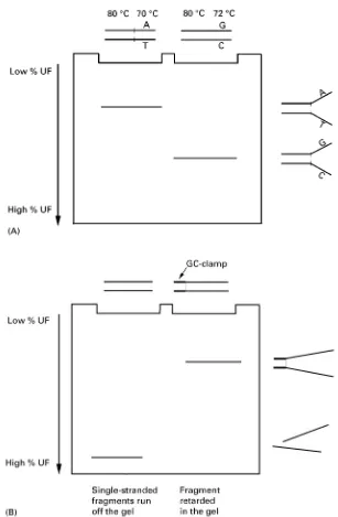
principle, in combination with PCR ampliRcation to prepare the target sequences. In DGGE, DNA frag-ments are subjected to electrophoresis in a polyac-rylamide gel against a gradient of ever higher temper-ature or chemical denaturants (i.e. a mixture of urea and formamide). Unlike nucleotide sequencing, DGGE detects mutations, including base pair substi-tutions and small insertions and deletions, on the basis of differences in the melting temperature of the target fragments. A given DNA fragment com-prises one or more domains, each representing a stretch of between 50 and 300 base pairs with equal melting temperature (the temperature at which each base pair has a 50% probability of being in either the helical or the denatured state). Since the stability of each domain depends on its sequence, mutational differences among different fragments are revealed as migrational differences in the gel (Figure 1A).
Figure 1 Principles of denaturing gradient gel electrophoresis. (A) Single base changes affect the melting temperature of a fragment, which results in a gel shift. (B) After complete denaturation, single-stranded fragments will run off the gel; the addition of a GC-clamp to the target fragment prevents complete denaturation and therefore fragments will be retarded in the gel. (C) The addition of a GC-clamp to a multiple-domain fragment can make the fragment behave as a single-domain fragment. Continuous line: target fragment without a GC-clamp. Dashed line: target fragment, including a 40 bp GC-clamp. (D) The introduction of a heteroduplex cycle at the end of PCR amplification of target fragments facilitates detection of heterozygous mutations as four molecules: two homoduplexes and two (early-melting) heteroduplexes.
melting domain. In general, target fragments in DGGE have an average size of 275 bp, including a GC-clamp and PCR primer sequences.
The sensitivity of DGGE for detecting variants is further enhanced by the introduction of a heterodup-lexing step using one round of denaturation/ renatura-tion, usually at the end of PCR ampliRcation of the target fragment. In this manner, a heterozygous
Figure 1 Continued
Although DGGE has the crucial advantage of hav-ing virtually 100% sensitivity in detecthav-ing mutations, it has typically been applied in a serial fashion, e.g. on a fragment-by-fragment basis. For analysing large genes or multiple genes this is not practical. A solu-tion for this problem, which we adopted, is to apply the DGGE principle in the format as it was originally described, i.e. a two-dimensional system of separ-ation by size followed by DGGE. Successful imple-mentation of such a two-dimensional DNA elec-trophoresis system in mutation scanning of large genes requires an efRcient multiplex PCR proto-col. Indeed, without the possibility to PCR-amplify multiple target fragments (i.e. typically 10 or more) in one single reaction, the application of a parallel
analysis system offers only a limited advantage. Multiplex PCR systems for genes and genetic markers are now becoming available and it has been demon-strated that as many as 26 fragments can be co-ampliRed in one single tube under the same reaction conditions.
Two-Dimensional DNA Electrophoresis
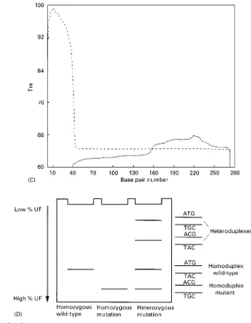
Figure 2 Schematic depiction of a TDGS test. All exons are amplified in an extensive multiplex reaction, and the fragments are resolved by size separation, followed by separation in a gradient of denaturants. Heterozygous mutations would show up as four spots instead of one.
denaturants. This opens up the possibility to analyse multiple fragments for all possible mutations under the same set of experimental conditions. The total number of target fragments that can be analysed simultaneously depends on the resolution of the gel system used. Although high resolution can be ob-tained by using one-dimensional denaturing gradient gels, two-dimensional separation allows characteriza-tion of each fragment on the basis of two independent criteria, size and melting temperature. In practice, a fragment mixture corresponding to all exons of a gene is electrophoresed in a nondenaturing size gel. Fragments are further sorted out in a denaturing gradient gel as the second dimension (Figure 2). By using the two-dimensional system, it is possible to visualize completely all fragments corresponding to
an entire gene for a particular DNA sample and immediately recognize each exon and variants there-in. This has been demonstrated for several large hu-man disease genes, including CFTR, RB1, MLH1, TP53, TSC1, BRCA1, as well as for a part of the mitochondrial genome.
Design Software and Instrumentation
for TDGS Tests
Computer-Automated Design of Target Fragments for PCR and DGGE
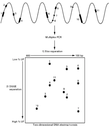
Figure 3 Two automatic dual-gel TDGS systems.
single-domain fragments which can be resolved under one set of electrophoretic conditions. To design com-plete gene tests for mutational analysis by TDGS, an automated generally applicable computer program was developed, which was based on a commercially available primer design program (Primer Designer 3; ScientiRc and Educational Software, State Line, PA), the melting routine MELT87 and a newly generated spot distribution routine. After entering a gene’s cod-ing sequence as exons with their Sanking intronic sequences, a rank of suitable PCR primers for each exon is designed by the PCR design subroutine. Next, the best primer pair is used in the melt subroutine to check for a one-domain target fragment. The program uses different GC-clamps at either the 5 or 3 end of the target fragment and, if necessary, additional small GC- or AT-clamps at either side of the target fragment. If it is impossible to design a one-domain fragment, the next optimal primer pair is tested, and so on. If a primer pair suitable to create a one-domain fragment cannot be found, the exon is split.
As soon as primers fulRl PCR and melting criteria, the fragment is positioned according to its size (x) and melting (y) coordinates. The spot distribution routine then checks for possible overlap. The output Rle of the program is a complete list of primers to be used in TDGS. (Questions regarding the use of the TDGS software should be directed to Accelerated Genomics, Concord, NH, http://www.accelerated genomics.com Tel.: (210) 616-5910; fax: (210) 692-7502.)
Electrophoresis
For two-dimensional DNA electrophoresis, originally two different gels were used for the R
rst-dimen-sion (separation according to size) and the subsequent second-dimension separation of these fragments by DGGE on the basis of their melting temperature. The Rrst-dimension separation was carried out in poly-acrylamide slab gels, which required staining of the gel to visualize the one-dimension separation pattern before this could be excised and transferred to the second-dimension denaturing gradient gel. Alternatively, tube gels have been employed for size separation, which obviated the need for gel staining and lane excision. However, routine application of TDGS requires standardization and automation, which is incompatible with the labour-intensive step of manual interference between theRrst- and second-dimension separation.
Recently, we developed a simple automated two-dimension instrument, which is based on an existing vertical electrophoresis system with an isolated hori-zontal unit on top (Figure 3). This top unit consists of two outer chambers and one middle chamber. The necessary contacts between the outer buffer chambers and the gel are provided by two strategi-cally located openings in the inner glass plate.
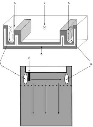
Figure 4 Schematic depiction of the automated two-dimensional electrophoresis unit. Buffer chambers for the first-dimension separation (A) are connected with the gel through openings in the (inner) glass plate (B). During the first-dimension electrophoresis, the middle buffer chamber (C) is isolated from the outer chambers. For the second-dimension run, buffer chamber C is flooded with buffer and the upper electrode is turned on in conjunction with a positive electrode in the lower reservoir (not shown in this figure). The gel cassette is sealed to the top unit with a serpentine silicone gasket (D), and sample is loaded in the single slot (E). The dashed line indicates the beginning of the gradient of urea/formamide.
electrophoresis is carried out vertically in the denaturing gradient gel. All components of the automatic TDGS electrophoresis system are depicted inFigure 6.
Gradient gels can be poured, up to nine at a time, using a simple linear gradient maker in combination with a multiple gel caster. The exact gradient that is to be applied is dependent on the GC-content of the gene(s) of interest and is determined by the TDGS primer designer software.
Miniaturization
Miniaturization of gene analysis systems, such as the TDGS system described here, offers two major advantages: increased speed and lower cost.
Figure 5 The entire automatic TDGS system. In this version of the system, four gels can be run simultaneously submerged in a buffer tank, which is equipped with a heater/stirrer to provide for a constant temperature. For more information, see http://www.cbssci.com
are 100 V, 5 h for the size separation and 100 V, 16 h for the second-dimension separation. Increasing the voltage increases the heat production, which nega-tively affects gel resolution. An obvious strategy is the use of thinner gels, which facilitate rapid heat dissipation into the surrounding buffer and thereby allow increasing the voltage while maintain-ing a good resolution. Currently, gels as thin as 0.35 mm are now run at 500 V, 0.8 h for the size separation and 500 V, 3.5 h for the second-dimension separation.
Cost The cost factor is of major importance for the large scale implementation of genetic testing. Since this is determined to a major extent by reagent and material cost, as well as space, miniaturization of analytical systems is of crucial importance. Miniatur-ization of TDGS results in thinner and smaller gels, which require less sample (smaller PCR volumes can now be applied) and lower gel and buffer amounts. Moreover, they take up less space. Instead of the current 17;22 cm format, two-dimensional patterns have already been produced on 10;10 cm mini-gels, and it is not unreasonable to expect that ultimately electrophoresis will be carried out on glass slides.
Detection of TDGS Patterns
After electrophoresis, the two-dimensional DNA fragment patterns can be visualized by incubating the gels with DNA staining dyes. Examples are ethidium bromide or the more sensitive dye Sybr-green. Pat-terns are photographed under UV light and evaluated
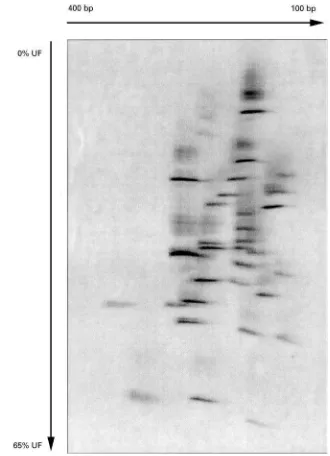
for the occurrence of variations (in the form of four spot patterns; see Figure 1D). An example of a TDGS pattern is shown inFigure 7, depicting theRB1gene, containing a mutation in exon 2.
However, for large scale application of TDGS, dye primer technology for the in-gel detection of two-dimensional spot patterns is an obvious strategy. Test results indicate similar two-dimensional patterns and sensitivity forSuorescein-labelled primers compared to Sybr-green-stained gels.
Introduction of Suorescent detection offers two advantages over gel staining. First, the reduction in labour is considerable and loss of gels due to breakage is prevented. Second, since there is no need to release the gel from between the glass plates it has become possible to use thinner gels, which will allow shorter electrophoresis times (see above). To increase the efRciency even further it is possible to label different samples with different Suorophores. CurrentSuorescence imagers have the option to ana-lyse multiplesSuorophores in the same gel.
Future Developments
Routine application of TDGS requires standardiz-ation and further streamlining of the procedure. Ulti-mately, one could envisage a fully automated system of PCR ampliRcation, sample loading, electrophor-esis, scanning of gels by a Suorescent imager, fol-lowed by online interpretation of gels by image analy-sis systems.
Figure 6 Line drawing of all the components of a 4-gel automatic TDGS system.
diminished by PCR robotics. Such instruments have now become widely available and, in combination with an ongoing effort to increase multiplex groups, are expected to increase greatly the front-end throughput of genetic testing. Multiple two-dimen-sional gels can be stacked for simultaneous elec-trophoresis of manifold samples. A simple robot arm could load the gel sandwiches into the Suorescent imager for quick gel scanning. Finally, while the
Figure 7 Empirical TDGS pattern of the retinoblastoma susceptibility geneRB1, containing a mutation in exon 2.
conRguration. Such software should also be capable of storing two-dimensional patterns and link subsets of them in particular experiments requiring compari-sons of large numbers of individuals. It could also provide for a sample tracking system.
Further Reading
Cotton RGH (1997)Mutation Detection. Oxford: Oxford University Press.
Dhanda RK, Smith WM, Scott CBet al. (1998) A simple system for automated two-dimensional electrophoresis: applications to genetic testing. Genetic Testing 2: 67}70.
Eng C and Vijg J (1997) Genetic testing: the problems and the promise.Nature Biotechnology15: 422}426. Fischer SG and Lerman LS (1979) Length-independent
sep-aration of DNA restriction fragments in two-dimen-sional gel electrophoresis.Cell16: 191}200.
Lerman LS and Silverstein K (1987) Computational simula-tion of DNA melting and its applicasimula-tion to denaturing gradient gel electrophoresis. Methods in Enzymology 155: 501}527.
van Orsouw NJ, Li D, van der Vlies Pet al. (1996) Muta-tional scanning of large genes by extensive PCR multi-plexing and two-dimensional electrophoresis: applica-tion to the RB1 gene. Human Molecular Genetics 5: 755}761.
van Orsouw NJ, Dhanda RK, Rines DRet al. (1998) Rapid design of denaturing gradient-based two-dimensional
electrophoretic gene mutational scanning tests.Nucleic Acids Research10: 2398}2406.
Vijg J and van Orsouw NJ (1999) Two-dimensional gene scanning: exploring human genetic variability. Electro-phoresis20: 1239}1249.
GEOCHEMICAL ANALYSIS:
GAS CHROMATOGRAPHY AND GC-MS
R. P. Philp, University of Oklahoma, Norman, OK, USA
Copyright^ 2000 Academic Press
Introduction
Geochemical analysis, and more speciRcally chromatography, is concerned with samples derived from two different sources: those of relatively recent origin, related to environmental problems; and those of a much greater geological age, related to fossil fuel exploration and exploitation. The chromatographic techniques utilized to analyse and characterize such samples are virtually identical re-gardless of the age and origin of the sample. The extracts from geochemical samples, whether they are rocks, soils, crude oil spills, contaminated wildlife or spills of reRned products, are very complex mixtures of a wide variety of organic compounds. Compounds derived from fossil fuels typically will be complex mixtures of hydrocarbons, and the environmental samples from more recent sediments probably will contain a variety of other compounds such as chlorin-ated compounds, pesticides or herbicides. In view of the similarities of the techniques used for analysing the samples from these different sources, the ma-jority of examples used in this article to illustrate the techniques will be based on the characterization of fossil fuel samples.
The major goal of any geochemical analysis is to take a sample and, through a variety of fractionations and analytical techniques, reach a point where either the presence or absence of speciRc target compounds can be determined, orRngerprints for speciRc classes of compounds can be obtained and used for correla-tion purposes. Applicacorrela-tions related to petroleum exploration might use suchRngerprints for oil}source rock or oil}oil correlation studies, whereas in
envir-onmental studies one is more concerned with cor-relating a spilled product with its original source material or trying to evaluate the extent of removal during clean-up procedures.