R E S E A R C H
Open Access
Characterisation and manipulation of docetaxel
resistant prostate cancer cell lines
Amanda J O
’
Neill
1*†, Maria Prencipe
1†, Catherine Dowling
1†, Yue Fan
1, Laoighse Mulrane
2, William M Gallagher
2,
Darran O
’
Connor
2, Robert O
’
Connor
3, Aoife Devery
3, Claire Corcoran
4, Sweta Rani
4, Lorraine O
’
Driscoll
4,
John M Fitzpatrick
1and R William G Watson
1Abstract
Background:There is no effective treatment strategy for advanced castration-resistant prostate cancer. Although Docetaxel (Taxotere®) represents the most active chemotherapeutic agent it only gives a modest survival
advantage with most patients eventually progressing because of inherent or acquired drug resistance. The aims of this study were to further investigate the mechanisms of resistance to Docetaxel. Three Docetaxel resistant sub-lines were generated and confirmed to be resistant to the apoptotic and anti-proliferative effects of increasing concentrations of Docetaxel.
Results:The resistant DU-145 R and 22RV1 R had expression of P-glycoprotein and its inhibition with Elacridar partially and totally reversed the resistant phenotype in the two cell lines respectively, which was not seen in the PC-3 resistant sublines. Resistance was also not mediated in the PC-3 cells by cellular senescence or autophagy but multiple changes in pro- and anti-apoptotic genes and proteins were demonstrated. Even though there were lower basal levels of NF-B activity in the PC-3 D12 cells compared to the Parental PC-3, docetaxel induced higher NF-B activity and IB phosphorylation at 3 and 6 hours with only minor changes in the DU-145 cells. Inhibition of NF-B with the BAY 11-7082 inhibitor reversed the resistance to Docetaxel.
Conclusion:This study confirms that multiple mechanisms contribute to Docetaxel resistance and the central transcription factor NF-B plays an immensely important role in determining docetaxel-resistance which may represent an appropriate therapeutic target.
Keywords:Docetaxel, Prostate, NF-?κ?B, Apoptosis, Viability
Background
Unfortunately there is no effective treatment strategy for advanced castration-resistant prostate cancer [1,2]. Although Docetaxel (Taxotere®) currently represents the most active chemotherapeutic agent it only gives a modest survival advantage with most patients eventually progressing because of inherent or acquired drug resis-tance. A number of mechanisms have been proposed to contribute to this resistance. Firstly, the majority of prostate tumours are slow growing even in metastatic disease and thus are unlikely to respond to drugs that
are S phase-dependent [3]. Secondly, failure of che-motherapy may be caused by reduced intracellular con-centrations of a drug through either increased efflux or decreased intake secondary to alterations in drug trans-porters, particularly P-glycoprotein (P-gp). Multidrug resistance (MDR) mechanisms including increased expression of the P-gp or increased cellular metabolism of drug detoxifying proteins, such as glutathione-S-transferase, have been shown to protect the cancer cells against cytotoxic drugs [4]. Thirdly, alterations in b -tublin isotypes with different kinetics of microtubule formation have been shown to contribute to resistance. With an increase in isotypes III and IV correlating with Docetaxel resistancein vitro[5]. Fourthly, mutations in tumour suppressor proteins, such as loss of PTEN which is a common event occurring in about 60% of
* Correspondence: [email protected]
†Contributed equally
1UCD School of Medicine and Medical Science, UCD Conway Institute of
Biomolecular and Biomedical Research, University College Dublin, Dublin, Ireland
Full list of author information is available at the end of the article
prostate cancer patients results in the activation of the phosphatidylinositol 3’-kinase (PI3K) signal transduction cascade resulting in increased cellular proliferation and survival mediated by AKT [6]. Finally, as the prostate cancer phenotype progresses there is the expression of survival factors that inhibits the apoptotic cell death pathway [7], mediated in part by the activation of AKT and other survival signalling pathways. Studies by our group and others have identified elevated protein levels of Bcl-2 [7], Inhibitors of Apoptosis proteins (cIAP-1, cIAP-2, XIAP and Survivin) [8], Clusterin and Heat Shock Proteins [9], as important anti-apoptotic proteins in the development of resistance to a number of apopto-tic triggers. However the identification and manipulation of these multiple mechanisms represents a significant challenge as targeting individual proteins has little clini-cal impact. This was demonstrated in a recent phase II clinical trial with oblimersen sodium, a Bcl-2 antisense oligonucleotide and Docetaxel which did not achieve its primary endpoint of reducing PSA and was associated with increased toxicity [10]. However strategies to block multiple Bcl-2 family members are under way with AT-101, a small molecular inhibitor of Bcl-2, Bcl-xl, Bcl-w and Mcl-1 (clinicaltrials.gov ID: NCT00571675).
Another approach to block multiple downstream genes is to inhibit central transcription factors. There is increasing evidence that inflammation drives the devel-opment and progression of prostate cancer [11]. Nuclear factor kappa B (NF-B) is a central transcription factors activated by inflammation and other cells stresses including paclitaxel [12]. Inhibition of NF-B with CAPE increases caspase dependent cell death in PC-3 cells mediated via a reduction in IAP expression [12]. Indirect inhibition of NF-B with an IKK complex inhi-bitor enhances Docetaxel induced apoptosis in PC-3 and DU-145 cells [13].
This study was undertaken to further investigate the mechanisms of resistance to Docetaxel. A number of Docetaxel resistant sub-lines were generated in the androgen-independent (PC-3, DU-145) and sensitive (22RV1) cell lines. Resistance in the 22RV1 R cells was explained via over expression of P-gp which could be reversed by its inhibition. DU-145 cells have lower levels of P-gp and the resistance was partially blocked by Ela-cridar. PC-3 cells had no detectable levels of P-gp and Elacridar had no effect on resistance to Docetaxel. Resis-tance was also not mediated by cellular senescence or autophagy in the PC-3 cells, but multiple changes in pro- and anti-apoptotic genes and proteins were demon-strated. Even though there were lower basal levels of NF-B activity in the PC-3 D12 cells compared to the Parental PC-3, docetaxel induced higher levels of NF-B activity and IB phosphorylation with only minor changes in the DU-145 cells. Inhibition of NF-B with
the BAY 11-7082 inhibitor reversed the resistance to Docetaxel.
Materials and methods
Cell culture and resistant cell line development
The human prostate cancer cell lines PC-3, DU-145 and 22RV1 were purchased from the American Type Culture Collection (ATCC) and maintained in RPMI-1640 med-ium supplemented with 10% Fetal Bovine Serum (FBS), 50 U/ml penicillin/50μg/ml streptomycin and 2 mM L-glutamine (Invitrogen).
PC-3 resistant sub-lines were generated by initially treating with Docetaxel (Sigma) at 4 nM and 8 nM (sus-pended in dimethyl sulfoxide (DMSO) (Fluke)) in 75 cm2 flasks for 48 hours. After treatment, the surviving cells were re-seeded into new flasks and allowed to recover for 2-3 weeks. After 5 (at 8 nM) and 7 (at 4 nM) treatments, the dose of docetaxel was increased from 4 nM and 8 nM to 8 nM and 12 nM respectively. The cells underwent a total of 18 treatment cycles at 8 nM and 12 treatment cycles at 12 nM, (cell passage numbers 21-55). Following each treatment they were allowed to fully recover before assessing their resistance to docetaxel and any experimental work. As the passage number of these treated cells increased over time, a sub-set of PC-3 cells were aged alongside these cells as an appropriate control to ensure that the effects seen were due to resistance rather than due to an ageing effect of the PC-3 cells (PC-3 Ag). In all subsequent experiments, the resistant cells are referred to as PC-3 D8 and PC-3 D12 (reflecting the final treatment doses), and the age-ing control cells are referred to as P-C3 Ag. Batches of cells were frozen down and all experiments were carried out on similar passages.
Docetaxel resistant cell lines (DU-145 R; 22RV1 R) were developed over a period of 6 months by stepwise increased concentrations of docetaxel. Cells were con-tinuously maintained in docetaxel, with treatments beginning at the initial IC50of the respective parent cell
lines. Media containing docetaxel was changed every 2-3 days. As cells displayed resistance to treatments of doce-taxel the concentration was subsequently increased with final treatment doses of 100 nM. Resistance was judged based on decreased cell death and increased prolifera-tion of cells. Age-matched parent cells (DU-145 Ag; 22RV1 Ag) which did not receive treatment were also maintained in culture. Batches of cells were frozen down and all experiments were carried out on similar passages.
Quantification of apoptosis and viability
Cells were harvested by trypsinisation, permeabilised with a hypotonic fluorochrome solution (50 mg/ml PI, 3.4 mM sodium citrate, 1 mM Tris, 0.1 mM EDTA, and 0.1% Triton X-100) and incubated on ice for 10 minutes prior to analysis. Samples were run on a Beck-man-Coulter FC-500 Cytometer. Ten thousand events were gated on PI intensity and analysed using Mplus software.
NF-B Inhibitor
Cells (100,000 cells/well) were pre-treated with the
B inhibitor, BAY 11-7082 (Merck), (5 μM) for 24 hours after which they were treated with docetaxel (20 nM) for a further 48 hours before been assessed for apoptosis as previously described above.
P-glycoprotein Inhibitor
Cells (100,000 cells/well) were pre-treated with the P-glycoprotein inhibitor, Elacridar (DCU), (0.25 & 0.5 μM) for 24 hours after which they were treated with doce-taxel (20 nM) for a further 48 hours before been assessed for apoptosis as previously described above.
3-(4,5)-dimethylthiazol-2-yl-2,5-diphenyltetrazolium bromide (MTT) assay cell viability assay
Cell viability was assessed by MTT cell staining as pre-viously described [15]. Ten thousand cells/well were cul-tured in a 96-well plate. Twenty-four hours later, cells were treated with several concentrations of Docetaxel (10 nM, 20 nM, 50 nM and 100 nM) for 24, 48 and 72 hours. MTT (50μl of a 5 mg/ml in PBS; Sigma-Aldrich) was added to each well and the cells were incubated in a CO2 incubator at 37°C for 5 hours. Following media
removal, the MTT-formazan formed by metabolically viable cells was dissolved in 200 μl of DMSO (Sigma-Aldrich) and the absorbance was measured in a plate reader at 550 nm.
Senescence-associated-ß-galactosidase activity
Senescence was assessed by staining cells forb -galacto-sidase expression as previously described [16]. Briefly, 150,000 cells were seeded in 6 well/plates, 24 hours later cells were treated with several concentrations of Docetaxel for specific times as determined in the experi-mental design. The cells were then washed twice with PBS, fixed with 2% formaldehyde (Sigma) and 0.2% glu-taraldehyde (Sigma-Aldrich) in water for 10 minutes and washed again in 2 × PBS washes. Cells were stained with X-gal staining solution (1 mg/ml X-gal, 40 mmol/l citric acid/sodium phosphate pH 6, 5 mmol/l potassium ferrocyanide, 150 mmol/l NaCl, 2 mmol/l MgCl2) for 24
hours in a CO2 incubator at 37°C and then rinsed in 2
× washes of PBS and counted using a phase contrast microscope (Olympus CK2). Senescent cells were expressed as a percentage of the total number of cells counted (300 cells/well).
RNA isolation, cDNA synthesis and Real-time RT-PCR analysis using TaqMan Low Density Arrays
Total RNA was extracted using Trizol reagent (Invitro-gen, Paisley, UK) using standard procedures as pre-viously described [17]. The RNA was used to generate cDNA as previously described [17]. PCR amplification of cDNA template was performed in a thermal cycler (Perkin Elmer 7700).
Pre-designed TaqMan probe and primer sets for target genes were chosen from an on-line catalogue (Applied Biosystems). Once selected, the sets were factory-loaded into the 384 wells of TaqMan Low Density Arrays (LDAs). The array format was customized on-line with one replicate per target gene. Expression levels of target genes were normalised to 18S rRNA (Additional file 1 lists the 95 genes chosen for inclusion, and their func-tion). Samples were analyzed using the 7900HT system with a TaqMan LDA Upgrade (Applied Biosystems), according to the manufacturer’s instructions. In short, single-stranded cDNA (to final concentration of 100 ng as calculated from starting RNA) was combined with water and TaqMan Universal PCR Master Mix, follow-ing by loadfollow-ing 100μl of each sample per port. Thermal cycling conditions were as follows: 50°C for 2 min, 94°C for 10 min, 97°C for 30 s, and 59.7°C for 1 min. Gene expression values were calculated based on the ΔΔCt method, where one sample was designated the calibra-tor, through which all other samples were analysed. Briefly, ΔCt represents the threshold cycle (Ct) of the target minus that of 18S rRNA andΔΔCt represents the
ΔCt of each target minus that of the calibrator. Relative quantities were determined using the equation 2-ΔΔCt. For the calibrator sample (i.e. normal LCM tissue), the equation is relative quantity = 2-0, which is 1; therefore, every other sample is expressed relative to this [18].
Total cellular protein isolation and western blotting Whole cell lysates were extracted from treated cells grown to 90% confluence on T75 flasks and 6-well plates. Cells were washed in cold PBS (1100 rpm, 1 min, 4°C in a microcentrifuge) and resuspended in NP-40, Tris 10 mM pH 8.0, 60 mM KCl, 1 mM EDTA pH 8.0, 1.0 mM DTT, 10 μl/ml Protease Inhibitor Cocktail (Sigma P8340)/1 ml of lysis buffer and 10 mM PMSF. Samples were then placed on ice for 10 mins and the cell lysate collected after centrifugation (13000 rpm 5 mins at 4°C). Phosphorylated proteins were isolated using the same protocol above with the addition of 10
μl Phosphatase Inhibitor Cocktail (Sigma P2850)/1 ml of lysis buffer [18].
being trans-blotted onto Immobilin P (Millipore) mem-branes as previously described [14]. Western blotting was performed using antibodies to Bax (1/1000, Upstate Biotechnology), Bcl-xL (1/1000), Bcl-2 (1/500, BD Bios-ciences), Bid (1/1000, Biosource), Clusterin (1/1000), Id-1 (Id-1/200, Santa Cruz), LC3 (Id-1/Id-1000) P-gp antibody (Mdr-1 D11, 1/200, Santa Cruz), HSP 90 (1/500, Stress-Marq Biosciences Inc.) andb-Actin (1/5000, Sigma), fol-lowed by incubation with the appropriate horseradish peroxidase-conjugated secondary antibodies (Cell Signal-ing). Autophagy was assessed by western blotting, using an antibody against the autophagy marker LC3 (1/1000, Sigma-Aldrich). As a positive control for the induction of autophagy, cells were starved for 2 hours in Earl’s balanced salt solution (EBSS). Bafilomycin A1 (20 nM, Sigma) was used as an inhibitor of autophagy. Signals were detected using ECL™(Pierce).
Assessment of NK-B
TransAM™NF-B Family Transcription Factor Assay Kit NF-B activity in nuclear extracts in the cell lines was determined using the the TransAM™ NF-B Family Transcription Factor Assay Kit (Active Motif, Belgium). Cells were grown to confluency and nuclear extracts prepared as per the TransAM™ Assay Kit manual. Briefly, nuclear extracts were suspended in TransAM lysis buffer and nuclear proteins (12 μg total protein) were incubated with immobilized oligonucleotides con-taining the NF-B consensus DNA-binding site (5’ -GGGACTTTCC-3’) for 1 hour at room temperature. After washing, 100μl of one of the diluted NFB anti-bodies (1:1000 antibody in 1X Antibody Binding Buffer) was added to each well being used, including blank wells for 1 hour at room temperature. After 3 washes, 100 μl of horseradish-peroxidase-conjugated secondary antibody (1:1000 dilutions) were added to each well for 1 hour at room temperature. The colourimetric sub-strate was then added after 3 subsequent washes, and the plate was allowed to develop for up to 5 minutes. Following this, the reaction was stopped and absorbance read at 450 nm on a SpectraMax M2 plate reader (Molecular Devices, CA, USA). Relative activation of the NF-B subunits between the three cell lines could then be plotted, with an increase in absorbance being indica-tive of increased activation.
NF-B Reporter assay
NF-B transcriptional activity was assessed using a plas-mid containing the luciferase reporter gene regulated by five copies of an NF-B responsive element (Promega). A TK-Renilla luciferase plasmid was used as a transfec-tion efficiency control. Plasmids were co-transfected using GeneJuice® Transfection Reagent (Novagen) fol-lowing the manufacturer’s instructions. After 6 hours,
cells were treated with or without 50 nM of docetaxel, for 3, 6 and 24 hours. The luciferase and renilla activ-ities were measured using a Dual-Luciferase® reporter assay (Promega). The NF-B transcriptional activity was expressed as fold change of Relative Luciferase Units (ratio between luciferase light values divided by the renilla light values), taking the untreated controls as the baseline.
Statistical analysis
Statistical analysis was carried out using independent samples student t-tests. Results were considered statisti-cally significant where p < 0.05 and results are expressed as mean ± the standard deviation.
Results
Effects of Docetaxel on (A) Apoptosis and (B) Proliferation in the Docetaxel resistant sublines
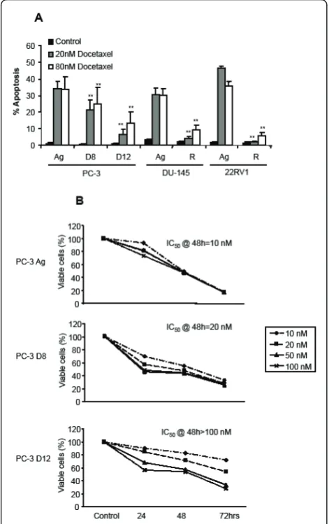
Figure 1A demonstrates the apoptotic effects of two dif-ferent concentrations of Docetaxel (20 nM and 80 nM) for 48 hours on the four different Docetaxel resistant sublines. PC-3 D8 and PC-3 D12 demonstrated partial but increasing resistance to docetaxel treatment over the different doses, when compared to the PC-3 Ag. The DU-145 R and 22RV1 R showed significantly more resistance compared to the aged matched control cells (DU-145 Ag & 22RV1 Ag). Cell viability was then deter-mined by the MTT assay (B), following treatment with docetaxel (10 nM, 20 nM, 50 nM and 100 nM) for 24, 48 and 72 hours. The PC-3 Ag cells had an IC50@ 48
hrs = 10 nM, the PC-3 D8 an IC50@ 48 hrs = 20 nM
and the PC-3 D12 an IC50@ 48 hrs = 100 nM following
treatment with Docetaxel (Figure 1B). This confirmed that the PC-3 D8 and PC-3 D12 sublines had a resis-tance to Docetaxel treatment.
P-glycoprotein (P-gp) expression in the Docetaxel resistant sub-lines
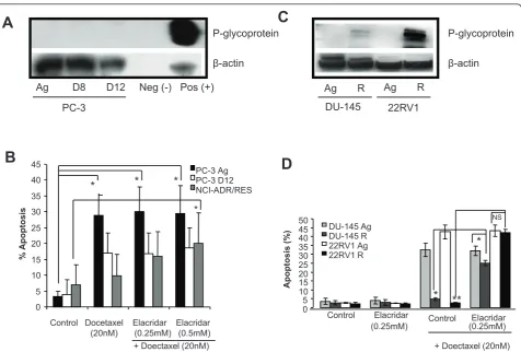
We next wanted to investigate the mechanisms respon-sible for Docetaxel resistance. We firstly examined the expression of the classical drug pump, P-gp (Mdr-1) in the PC-3 D8 and PC-3 D12 sublines compared to the PC-3 Ag subline. Figure 2A clearly shows no expression of P-gp in any of the PC-3 sublines, when compared to the P-gp positive cell line (DLKP-A, Figure 2A), and the DLKP negative cell line, as previously described [19].
following treatment with Docetaxel (20 nM) after 48 hours (Figure 2B).
Similar experiments were carried out with the DU-145 R and 22RV1 R sublines. Western blotting demonstrated expression of P-gp in the DU-145 R and 22RV1 R sub-lines with higher expression in the 22RV1 R (Figure 2C). Elacridar treatment (0.25 and 0.5μM) also partially reversed the resistance to apoptosis in the DU-145 R cells and totally reversed the resistance in the 22RV1 R
sublines following treatment with Docetaxel (20 nM) for 48 hours (Figure 2D). As the resistance to Docetaxel induced apoptosis may be partially explained by the over expression of P-gp in the DU-145 R and 22RV1 R cells we focused on the PC-3 D8 and PC-3 D12 sublines whose resistance was not P-gp dependent.
Cellular senescence and autophagy as mechanisms of Docetaxel resistance
Senescent cells demonstrate resistance to apoptosis and express b-galactosidase. The effects of Docetaxel treat-ment (100 nM for 48 hours) on PC-3 Ag, PC-3 D8 and PC-3 D12 cellular senescence is demonstrated in Figure 3 which shows no significant increase inb-galactosidase staining.
Autophagy (macroautophagy) is also a well conserved mechanism by which cells adapt to stress such as starva-tion [22]. This complex cellular process has recently been associated with resistance to cancer therapies [23]. Total cellular protein was extracted from the different cell lines following Docetaxel treatment (50 nM) and assessed for the expression of LC3 II, a protein asso-ciated with autophagosome formation and a marker of autophagy [24]. While the resistant PC-3 D12 sub-lines showed a higher baseline expression of LC3I, the pre-cursor of LC3II, no consistent difference in the baseline expression levels of LC3II was demonstrated between the PC-3 Ag and the resistant PC-3 D12 subline (Figure 4) following treatment.
Altered expression of apoptotic related genes in the Docetaxel resistant sublines
Having ruled out P-gp, senescence and autophagy as possible mechanisms of resistance in the PC-3 resistant sublines we next investigated other mechanisms and focused on the alteration in genes and proteins which regulate cellular apoptosis. Custom designed Low Den-sity Arrays (LDA) which included the probes for the IAPs, death receptors, death ligands, and signalling molecules as well as genes involved in cell cycle regula-tion, DNA damage and repair and chemotherapy resis-tance were developed. Additional file 1, lists the 95 genes chosen for inclusion and their function.
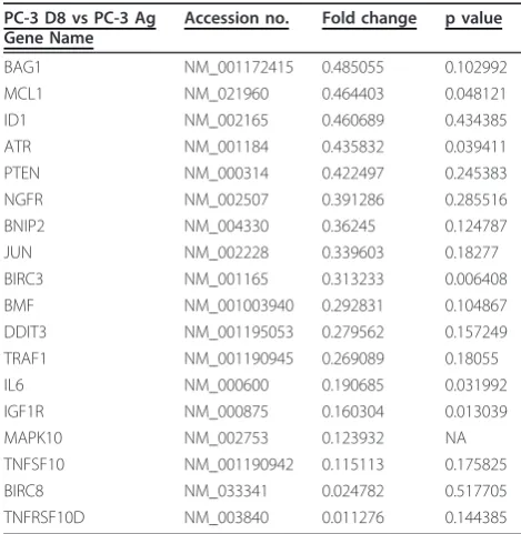
[image:5.595.56.292.90.465.2]Table 1 demonstrates the genes which were increased in the PC-3 D8 subline compared to the PC-3 Ag cells. The majority of these genes are anti-apoptotic including BIRC7 (Livin-a member of the IAP family), Bcl2-A1 (Bcl-2 member), Foxo1A and HSP 90. Clusterin, a mole-cular chaperone was also found to be increased. TNF receptor family member 10C, which is known to inhibit TRAIL-induced apoptosis, was also increased in this subline. Nibrin and p73 which are involved in DNA damage and repair were also up-regulated. Table 2 demonstrates the genes which were down-regulated in
the PC-3 D8 subline compared to the PC-3 Ag cells. A number of genes involved in the induction of apoptosis were shown to be changed. These include; FOXO1, NGFR, TRAF-1, and TRAF-2. Surprisingly a number of anti-apoptotic genes were also decreased including; MCL-1 and BIRC3.
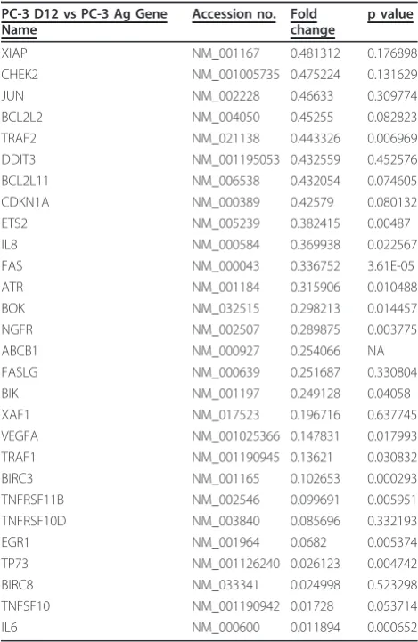
Genes increased in the PC3-D12 subline compared to the PC-3 Ag cells are represented in Table 3. Many of the increased genes are anti-apoptotic genes such as BIRC1, BCL2-A1, FOXO1A, NOL3 and Clusterin. Genes involved in DNA damage and repair such as Nibrin, Chek-1 and ATM were also increased. Decreased gene expression in the PC3-D12 subline com-pared to the PC-3 Ag cells is demonstrated in Table 4. Many of these genes are those involved in the induction
of apoptosis. These include BOK, NGFR, Fas, FasLG, TNF receptor member 11b, TRAF-1 and TRAIL. A number of genes involved in cell cycle regulation and DNA damage detection were also decreased. These include Chek2, p21, ETS2 and ATR.
Validation of altered gene expression
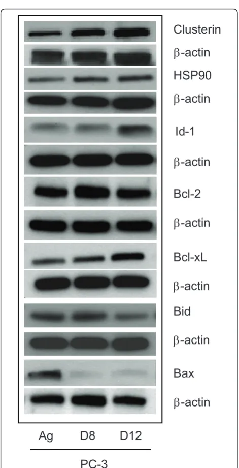
We next wanted to validate specific genes at the protein level. Total cellular protein was extracted from the PC-3 Ag, PC-3 D8 and PC-3 D12 and assessed by western blotting for Clusterin, Id-1, Bcl-2, Bcl-xL, Bid and Bax. As demonstrated in Figure 5, Id-1 expression was increased in the PC-3 D12 subline only, while the pro-tein expression of Bcl-2 was increased in PC-3 D8 but not in PC-3 D12. Anti-apoptotic Bcl-xL was increased P-glycoprotein
ȕ-actin
A
Neg (-) Pos (+)
Ag D8 D12
PC-3
C
Ag R DU-145
Ag R
22RV1
NS
**
*
*
0 5 10 15 20 25 30 35 40 45 50
Apoptosis (%
)
Control Elacridar
(0.25mM) Elacridar
D
DU-145 Ag DU-145 R 22RV1 Ag 22RV1 R
+ Doectaxel (20nM) Control
(0.25mM) P-glycoprotein
ȕ-actin
B
0 5 10 15 20 25 30 35 40 45
%
Apoptosis
Control Docetaxel Elacridar Elacridar (20nM) (0.25mM) (0.5mM)
+ Doectaxel (20nM) PC-3 Ag PC-3 D12 NCI-ADR/RES
*
*
*
[image:6.595.61.538.86.408.2]*
in both sublines. Pro-apoptotic protein Bid was decreased in PC-3 D12 while Bax was decreased in both sublines. Clusterin was significantly increased in both sublines. The molecular chaperone HSP90 was also increased in both sublines (Figure 5).
Docetaxel treatment increases NF-B activity in the PC-3 D12 resistant PC-3 subline
As NF-B regulated the expression of a number of the apoptotic genes listed in Tables 1, 2, 3 and 4, we next Control 100 nM Docetaxel
PC-3 Ag
PC-3 D8
PC-3 D12
0.4% (2/485) 0.6% (2/318)
0.7% (2/276) 1% (1/100)
[image:7.595.57.291.86.320.2]1.1% (3/267) 1.4% (5/350)
Figure 3 Cellular sensecence in response to Docetaxel treatment. The PC-3 Ag, PC-3 D8 and PC-3 D12 cells were plated in 6 well plates at 300,000/well and grown for 24 hours. The cells were then treated with Docetaxel (100 nM) for 48 hours and fixed with 2% formaldehyde and 0.2% glutaraldehyde. Senescent cells were then assessed by staining cells forb-galactosidase expression. Positive cells were counted and expressed as a percentage of the total number of cells in the well. Pictures are a representation of 5 independent experiments.
LC3 I LC3 II
ȕ-actin
LC3 I LC3 II
ȕ-actin
18 KD 16 KD 46 KD
18 KD 16 KD 46 KD C 1 3 6 12 24 R S S+B PC-3 Ag
PC-3 D12
[image:7.595.303.539.111.303.2]Docetaxel (50 nM)
[image:7.595.303.538.472.713.2]Figure 4Autophagy as a mechanisms of Docetaxel resistance. Following treatment of the Aged matched controls (Upper panel) and PC-3 D12 resistant sublines (Bottom panel) with 50 nM of Docetaxel for 1, 3, 6, 12 and 24 hrs, total cellular protein was extracted and assessed for LC3II by western blotting using the relevant anti-body and conditions as described in the methods section. For positive controls cells were either treated with Rapamycin (R, 0.2μM) for 4 hrs, Starvation (S, the cells were starved in EBSS (Earls Balanced Salt Solution) medium for 2 hrs), or Starvation plus Bafilomycin (S+B, 20 nM) for 2 hrs.b-actin the house keeping protein was used as loading control. Blots shown are representative of three independent experiments.
Table 1 Genes increased in the PC-3 D8 Docetaxel Resistant Cell line
PC-3 D8 vs PC-3 Ag Gene Name
Accession no. Fold change
p value
BIK NM_001197 4.606724005 0.024378 FOXO1 NM_002015 4.092077589 0.006372 NAIP NM_004536 3.895819042 NA HSP90AA1 NM_001017963 2.255183574 0.116769 IL8 NM_000584 2.194814925 0.060076 BCL2A1 NM_001114735 2.003718027 0.018472 NBN NM_002485 1.994366021 0.011912 TNFRSF10C NM_003841 1.931533233 0.288353 TP73 NM_001126240 1.785086313 0.082871 BNIP3L NM_004331 1.747205486 0.03454 CLU NM_001171138 1.740002897 0.304795 APAF1 NM_001160 1.673345235 0.38724 FASLG NM_000639 1.563978305 0.73945 BNIP3 NM_004052 1.50139036 0.173556 Pre-designed TaqMan probe and primer sets for target genes were chosen from an on-line catalogue (Applied Biosystems). Expression levels of target genes were normalised to concentration of 18S rRNA (Additional File 1 lists the 95 genes chosen for inclusion and their function). Total mRNA was extracted from the PC-3 Ag and PC-3 D8 cells and used on custom designed Low Density Arrays (LDA) to determine which genes were increased (fold increase > 1.5) in the Docetaxel resistanct PC-3 D8 subline compared to the PC-3 Ag. n = 3.
Table 2 Genes decreased in the PC-3 D8 Docetaxel Resistant Cell line
PC-3 D8 vs PC-3 Ag Gene Name
Accession no. Fold change p value
[image:7.595.58.291.511.604.2]wanted to determine whether NF-B played a role in the resistance to Docetaxel. We firstly assessed the base-line transcriptional activity in the PC-3 resistant subbase-line using a luciferase assay. Interestingly, NF-B transcrip-tional activity was significantly decreased (p < 0.001) in the resistant PC-3 D12 subline compared to the PC-3 Ag cells (Figure 6A). The DU145 cell lines showed a similar trend with decreased NF-B transcriptional activity in the resistant subline (DU-145 R) compared to the parental (DU-145 Ag); while there was no difference in NF-B activity in the aged (22RV1 Ag) matched and resistant 22RV1 (22RV1 R), being the baseline NF-B activity in these cells was very low in comparison to the
PC-3 and DU145 cells (data not shown). NF-B
decreased activity in the resistant PC-3 sublines was confirmed by the TransAM ELISA based assay which showed a significant decrease in the activity of p52 (p < 0.01) and RelB (p < 0.05) subunits, in both the PC-3 D8 and PC-3 D12 resistant cells compared to the PC-3 Ag (Figure 6B).
We then assessed the effects of Docetaxel treatment on NF-B transcriptional activity in the resistant sub-line. Following Docetaxel treatment there was signifi-cantly increased transcriptional activity in the PC-3 D12 resistant subline compared to the PC-3 Ag cells (Figure 7A). There was no increase in the DU-145 R subline (Figure 7A), and there was no NF-B activity in the 22RV1 cells neither in response to docetaxel or to the classical NF-B inducers such as TNFa or LPS (data not shown). Activation of NF-B in the resistant PC-3 D12 subline compared to the PC-3 Ag cells was
confirmed by western blotting showing higher levels of phospho-IBa in the PC-3 D12 compared to the PC-3 Ag, following 3-6 h of docetaxel treatment (Figure 7B). NF-B was functionally confirmed to be centrally involved in resistance to Docetaxel using the NF-B inhibitor (BAY 11-7082, Merck) which reversed resis-tance in both the PC-3 D8 and PC-3 D12 sublines (Figure 8).
Discussion
[image:8.595.304.539.110.471.2]The treatment of advanced prostate cancer represents a significant challenge due to the accumulation of multi-ple mechanisms of drug resistance. Resistance to Doce-taxel treatment includes changes in classical multiple drug resistant pathways, expression of differentb-tublin isotypes, mutations in tumour suppressor proteins and altered expression of pro- and anti-apoptotic proteins [25]. We have generated three models of docetaxel-resistance in prostate cancer cells which may mimic the Table 3 Genes increased in the PC-3 D12 Docetaxel
Resistant Cell line
PC-3 D12 vs PC-3 Ag Gene Name
Accession no. Fold change
p value
[image:8.595.57.291.111.279.2]NAIP NM_004536 28.14023 NA BCL2A1 NM_001114735 9.956213 7.84E-06 CLU NM_001171138 6.138126 0.019757 TNFRSF1B NM_001066 4.568718 NA ATM NM_000051 2.884063 0.013665 FOXO1 NM_002015 2.746056 0.0167 MAP2K4 NM_003010 2.456245 0.13486 APAF1 NM_001160 2.264102 0.34641 NOL3 NM_001185057 1.946074 0.101148 CHEK1 NM_001114121 1.554884 0.433367 BNIP3L NM_004331 1.514918 0.113779 NBN NM_002485 1.505995 0.051555 Total mRNA was extracted from the PC-3 Ag and PC-3 D12 cells and used on custom designed Low Density Arrays (LDA) which included the transcriptional profile probes for the IAPs, death receptors, death ligands, and signalling molecules as well as genes involved in cell cycle regulation, DNA damage and repair and chemotherapy resistance. Additional File 1, lists the 95 genes chosen for inclusion, and their function. Genes whose expression has increased (fold increase > 1.5) in the Docetaxel resistanct PC-3 D12 subline compared to the PC-3 Ag. n = 3.
Table 4 Genes decreased in the PC-3 D12 Docetaxel Resistant Cell line
PC-3 D12 vs PC-3 Ag Gene Name
Accession no. Fold change
p value
situation in the clinical setting and used them to understand the complexity of mechanisms leading to treatment resistance. Understanding these pathways will be essential if we want to better impact on the outcome of these patients and identify additional co-treatments which will manipulate this resistance and develop personalised treatments. We demonstrated
that all four sublines are resistant to increasing con-centrations of Docetaxel as assessed by apoptosis and viability assays.
One of the main mechanisms of resistance to Doce-taxel is the over expression of P-gp [4] which would reduce intracellular concentrations of the drug through increased efflux. Chemoresistant PC-3 cells do not over express P-gp, while resistant DU145 cells exhibit both over expression of the protein and restored chemosensi-tivity with P-gp inhibition [5,26]. This has been the basis for the development of cabazitaxel which can over-come P-gp over expression [27] and is now used as a second line therapy for patients who fail Docetaxel treatments. In our study we demonstrated that the resis-tance in the PC-3 D8 and PC-3 D12 sublines was inde-pendent of P-gp over expression as there were no detectable levels and no effects of the P-gp inhibitor Ela-cridar on their resistance. However the DU-145 and 22RV1 cells expressed increasing levels of P-gp and Ela-cridar totally reversed the resistance in the 22RV1 and partially in the DU145 resistant sublines. Based on these results we focused on the PC-3 cells to understand the mechanisms of resistance independent of P-gp.
Bcl-xL
Bax
Id-1
Bcl-2
Bid
E
-actin
E
-actin
E
-actin
E
-actin
E
-actin
Clusterin
HSP90
E
-actin
E
-actin
Ag
D8
D12
[image:9.595.56.292.81.542.2]PC-3
Figure 5Validation of Low Density Arrays. Total cellular protein was extracted from the PC-3 sublines (PC-3 Ag, PC-3 D8 & PC-3 D12) and 50μg was loaded onto a western blot to assess the expression of Clusterin, HSP90, Id-1, Bcl-2, Bcl-xL, Bax, and Bid using their respective antibodies as described in the Method section. The house keeping proteinb-actin was used as a loading control. Blots shown are representative of three independent experiments.
A
**
0 5 10 15 20 25
RLU
PC-3 Ag PC-3 D12
* * * *
B
0 0.5
1 1.5
2 2.5
3
PC-3 Ag PC-3 D8 PC-3 D12
p50 p65 p52 Rel B cRel
Abs @
450nm
[image:9.595.304.540.89.322.2]We next investigated cellular senescence as a possible mechanism of resistance. Premature cellular senescence, a phenomenon by which cells stop dividing and acquire a typical morphology and an altered gene expression
profile in response to stress [3], has recently been asso-ciated with chemotherapy resistance. We performed a ß-galactosidase assay to assess cellular senescence fol-lowing docetaxel treatment. Although ß-galactosidase
NF
N
B inhibitor (5
μ
M)
*
*
0
10
20
30
40
50
%
Apoptosis
Control
Docetaxel (20nM)
PC-3 Ag
PC-3 D8
PC-3 D12
[image:10.595.58.540.87.380.2]-
+
-
+
-
+
Figure 8NF-B inhibitor studies following docetaxel treatment. The PC-3 sublines where pre-treated with the NFB inhibitor (BAY 11-7082, 5μM) for 24 hours followed by Docetaxel (20 nM) for a further 48 hours and assessed for apoptosis by flow cytometry. Graphs represent the results of 3 independent experiments. Mean values were compared using t-test assuming equal variances. * p < 0.05.
C 3 6 24hr C 3 6 24h
B
P-IkBĮ(40 kDa)
IkBĮ(35 kDa)
ȕ-actin(46 kDa)
PC-3 Ag PC-3 D12
P-IkBĮ(40 kDa)
IkBĮ(35 kDa)
ȕ-actin(46 kDa)
DU-145 Ag DU-145 R C 3 6 24hr C 3 6 24h
PC-3 Ag PC-3 D12 DU-145 Ag DU-145R
Fold change
*
*
*
**
**
A
[image:10.595.58.539.525.694.2]Control 3h Doc 50nM 6h Doc 50nM 24h Doc 50nM
staining showed a modest increase in the percentage of senescent cells following docetaxel treatment, this increase was considered to be insufficient to explain the resistance of the PC-3 D8 and PC-3 D12 sublines to docetaxel.
Autophagy (macroautophagy), a well conserved mechanism by which cells adapt to stress such as starva-tion, has also been recently associated with resistance to cancer therapies [23]. Therefore we investigated whether increased autophagy was involved in the resistance to docetaxel in the PC-3 D12 subline. Although western blotting showed increased expression of the autophagic marker LC3II in the PC-3 D12 compared to the PC-3 Ag cells following docetaxel treatment, this was only a modest effect. Interestingly, the resistant PC-3 D12 sub-line showed a higher basesub-line expression of LC3I, the precursor of LC3II, which warrants further investigation. We next undertook to assess alterations in the apop-totic phenotype of the resistant cells in order to under-stand the mechanisms involved. Custom-designed LDA containing real-time PCR assays for all the key apoptotic genes including the IAPs, death receptors, death ligands, and signalling molecules as well as genes involved in cell cycle regulation, DNA damage and repair were developed. The LDA results showed a complex interplay between changes in the expression of both pro- and anti-apoptotic proteins. From our study, genes which were shown to be altered in both the docetaxel-resistant sublines (PC-3 D8 and PC-3 D12) included; FOXO1, NGFR, TRAF-1, Mcl-1, BIRC3, BIRC1, Bcl-2-A1, NOL3, Clusterin, BOK, NGFR, Fas, FasLG, TNF receptor mem-ber 11b and TRAIL.
Trougakoset aldemonstrated that secreted clusterin (sCLU) knockdown in human prostate cancer cells induces significant reduction of cellular growth and higher rates of spontaneous endogenous apoptosis [28]. sCLU is a cytoprotective chaperone that stabilizes con-formations of proteins during times of cellular stress, thereby inhibiting protein aggregation and precipitation [29]. Additionally, sCLU can interact with and inhibit activated Bax, thereby inhibiting cytochrome C release and caspase activation [30]. In prostate cancer, sCLU levels have previously been correlated with Gleason grade [31]. A recent publication identified sCLU to be over-expressed in a docetaxel-resistant PC-3 subline and reported that knockdown of sClu chemosensitizes this cell line to taxane and mitoxantrone based chemother-apy bothin vitroand in vivo[32].
Bcl-2 related protein A1 (Bcl2-A1) or BLF1 is a mem-ber of the Bcl-2 family. Although it is regulated differ-ently from Bcl-2, it has similar anti-apoptotic activity. A recent publication demonstrated that Bcl2-A1 mediates resistance to doxorubicin in haematopoietic cell lines [33].
Modur et al found that FOXO1A was highly
expressed in normal prostate. They also noted that in PTEN-deficient prostate carcinoma cell lines, FOXO1A was cytoplasmically sequestered and inactive and expression of TRAIL, a pro-apoptotic effector, was decreased. They determined that TRAIL is a direct tar-get of FOXO1A, and they hypothesized that the loss of PTEN contributes to increased tumour cell survival through decreased transcriptional activity of FOXO1A and FOXO3A followed by decreased TRAIL expression and apoptosis [34].
BNIP3L which is also elevated in both sublines, plays a pro-survival role in apoptosis. Yasuda et al showed that in vitro translated BNIP3L binds specifically and directly with GST fusion proteins of various Bcl-2 family anti-apoptosis proteins, suggesting that, like BNIP3, BNIP3L may antagonize the activity of Bcl-2 family anti-apoptosis proteins [35]. In this setting, BNIP3L may be increased in our resistant sublines in an attempt to antagonize the very elevated levels of Bcl2-A1.
Genes down-regulated in both sublines include TRAIL, NGFR, TNF member 10D and ATR among others (see Tables 1, 2, 3 and 4 results). As PC-3 cells are highly resistant to TRAIL, it is unsurprising that levels of pro-apoptotic TRAIL are further decreased in the resistant sublines. Tumour necrosis factor superfam-ily member 10D (TNF member 10D) has been shown to act as a decoy receptor for TRAIL (TRAIL R4). Transi-ent over-expression of TRAIL R4 in cells normally sen-sitive to TRAIL-mediated apoptosis conferred complete protection, and Degli-Esposti et al suggested that one function of TRAIL R4 may be the inhibition of TRAIL cytotoxicity [36]. As a large number of genes were either increased or decreased in the docetaxel-resistant sub-lines, targeting these individually may not lead to altera-tions in the resistant phenotype but understanding the central signalling pathways and transcription factors would represent a more appropriate therapeutic target-ing approach.
Despite a decrease in the baseline expression levels of the NF-B subunits p52 and RelB and transcriptional activity, when the cells were treated with Docetaxel there was an increase in NF-B transcriptional activity. This led us to the inhibition of NF-B activity using the NF-B inhibitor BAY11-7082 prior to treatment with Docetaxel which resulted in significant reversal of Doce-taxel resistance.
Conclusion
A mounting body of evidence implicates dysregulation of cell-survival signalling pathways in the pathogenesis of castration-resistant prostate cancer. What is not clear is which are the most salient targets in these pathways that when inhibited will lead to reversal of resistance and the most effective clinical outcome. An improved understanding of the molecular mechanisms regulating castration-resistant prostate cancer will drive both the identification of appropriate treatment strategies and biomarkers to inform such strategies. Our current results indicate that NF-B plays an important role in determining Docetaxel resistance and attempts to sensi-tise resistant prostate cancer cells to Docetaxel therapy should target this transcription factor.
Additional material
Additional file 1: List of genes on the Low Density Arrays. This table contains the list of 95 genes contained on the Low Density Arrays including gene name, gene symbol and gene function for each gene included on the array.
Abbreviations
MDR: multi drug resistance; P-gp: p-glycoprotein; PI3K: phosphatidylinositol 3’-kinase
Acknowledgements
We acknowledge the technical support of Dr Alfonso Blanco in the Flow Cytometry core facility and Ms. Catherine Moss in the Transcriptomics core facility of the UCD Conway Institute of Biomolecular and Biomedical Research. The work was funded by a Science Foundation Ireland, Strategic Research Cluster award to Molecular Therapeutics for Cancer Ireland (award 08/SRC/B1410).
Author details
1UCD School of Medicine and Medical Science, UCD Conway Institute of
Biomolecular and Biomedical Research, University College Dublin, Dublin, Ireland.2UCD School of Biomolecular and Biomedical Science, UCD Conway
Institute of Biomolecular and Biomedical Research, University College Dublin, Dublin, Ireland.3National Institute for Cellular Biotechnology, Dublin City
University, Ireland.4School of Pharmacy and Pharmaceutical Sciences, Trinity College Dublin, Dublin, Ireland.
Authors’contributions
AJON undertook the low density arrays and western blotting validation assays as well as preparing and writing the manuscript. MP undertook the NF-κB experiments as well as preparing and writing the manuscript. CD made the Docetaxel resitant PC-3 cell lines and performed the viability and apoptosis experiments. AJON, MP and CD contributed equally to the delivery of the manuscript. YF did the statistical analysis for the manuscript.
LM, WMG, and DOC undertook the NFκB Trans Am kit experiments. ROC and AD examined the P-gp protein expression in the cell lines and provided the inhibitor. CC, SR and LOD made the DU-145 and 22RV1 Docetaxel resistant cell lines. JMF and RWGW conceived the study. RWGW was central in the design, coordination and manuscript editing. All authors have read and approved the final manuscript.
Competing interests
The authors declare that they have no competing interests.
Received: 18 July 2011 Accepted: 7 October 2011 Published: 7 October 2011
References
1. Mostaghel EA, Page ST, Lin DW, Fazli L, Coleman IM, True LD, Knudsen B, Hess DL, Nelson CC, Matsumoto AM,et al:Intraprostatic androgens and androgen-regulated gene expression persist after testosterone suppression: therapeutic implications for castration-resistant prostate cancer.Cancer Res2007,67:5033-5041.
2. Scher HI, Halabi S, Tannock I, Morris M, Sternberg CN, Carducci MA, Eisenberger MA, Higano C, Bubley GJ, Dreicer R,et al:Design and end points of clinical trials for patients with progressive prostate cancer and castrate levels of testosterone: recommendations of the Prostate Cancer Clinical Trials Working Group.J Clin Oncol2008,26:1148-1159.
3. Raghavan D, Koczwara B, Javle M:Evolving strategies of cytotoxic chemotherapy for advanced prostate cancer.Eur J Cancer1997,
33:566-574.
4. van Brussel JP, Mickisch GH:Multidrug resistance in prostate cancer.
Onkologie2003,26:175-181.
5. Makarovskiy AN, Siryaporn E, Hixson DC, Akerley W:Survival of docetaxel-resistant prostate cancer cells in vitro depends on phenotype alterations and continuity of drug exposure.Cell Mol Life Sci2002,59:1198-1211. 6. Shen MM, Abate-Shen C:Pten inactivation and the emergence of
androgen-independent prostate cancer.Cancer Res2007,67:6535-6538. 7. Feldman BJ, Feldman D:The development of androgen-independent
prostate cancer.Nat Rev Cancer2001,1:34-45.
8. McEleny K, Coffey R, Morrissey C, Williamson K, Zangemeister-Wittke U, Fitzpatrick JM, Watson RW:An antisense oligonucleotide to cIAP-1 sensitizes prostate cancer cells to fas and TNFalpha mediated apoptosis.
Prostate2004,59:419-425.
9. Gibbons NB, Watson RW, Coffey RN, Brady HP, Fitzpatrick JM:Heat-shock proteins inhibit induction of prostate cancer cell apoptosis.Prostate2000,
45:58-65.
10. Sternberg CN, Dumez H, Van Poppel H, Skoneczna I, Sella A, Daugaard G, Gil T, Graham J, Carpentier P, Calabro F,et al:Docetaxel plus oblimersen sodium (Bcl-2 antisense oligonucleotide): an EORTC multicenter, randomized phase II study in patients with castration-resistant prostate cancer.Ann Oncol2009,20:1264-1269.
11. De Marzo AM, Platz EA, Sutcliffe S, Xu J, Gronberg H, Drake CG, Nakai Y, Isaacs WB, Nelson WG:Inflammation in prostate carcinogenesis.Nat Rev Cancer2007,7:256-269.
12. McEleny K, Coffey R, Morrissey C, Fitzpatrick JM, Watson RW:Caffeic acid phenethyl ester-induced PC-3 cell apoptosis is caspase-dependent and mediated through the loss of inhibitors of apoptosis proteins.BJU Int
2004,94:402-406.
13. Domingo-Domenech J, Oliva C, Rovira A, Codony-Servat J, Bosch M, Filella X, Montagut C, Tapia M, Campas C, Dang L,et al:Interleukin 6, a nuclear factor-kappaB target, predicts resistance to docetaxel in hormone-independent prostate cancer and nuclear factor-kappaB inhibition by PS-1145 enhances docetaxel antitumor activity.Clin Cancer Res2006,12:5578-5586.
14. Gill C, Dowling C, O’Neill AJ, Watson RW:Effects of cIAP-1, cIAP-2 and XIAP triple knockdown on prostate cancer cell susceptibility to apoptosis, cell survival and proliferation.Mol Cancer2009,8:39. 15. Prencipe M, Fitzpatrick P, Gorman S, Tosetto M, Klinger R, Furlong F,
Harrison M, O’Connor D, Roninson IB, O’Sullivan J, McCann A:Cellular senescence induced by aberrant MAD2 levels impacts on paclitaxel responsiveness in vitro.Br J Cancer2009,101:1900-1908.
senescent human cells in culture and in aging skin in vivo.Proc Natl Acad Sci USA1995,92:9363-9367.
17. Maria McCrohan A, Morrissey C, O’Keane C, Mulligan N, Watson C, Smith J, Fitzpatrick JM, Watson RW:Effects of the dual 5 alpha-reductase inhibitor dutasteride on apoptosis in primary cultures of prostate cancer epithelial cells and cell lines.Cancer2006,106:2743-2752.
18. Walsh S, Gill C, O’Neill A, Fitzpatrick JM, Watson RW:Hypoxia increases normal prostate epithelial cell resistance to receptor-mediated apoptosis via AKT activation.Int J Cancer2009,124:1871-1878.
19. Collins DM, Crown J, O’Donovan N, Devery A, O’Sullivan F, O’Driscoll L, Clynes M, O’Connor R:Tyrosine kinase inhibitors potentiate the cytotoxicity of MDR-substrate anticancer agents independent of growth factor receptor status in lung cancer cell lines.Invest New Drugs
28:433-444.
20. Breedveld P, Beijnen JH, Schellens JH:Use of P-glycoprotein and BCRP inhibitors to improve oral bioavailability and CNS penetration of anticancer drugs.Trends Pharmacol Sci2006,27:17-24.
21. Hyafil F, Vergely C, Du Vignaud P, Grand-Perret T:In vitro and in vivo reversal of multidrug resistance by GF120918, an acridonecarboxamide derivative.Cancer Res1209,53:4595-4602.
22. Nakatogawa H, Suzuki K, Kamada Y, Ohsumi Y:Dynamics and diversity in autophagy mechanisms: lessons from yeast.Nat Rev Mol Cell Biol2009,
10:458-467.
23. Morselli E, Galluzzi L, Kepp O, Vicencio JM, Criollo A, Maiuri MC, Kroemer G:
Anti- and pro-tumor functions of autophagy.Biochim Biophys Acta2009,
1793:1524-1532.
24. Klionsky DJ, Abeliovich H, Agostinis P, Agrawal DK, Aliev G, Askew DS, Baba M, Baehrecke EH, Bahr BA, Ballabio A,et al:Guidelines for the use and interpretation of assays for monitoring autophagy in higher eukaryotes.Autophagy2008,4:151-175.
25. Mahon KL, Henshall SM, Sutherland RL, Horvath LG:Pathways of chemotherapy resistance in castration-resistant prostate cancer.Endocr Relat Cancer2011,18:103-123.
26. Takeda M, Mizokami A, Mamiya K, Li YQ, Zhang J, Keller ET, Namiki M:The establishment of two paclitaxel-resistant prostate cancer cell lines and the mechanisms of paclitaxel resistance with two cell lines.Prostate
2007,67:955-967.
27. de Bono JS, Oudard S, Ozguroglu M, Hansen S, Machiels JP, Kocak I, Gravis G, Bodrogi I, Mackenzie MJ, Shen L,et al:Prednisone plus cabazitaxel or mitoxantrone for metastatic castration-resistant prostate cancer progressing after docetaxel treatment: a randomised open-label trial.Lancet376:1147-1154.
28. Trougakos IP, So A, Jansen B, Gleave ME, Gonos ES:Silencing expression of the clusterin/apolipoprotein j gene in human cancer cells using small interfering RNA induces spontaneous apoptosis, reduced growth ability, and cell sensitization to genotoxic and oxidative stress.Cancer Res2004,
64:1834-1842.
29. Shannan B, Seifert M, Leskov K, Willis J, Boothman D, Tilgen W, Reichrath J:
Challenge and promise: roles for clusterin in pathogenesis, progression and therapy of cancer.Cell Death Differ2006,13:12-19.
30. Zhang H, Kim JK, Edwards CA, Xu Z, Taichman R, Wang CY:Clusterin inhibits apoptosis by interacting with activated Bax.Nat Cell Biol2005,
7:909-915.
31. Steinberg J, Oyasu R, Lang S, Sintich S, Rademaker A, Lee C, Kozlowski JM, Sensibar JA:Intracellular levels of SGP-2 (Clusterin) correlate with tumor grade in prostate cancer.Clin Cancer Res1997,3:1707-1711.
32. Sowery RD, Hadaschik BA, So AI, Zoubeidi A, Fazli L, Hurtado-Coll A, Gleave ME:Clusterin knockdown using the antisense oligonucleotide OGX-011 re-sensitizes docetaxel-refractory prostate cancer PC-3 cells to chemotherapy.BJU Int2008,102:389-397.
33. Simpson LA, Burwell EA, Thompson KA, Shahnaz S, Chen AR, Loeb DM:The antiapoptotic gene A1/BFL1 is a WT1 target gene that mediates granulocytic differentiation and resistance to chemotherapy.Blood2006,
107:4695-4702.
34. Modur V, Nagarajan R, Evers BM, Milbrandt J:FOXO proteins regulate tumor necrosis factor-related apoptosis inducing ligand expression. Implications for PTEN mutation in prostate cancer.J Biol Chem2002,
277:47928-47937.
35. Yasuda M, Han JW, Dionne CA, Boyd JM, Chinnadurai G:BNIP3alpha: a human homolog of mitochondrial proapoptotic protein BNIP3.Cancer Res1999,59:533-537.
36. Degli-Esposti MA, Dougall WC, Smolak PJ, Waugh JY, Smith CA, Goodwin RG:The novel receptor TRAIL-R4 induces NF-kappaB and protects against TRAIL-mediated apoptosis, yet retains an incomplete death domain.Immunity1997,7:813-820.
37. Karin M, Yamamoto Y, Wang QM:The IKK NF-kappa B system: a treasure trove for drug development.Nat Rev Drug Discov2004,3:17-26. 38. Pu YS, Hour TC, Chuang SE, Cheng AL, Lai MK, Kuo ML:Interleukin-6 is
responsible for drug resistance and anti-apoptotic effects in prostatic cancer cells.Prostate2004,60:120-129.
39. Venkatraman M, Anto RJ, Nair A, Varghese M, Karunagaran D:Biological and chemical inhibitors of NF-kappaB sensitize SiHa cells to cisplatin-induced apoptosis.Mol Carcinog2005,44:51-59.
40. Hour TC, Chen J, Huang CY, Guan JY, Lu SH, Pu YS:Curcumin enhances cytotoxicity of chemotherapeutic agents in prostate cancer cells by inducing p21(WAF1/CIP1) and C/EBPbeta expressions and suppressing NF-kappaB activation.Prostate2002,51:211-218.
41. Li Y, Ahmed F, Ali S, Philip PA, Kucuk O, Sarkar FH:Inactivation of nuclear factor kappaB by soy isoflavone genistein contributes to increased apoptosis induced by chemotherapeutic agents in human cancer cells.
Cancer Res2005,65:6934-6942. doi:10.1186/1476-4598-10-126
Cite this article as:O’Neillet al.:Characterisation and manipulation of docetaxel resistant prostate cancer cell lines.Molecular Cancer2011
10:126.
Submit your next manuscript to BioMed Central and take full advantage of:
• Convenient online submission
• Thorough peer review
• No space constraints or color figure charges
• Immediate publication on acceptance
• Inclusion in PubMed, CAS, Scopus and Google Scholar
• Research which is freely available for redistribution