Effect of serum-containing and serum-free culture
medium-mediated activation of matrix metalloproteinases
on embryonic developmental competence
Sang Hwan Kim
1,
Jong Taek Yoon
1,2*
1Institute of Genetic Engineering, Hankyong National University, Anseong Gyeonggi-do, Republic of Korea
2Department of Animal Life Research, Hankyong National University, Anseong Gyeonggi-do, Republic of Korea
*Corresponding author: jtyoon@hknu.ac.kr
Citation: Kim S.H., Yoon J.T. (2019): Effect of serum-containing and serum-free culture medium-mediated activation of matrix metalloproteinases on embryonic developmental competence. Czech J. Anim. Sci., 64, 473–482.
Abstract:In this study, we examined whether serum-free and serum-containing media affect matrix
metalloprotein-ase (MMP) activity with respect to embryonic development, and whether MMP expression is correlated with the development of in vitro fertilized eggs. When oocytes were cultured in serum-free medium (containing polyvinylpyr-rolidone) and serum (foetal bovine serum)-containing medium, the generation of meiosis 2 (MII) oocytes was 76% and 87.5%, respectively (P < 0.05). After in vitro fertilization using mature oocytes, we observed 39.72% and 64.05% of cleaved oocytes in serum-free and serum-containing groups, respectively (P < 0.05). Our analysis revealed dif-ferential expression and activity of MMPs. The serum-containing group showed high MMP-9 activity during oocyte maturation and development of in vitro produced embryos, with particularly high activity in the inner cell mass zone of the embryos. Therefore, this study suggests that the presence or the absence of serum will affect the activity of MMPs, which can be used to measure the rate of embryonic development.
Keywords: embryo; matrix metalloproteinase; polyvinylpyrrolidone; in vitro produced; bovine
The system used for embryo culture is important for embryonic development and offspring produc-tion during in vitro fertilization. In particular, a medium supplemented with serum to promote the development and survival of normal embry-os contains unknown growth-promoting factors (Yamashita and Hoshi 1996), and can prevent oo-cyte adhesion and hardening in vitro (Kane 1987). The serum can inhibit embryo development, induce malformations, and cause immunological problems (Chatot et al. 1984; Ogawa et al. 1987). To resolve these issues, polyvinylpyrrolidone (PVP) is used to stimulate the cell division during embryonic development (Kim et al. 1996). PVP can be used as a promoter of nitrogen fixation and protein
activ-Table 1. Experimental design and processing group
Experiment Media
IVM (24 h)* sperm capacitation IVF (18 h) IVC (144 h)
1 TCM-199 + 0.1% PVPTCM-199 + 10% FBS – – –
2 TCM-199 + 10% FBS SBO + 0.1 µM calcium + 1.5 U/ml heparin+
5 mM caffeine
FBO + 1.5 U/ml heparin – TCM-199 + 0.1% PVP
3 TCM-199 + 0.1% PVPTCM-199 + 10% FBS FBO + 1.5 U/ml heparin KSOM/aa + 0.1% PVPCR/aa + 10% FBS
IVM = in vitro maturation, IVF = in vitro fertilization, IVC = in vitro culture, TCM = tissue culture medium, FBS = fetal bovine serum, PVP = polyvinylpyrrolidone, SBO = sperm Brackett-Oliphant medium, FBO = fertilization Brackett-Oliphant medium, CR = Charles Rosenkrans amino acid medium, KSOM = potassium simplex optimized medium
1whole IVM group was supplemented with 2.5 µg/ml follicle stimulating hormone, 1 µg/ml estradiol-17β, and 50 μg/ml gentamycin
ity of matrix metalloproteinases (MMPs) during embryonic development (Mizumachi et al. 2018). Dynamic changes in the cytoplasm are crucial for embryonic development. MMPs promote the development of oocytes and induce changes in the intercellular extracellular matrix (ECM) during embryonic development. MMPs are involved in the development of ovarian follicles during ovarian development and ovulation (Imai et al. 2003). In particular, MMP-9 is implicated in the develop-ment of early embryos, and plays an important role in improving the quality and implantation of blastocysts (Gu et al. 2015). The activity of MMPs also influences the interaction between mature oocytes and spermatozoa during oocyte fertilization (Bilen et al. 2014). Additionally, using tissue inhibitors of metalloproteinases (TIMPs) affects embryonic development by stabilizing the ECM during the embryonic cell division (Yang et al. 2015; Atabakhsh et al. 2018). And mecha-nisms that act on cell remodelling or cell death during the embryonic cell division are formed along with cytoplasmic degradation, and Casp-3 activity is a very important factor (Song et al. 2011). Thus, the activity of Casp-3 can determine whether cells are dead or confirm the possibility of death, and can predict apoptosis after the action of MMPs (Lee et al. 2019). These studies indicate that the activity of MMPs and Casp-3 in serum-containing and serum-free media may therefore influence embryonic development. In this study, we investigated whether the activity of MMPs in serum-containing or serum-free media can affect embryonic development.
MATERIAL AND METHODS
[image:2.595.63.537.570.684.2]Sperm processing and in vitro fertilization. Frozen semen was thawed in warm water at 37°C for 15–20 s and centrifuged at 600 g for 25 min using 90% and 45% Percoll gradients (Sigma-Al-drich). Next, 1 ml was diluted with 3 ml sperm Brackett-Oliphant (SBO) medium and centrifuged again at 600 RCF for 5 min to wash the sperm. For in vitro fertilization using the prepared sperm, 13–15 mature oocytes were suspended in 25 µl of in vitro fertilization Brackett-Oliphant (IVF-BO) medium in a 60-mm culture dish and covered with mineral oil. The sperm pellet was resuspended in IVF-BO medium, and 2 × 10⁶ sperms were added
to the oocytes. The gametes were then incubated for 18 h in a 5% CO2 humid atmosphere incuba-tor at 39°C for in vitro fertilization (Brackett and Oliphant 1975; Momozawa and Fukuda 2003).
In vitro culture. For experiments using in vitro
culture, we prepared two types of in vitro culture media: the CR medium (Charles Rosenkrans amino acid medium) contained 10% FBS (Rosenkrans Jr. et al. 1993), and the modified potassium simplex optimized medium (KSOM) contained 0.1% PVP instead of BSA (Liu and Foote 1995). After 18 h of
in vitro fertilization, the cumulus cells and corona radiata cells were removed by pipetting and washed in TCM-199 medium. Ten presumptive zygotes with cumulus cells and corona radiata cells were added to a 30 µl drop of each type of prepared media and covered with mineral oil, while the incubation was conducted at 39°C for 7 days in a 5% CO2 humid atmosphere (10 fertilized eggs per each drop). The zygotes were cultured for 3 days, and were then transferred into freshly prepared culture media.
Gelatin zymography. For zymography and West-ern blots, total protein was extracted from mature oocytes, presumptive zygotes, and cultured blasto-cysts, using a Pro-Prep protein extraction solution (iNtRON, Republic of Korea) according to the manu-facturer’s instructions. Protein samples (50 µg) were added to loading buffer (0.06% bromophenol, 10% SDS, 2% glycerol), allowed to stand on ice for 5 min, and then subjected to electrophoresis for 90 min at 150 V; we used 12% SDS-PAGE gels containing 100 mg/ml gelatin A/B. The gels were washed with renaturation buffer (2.5% Triton X-100) twice for 20 min, then placed in zymography reaction buf-fer and incubated at 37°C for 18 h. Then, the gels were stained with 0.5% Coomassie blue R250 (Bio-Rad Laboratories, Inc., USA) staining solution for 1 h and destained with Destain Solution (Bio-Rad
Laboratories, Inc.) for measurement of white bands to MMP-2 and MMP-9 activity.
ELISA. The MMP-2 (ab78796; Abcam, UK), MMP-9, TIMP-2 (sc-9905), and TIMP-3 (sc-6836) (all Santa Cruz Biotechnology Inc., USA) primary antibodies were added to 96-well ELISA plates to analyse the expression levels of these proteins in protein sample and culture media; incubation was conducted at 4°C for 24 h. After washing twice with washing buffer (1 × PBS containing 2.5% Triton X-100), the well contents were blocked for 24 h at 4°C with 1% skim milk blocking solution. After washing with washing buffer, anti-rabbit (sc-2054) and anti-mouse (sc-2054 and sc-2031) (all Santa Cruz Biotechnology Inc.) secondary antibodies were added to each well, and the plates were incu-bated for 2 h with detection or substrate solution (R & D Systems, USA). The reaction was stopped with 1 M NH2SO4, and absorbance was measured at 450 nm. Protein levels were determined using a standard curve based on four parameters (y = (A – D)/((1 + (x/C)^B)^G) + D).
Western blotting. Proteins (50 µg) extracted from each sample were quantified using the Bradford assay (state manufacturer) according to the manufacturer’s instructions, separated by SDS-PAGE at 150 V for 1 h, and transferred to polyvinylidene difluoride (PVDF) membranes (0.2µm); the membranes were blocked with skim milk blocking buffer for 1 h. We used the following primary antibodies: β-actin (sc-47778), TIMP-2 (sc-9905), TIMP-3 (sc-6836), Casp-3, BCL-2 (sc-492) (all Santa Cruz Biotechnol-ogy Inc.) diluted at 1 : 5000. The membranes were incubated with appropriate primary antibodies at 4°C for 20 h, and were then washed three times (for 10 min each time) with TBS-T (1× Tris + 1× NaCl + 0.05% Tween 20) to remove unbound antibodies. As secondary antibodies, we used an-ti-rabbit (ab6721) or anti-mouse (ab6798) (both Abcam) antibodies diluted at 1 : 5000, which were incubated with the membranes at room temperature for 2 h; the membranes were then washed three times with TBS-T for 10 min per time. Consequently, the membranes were fluorescently developed for 5 min using ECL detection reagents, and exposed to X-ray film for 1 to 5 min. Protein expression levels were compared with that of β-actin and quantified us-ing the Alpha Innotech Version 4.0 program (San Leandro, USA).
para-formaldehyde overnight at 4°C, washed for 30 min in PBS, and permeabilised with 0.2% Triton X-100 for 30 min at room temperature (RT). De-hydration and permeabilisation were performed by freezing the samples at −20°C in 0.1% Triton X-100 in PBS. After blocking with 3% BSA in PBS, the samples were incubated with an antibody against the active forms of MMP-2 (ab78796-100), MMP-9 (ab58803) (both Abcam), TIMP-2, and TIMP-3 at 1 : 150 dilution. The samples were then washed and incubated with anti-rabbit (35560) and anti-mouse IgG (A11001) antibodies conjugated to Alexa Fluor 488 or Alexa Fluor 594 (Molecular Probes: Invitrogen, Canada). Nuclei were coun-terstained with 1 g/ml Hoechst 33258, and cover slips were mounted using a fluorescent mounting medium (Dako, USA). Images were acquired using an Olympus AX70 fluorescence microscope fit-ted with a CCD colour camera (Olympus, Japan). Statistical analysis. Data were analysed by t-tests and a generalised linear model method using SPSS Statistics 20 (SPSS, Republic of Korea).
RESULTS
Oocyte maturation in free and serum-containing media. Table 2 shows the results of maturation analysis using first-grade eggs in which cumulus cells were formed in serum-containing and serum-free media. The development of meio-sis 1 (MI) was significantly higher in serum-free medium, but the development of meiosis 2 (MII) was highest in serum-containing medium. These results indicate that the maturation of eggs in serum-containing medium was relatively higher than that in serum-free medium (P < 0.05).
Fertilized egg generation in serum-free and serum-containing media.The results for fertilized egg development in serum-free and
serum-contain-ing media are shown in Table 3. The development of serum-containing medium to final blastocyst was significantly higher, but the embryo devel-opment of serum-free medium (containing PVP) was relatively lowest. These results indicate that the blastocyst development in serum-containing medium was approximately significantly higher than that in serum-free medium (P < 0.05).
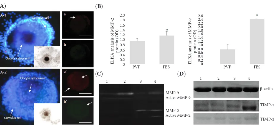
Influence of MMPs and TIMPs on in vitro oocyte maturation. We analysed the expression of MMPs and TIMPs in oocyte culture medium, and in oocytes matured in serum-containing and serum-free in vitro maturation medium. Our results indicate that MMPs were mainly expressed in the cytoplasm of cumulus cells; scant expression was detected in the cytoplasm of egg cells. In eggs matured in serum-containing in vitro maturation medium, MMP-9 was expressed at equal levels in cumulus cells and egg cytoplasm; however, the expression of MMP-2 was low in all the media examined (Figure 1A). ELISA, evaluating the concentration of MMP proteins in eggs, indicated that the expression of MMP-2 and -9 was significantly higher in the serum-containing group than in the serum-free group. Activity analysis indicated that MMP-9 activity was higher in the serum-containing medium and serum-containing medium-matured oocytes than in the serum-free group (Figure 1B). Zymography results indicated high activities of MMP-2 and MMP-9 in the me-dium and eggs of the serum-containing group, whereas high activity of MMP-2 was detected in the medium of the serum-free group (Figure 1C). Although we were unable to detect any expression of the TIMP inhibitors of MMPs in serum-containing and serum-free media, we observed a significantly higher expression of TIMP-2, which suppresses MMP-2, in the serum-containing group compared with that in the serum-containing group (Figure 1D).
[image:4.595.65.533.655.713.2]Analysis of the expression pattern of MMPs after in vitro fertilization. Figure 2 shows the
Table 2. Maturation rate of eggs after in-vitro maturation
Treatment IVM time (h) Oocytes n Oocytes n (%)
GVBD MI MII death
PVP 24 200 10 (5%) 28 (14%)* 152 (76%) 10 (5%)
FBS 24 210 0 25 (11.9%) 175(83.3%)* 10 (4.8%)
IVM = in vitro maturation, FBS = fetal bovine serum, PVP = polyvinyl-pyrroline, GVBD = germinal vesicle breakdown, MI = meiosis 1, MII = meiosis 2
expression and activity patterns of MMPs in em-bryos 18 h after in vitro fertilization of oocytes matured in serum-free and serum-containing media. The expression patterns of MMPs differed in fertilized eggs. In embryos generated by in vitro fertilization of oocytes matured in serum-free medium, MMP-2 was expressed in the cytoplasm. TIMPs were highly expressed in the cumulus cell layer, with TIMP-2 and -3 expression detected in the cytoplasm of oocytes. In eggs fertilized in serum-containing medium, TIMP-2 was ex-pressed throughout the cytoplasm of fertilized
eggs, whereas the expression of MMP-2 in the cytoplasm of fertilized eggs was very low. In con-trast, MMP-9 was expressed in the egg cytoplasm, whereas cytoplasmic expression of TIMP-3 was very low in the serum-free group (Figure 2A). The analysis of MMP activity in media and eggs indicated that the activity of MMPs was very low in media. The activity of MMP-9 was detected in serum-free medium; however, it was difficult to detect MMP-9 activity in proteins obtained from the eggs. In contrast, MMP-2 activity in the proteins of eggs from the serum-free group was considerably higher than that in the eggs from the serum-containing group. However, even in the serum-containing group, the expression of MMPs in the badge was low, whereas the expres-sion in the eggs was high. The activity of MMP-9 was particularly high, and was also detected in the proteins of fertilized eggs from the serum-containing group (Figure 2B, C).
[image:5.595.68.529.91.302.2]Distribution and expression patterns of MMPs and TIMPs in blastocysts. Figure 3 shows the
Figure 1. Analysis of matrix metalloproteinase (MMP) expression and activity after inducing maturation in vitro (A) MMP immunofluorescence analysis of (A-1) eggs matured in serum-free medium, (A-2) eggs matured in serum-con-taining medium; (a and a’) MMP-9 protein expression, (b and b’) MMP-2 protein expression. Green fluorescence shows the expression of MMP-2, and red fluorescence shows the expression of MMP-9. Embryonic nuclei were stained using Hoechst 33258; (B) ELISA of MMP-2 and -9 expression; (C) zymography of culture media and eggs (cumulus oocyte complexes); (D) Western blot analysis of tissue inhibitors of metalloproteinases TIMP-2 and -3 protein expression. Results for (B) and (C) (line 1: serum-free medium, line 2: serum-containing medium, line 3: serum-free in vitro matured oocytes, line 4: serum-containing in vitro matured oocytes). Data represent the mean ± SEM of five individual experiments; expression was normalized against that of β-actin used as internal standard. White arrows indicate the protein expression
[image:5.595.64.292.668.739.2]FBS = fetal bovine serum, PVP = polyvinylpyrrolidone *P < 0.05
Table 3. Emergence rate of blastocysts after extracorporeal development followingin-vitro fertilization
Medium Oocytes n
Embryos developed to cleavage/ blastocyst stage (n) cleavage blastocyst PVP 146 58 (39.72%) 28 (48.28%) FBS 153 98 (64.05%)* 60 (61.22%)*
*difference significant at P < 0.05
(B)
(C) (A)
MMP-9 Active MMP-9
MMP-2 Active MMP-2
PVP FBS PVP FBS
2.0 1.8 1.6 1.4 1.2 1.0 0.8 0.6 0.4 0.2 0
2.6 2.4 2.2 2.0 1.8 1.6 1.4 1.2 1.0 0.8 0.6 0.4 0.2 0
EL
ISA analu
si
s of MMP
-9
pr
ot
ein (OD)
EL
ISA analu
si
s of MMP
-2
pr
ot
ein (OD)
β-actin
TIMP-2
TIMP-3
1 2 3 4 1 2 3 4
results of analysis comparing the expression patterns of MMPs and TIMPs in blastocysts developed in serum-free and serum-containing media; these types of media were used for embryonic development of fer-tilized eggs. In blastocysts from the serum-containing group, MMPs and TIMPs were expressed in the inner cell mass. The expression of MMP-9 was generally higher than that of MMP-2, and the expression of TIMP-2 was generally higher than that of TIMP-3. In the serum-free group, the expression of MMP-2 was detected in the inner cells, whereas MMP-9 was expressed in trophoblast cells. The distribution and expression of TIMP-2 and TIMP-3 showed similar patterns. Thus, in the serum-containing group, we found that MMPs were expressed in the inner cell mass, whereas in the serum-free group, MMPs were expressed in trophoblast cells.
Analysis of MMP activity and expression pat-tern of apoptotic factors in blastocysts.Figure 4
[image:6.595.67.528.95.307.2]shows MMP activity, TIMP expression, and expression pattern of apoptosis-related fac-tors (Casp-3, BCL-2) in blastocysts cultured in serum-free and serum-containing media. MMP-9 was most highly expressed in the se-rum-containing medium. In contrast, we found that the expression of MMP-2 was higher in serum-free medium than in serum-contain-ing medium (Figure 4A, B). The expression of TIMPs contrasted with that of MMPs, with TIMP-3 being highly expressed in the serum-free group. However, we were unable to detect any expression of TIMP-2 in the serum-free group. The expression of Casp-3 (a cell-death factor in blastocysts) was detected in all the groups. Quantitative analysis indicated a high expres-sion of Casp-3 in the serum-free group and high expression of BCL-2 in the serum-containing group (Figure 4C).
Figure 2. Analysis of activity and expression of matrix metalloproteinases (MMPs) in fertilized eggs 18 h after in vitro
fertilization
(A) expression of MMPs and tissue inhibitors of metalloproteinases (TIMPs) in in vitro fertilized eggs in the serum-free group: expression of MMP-2 and TIMP-2 in in vitro fertilized eggs in the serum-containing group (A-1); expression of MMP-9 and TIMP-3 in serum used to culture in vitro fertilized eggs (A-2); expression of MMP-2 and TIMP-2 in in vitro fertilized eggs of the non-serum-containing group (A-3); expression of MMP-9 and TIMP-3 in in-vitro fertilized eggs of non-serum-containing group (A-4). (B) Zymography of culture medium and in vitro fertilized eggs: (1) eggs fertilized in vitro using serum-free culture medium; (2) eggs fertilized in vitro using serum-containing culture medium; (3) zygotes obtained using serum-free medium; (4) zygotes obtained using serum-containing medium. (C) ELISA of MMP-2, MMP-9, TIMP-2, and TIMP-3 expression in zygotes. Green fluorescence indicates the expression of MMPs, and red fluorescence shows the expression of TIMPs. The nuclei of fertilized eggs were stained using Hoechst 33258
FBS = fetal bovine serum, PVP = polyvinylpyrrolidone a–cP < 0.05
MMP-9 Active MMP-9
MMP-2 Active MMP-2 1 2 3 4
EL
ISA analy
si
s of pr
ot
ein
le
ve
l in z
ygot
es
2.0 1.8 1.6 1.4 1.2 1.0 0.8 0.6 0.4 0.2 0
MMP-2 TIMP-2 MMP-9 TIMP-2 PVP FBS
(B)
DISCUSSION
Our results indicate that the rate of matura-tion and blastocyst appearance differed markedly depending on culture in free or serum-containing media; patterns of MMP expression also differed with respect to the media used for culture. Previously, Lee et al. (2005) showed that MMP-9 was highly expressed in oocyte cytoplasm, and that probability of fertilization could be in-creased. Consistently, we observed high MMP-9 expression in mature oocytes cultured in serum-containing medium, and confirmed that the expres-sion of MMP-9 increased in the ovum cytoplasm as oocytes matured. However, the expression of MMP-2 was observed only in the cumulus cells of mature oocytes cultured in serum-containing medium, indicating that the activity of MMP-2 is important for the cumulus cell expansion during oocyte maturation. This result also indicates that
[image:7.595.71.530.104.359.2]serum plays an important role in the activity of MMPs contained in the sperm and egg during in vitro fertilization, as was shown previously by Kim et al. (2013). Our results indicate that from the induction of in vitro fertilization and until the start of embryonic development, MMP activity was maintained in the cumulus cell layer and ovum cytoplasm. Results also indicate that MMP-2 and MMP-9 showed contrasting expression patterns in cumulus cells and the ovum cytoplasm during embryonic development. These results also show that the expression of MMP-9 was increased in the embryonic cytoplasm, which is consistent with the findings of Whiteside et al. (2001), Imai et al. (2003), and Lee et al. (2005). We found that the expression of MMPs in the cytoplasm positively affected the cell division. Our results also indicate that the expression of MMP-9 in the inner cell mass of the blastocyst may promote embryonic development. However, we found that the expression of MMPs in
Figure 3. Expression site and surface analysis of matrix metalloproteinases (MMPs) and tissue inhibitors of metal-loproteinases (TIMPs) in blastocysts produced using serum-free and serum-containing media
free medium was lower than that in serum-containing medium. In particular, the expression of MMP-2 was increased in the cytoplasm after in vitro fertilization, and the activity of MMP-2 was higher than that in the serum. We also evaluated how us-ing PVP in serum-free medium influenced the rate of blastocyst generation. We found that the use of PVP affected the rate of blastocyst appearance; our results contrasted with those of Kim et al. (2004). In the development of bovine embryos, PVP can be used as a substitute for BSA to alleviate compositional and immunological problems associated with complex media containing antioxidants. However, PVP was not conducive to embryonic development (Hirao et al. 2004; Kim et al. 2004; Choi et al. 2007). In con-trast with the findings of Mizumachi et al. (2018), who investigated the effect of PVP on the growth and viability of cumulus cells during maturation of oocytes, our results show that using the serum for oocyte maturation in the ovaries increased the activity of MMPs (Hirao et al. 2004; Kim et al. 2004). MMP expression was significantly increased in the inner cell mass of blastocysts in the serum-containing
[image:8.595.74.520.94.355.2]group after in vitro fertilization; MMP expression in the trophoblast was insignificant (Horka et al. 2012; Yang et al. 2015; Mizumachi et al. 2018). However, in the serum-free medium supplemented with PVP, the expression of MMPs was increased in trophoblastic cells. These results indicate that the expression and activity of MMPs in the early blastocyst developmen-tal stage affect embryonic development, which was also shown by Yang et al. (2015). These results also show that the expression of MMPs in the inner cell mass of blastocysts in the serum-containing group influenced embryonic development. Momozawa and Fukuda (2011) reported an increased rate of development in bovine embryos obtained using complex media containing PVP. Our results show that the generation of blastocysts in serum-free medium (containing PVP) and serum-containing medium was 48.28% and 61.22%, respectively. Thus, our results on the activity of MMP-2 and -9, and on the locale of MMP expression in blastocysts, differ from those obtained in previous studies. The low incidence of blastocysts in the serum-free group was likely related to the activity of MMP-9, which does
Figure 4. Expression analysis of matrix metalloproteinases (MMPs), tissue inhibitors of metalloproteinases (TIMPs), and apoptosis factors in blastocysts generated in serum-free and serum-containing media
(A) zymography; (B) ELISA; (C) Western blot; (D) graph displaying quantification of Western blot results. Data represent the means ± SEM of five individual experiments; expression was normalised against that of β-actin used as internal standard FBS = fetal bovine serum, PVP = polyvinylpyrrolidone
*P < 0.05
MMP-9 Active MMP-9
MMP-2
Active MMP-2 Prot
ein e
xpr
ession analy
si
s
in embyo
s (me
an %)
3.5 3.0 2.5 2.0 1.5 1.0 0.5 0
MMP-9 MMP-2 (B)
(D)
β-actin
TIMP-2
TIMP-3
Casp-3
BCL-2 (A
(C)
PVP FBS PVP FBS
Caps-3 BCL-2 TIMP-3 TIMP-2
Pr
ot
ein e
xpr
ession analy
si
s
in embyo
s (me
an %)
80 70
60 50 40 30
20 10 0
*
*
* *
not affect oocyte cytoplasm and may be involved in apoptosis. We did not perform an immunolocal-ization analysis of apoptotic factors in this study. However, our Western blot analyses indicate that the expression of Casp-3 in the serum-free group was increased, and that the expression of BCL-2 was relatively high in the serum-containing group; this increased expression of BCL-2 may have been responsible for the reduced effects of Casp-3. The results of our present study indicate that the activity and expression site of MMP-2 and MMP-9 can affect the appearance of blastocysts during oocyte matura-tion and embryonic development. Similarly to the findings of Luddi et al. (2018), our results indicate that MMP-2 and MMP-9 participate in the wall rup-ture of cumulus cells and in oocyte maturation via collagen digestion, while TIMPs (TIMP-2: MMP-2 inhibitor, TIMP-3: MMP-9 inhibitor) modulate the activity of MMP-2 and -9 (Ogiwara et al. 2005). Our study suggests that the presence or the absence of MMPs, produced in culture medium and embryonic cells, can be used as an index to measure the rate of embryonic development.
CONCLUSION
Our results show that free and serum-containing media influence the expression and activity of MMPs. We confirmed that the activ-ity of MMP-9 increases in the inner cell mass of oocytes and blastocysts cultured in serum-containing medium, which was correlated with embryonic development. Furthermore, we found that MMP-2 activity in cumulus cells, and MMP-9 activity in oocyte cytoplasm, increased the rate of oocyte maturation during the early maturation stage. The activity of MMP-9 in the cytoplasm of developing blastocysts may promote the differen-tiation and development of embryonic cells. Thus, the activities of MMP-2 and MMP-9 at different sites in blastocysts play an important role in the development of bovine embryos.
REFERENCES
Ashwood-Smith M.J., Wardy C. (1971): Studies on the mo-lecular weight and cryoprotective properties of polyvinyl-pyrrolidone and dextran with bacteria and erythrocytes. Cryobiology, 8, 453–464.
Atabakhsh M., Khodadadi I., Amiri I., Mahjub H., Tavilani H. (2018): Activity of matrix metalloproteinase 2 and 9 in follicular fluid and seminal plasma and its relation to embryo quality and fertilization rate. Journal of Repro-duction and Infertility, 19, 140–145.
Bilen E., Tola E.N., Oral B., Doguc D.K., Gunyeli I., Kose S.A., Ilhan I. (2014): Do follicular fluid gelatinase levels affect fertilization rates and oocyte quality? Archives of Gynecology and Obstetrics, 290, 1265–1271.
Brackett B.G., Oliphant G. (1975): Capacitation of rab-bit spermatozoa in vitro. Biology of Reproduction, 12, 260–274.
Chatot C.L., Klein N.W., Clapper M.L., Resor S.R., Singer W.D., Russman B.S., Holmes G.L., Mattson R.H., Cramer J.A. (1984): Human serum teratogenicity studied by rat embryo culture: Epilepsy, anticonvulsant drugs and nutri-tion. Epilepsia, 25, 205–216.
Choi S.H., Ryu I.S., Kim I.H., Park S.B., Yeon S.H., Jin H.J., Suh S.W., Lee C.S., Son D.S. (1999): Effects of in vitro maturation condition on bovine IVF embryo develop-ment. Journal of Embryo Transfer, 14, 113–119.
Choi S.H., Cho S.R., Han M.H., Kim H.J., Choe C.Y., Son D.S., Chung Y.G., Kim S.K., Sohn S.H. (2007): Chromo-somal analysis of Hanwoo embryos by in vitro culture condition. Journal of Embryo Transfer, 22, 137–141. Dumoulin J.C., Bergers-Janssen J.M., Pieters M.H., Enginsu
M.E., Geraedts J.P., Evers J.L. (1994): The protective effects of polymers in the cryopreservation of human and mouse zonae pellucidae and embryos. Fertility and Sterility, 62, 793–798.
Gu J., Han C.H., Hu F.F., Wang Y.B., Cao Y.J. (2015): The correlation analysis of human embryonic MMP-9 secre-tion and embryo quality. European Review of Medical and Pharmacological Sciences, 19, 2354–2358.
Hirao Y., Itoh T., Shimizu M., Iga K., Aoyagi K., Kobayashi M., Kacchi M., Hoshi H., Takenouchi N. (2004): In vitro growth and development of bovine oocyte-granulosa cell complexes on the flat substratum: Effects of high polyvinylpyrrolidone concentration in culture medium. Biology of Reproduction, 70, 83–91.
Horka P., Malickova K., Jarosova R., Janatkova I., Zima T., Kalousova M. (2012): Matrix metalloproteinases in serum and the follicular fluid of women treated by in vitro fertilization. Journal of Assisted Reproduction and Genetics, 29, 1207–1212.
Kane M.J. (1987): Minimal nutrient requirements for culture of one-cell rabbit embryos. Biology of Reproduction, 37, 775–778.
Kim I.D., Kim S.N., Han S.K., Seok H.B. (2004): Effects of development and viability of pig oocytes matured in defined medium containing PVA, PVP and pFF. Korean Journal of Embryo Transfer, 19, 219–227.
Kim K., Mitsumizo N., Fujita K., Utsumi K. (1996): The effects of follicular fluid on in vitro maturation, oocyte fertilization and the development of bovine embryos. Theriogenology, 45, 787–799.
Kim S.H., Song Y.S., Hwang S.Y., Min K.S., Yoon J.T. (2013): Effects of hormones on the expression of matrix metal-loproteinases and their inhibitors in bovine spermato-zoa. Asian-Australasian Journal of Animal Sciences, 26, 334–342.
Lee D.M., Lee T.K., Song H., Kim C.H. (2005): The expres-sion of matrix metalloproteinase-9 in human follicular fluid is associated with in vitro fertilisation pregnancy. BJOG, 112, 946–951.
Lee J.Y., Kim S.H., Yoon J.T. (2019): Identifying biomark-ers of autophagy and apoptosis in transfected nuclear donor cells and transgenic cloned pig embryos. Annals of Animal Science, 19, 127–146.
Liu Z., Foote R.H. (1995): Development of bovine embryos in KSOM with added superoxide dismutase and taurine and with five and twenty percent O2. Biology of Repro-duction, 53, 786–790.
Luddi A., Gori M., Marrocco C., Capaldo A., Pavone V., Bi-anchi L., Boschi L., Morgante G., Piomboni P., de Leo V. (2018): Matrix metalloproteinases and their inhibitors in human cumulus and granulosa cells as biomarkers for oo-cyte quality estimation. Fertility and Sterility, 109, 930–939. Mizumachi S., Aritomi T., Sasaki K., Matsubara K., Hirao Y. (2018): Macromolecular crowded conditions strengthen contacts between mouse oocytes and companion granu-losa cells during in vitro growth. Journal of Reproduction and Development, 64, 153–160.
Momozawa K., Fukuda Y. (2003): Caffeine in fertilization medium is not essential for bovine IVF by fully capaci-tated spermatozoa. Journal of Reproduction and Devel-opment, 49, 507–512.
Momozawa K., Fukuda Y. (2011): Establishment of an ad-vanced chemically defined medium for early embryos de-rived from in vitro matured and fertilized bovine oocytes. Journal of Reproduction and Development, 57, 681–689. Ogawa T., Ono T., Marrss R.P. (1987): The effect of serum fractions on single-cell mouse embryos in vitro. Journal of In Vitro Fertilization and Embryo Transfer, 4, 153–158. Ogiwara K., Takano N., Shinohara M., Murakami M.,
Taka-hashi T. (2005): Gelatinase A and membrane-type matrix metalloproteinases 1 and 2 are responsible for follicle rupture during ovulation in the medaka. Proceedings of the National Academy of Sciences of the United States of America, 102, 8442–8447.
Rosenkrans Jr. C.F., Zeng G.Q., McNamara G.T., Schoff P.K., First N.L. (1993): Development of bovine embryos in vitro as affected by energy substrates. Biology of Re-production, 49, 459–462.
Song B.S., Kim J.S., Yoon S.B., Lee K.S., Koo D.B., Lee D.S., Choo Y.K., Huh J.W., Lee S.R., Kim S.U., Kim S.H., Kim H.M., Chang K.T. (2011): Inactivated Sendai-virus-mediated fusion improves early development of cloned bovine embryos by avoiding endoplasmic-reticulum-stress-associated apoptosis. Reproduction, Fertility and Development, 23, 826–836.
Whiteside E.J., Kan M., Jackson M.M., Thompson J.G., McNaughton C., Herington A.C., Harvey M.B. (2001): Urokinase-type plasminogen activator (uPA) and matrix metalloproteinase-9 (MMP-9) expression and activity during early embryo development in the cow. Anatomy and Embryology, 204, 477–483.
Yamashita S., Hoshi H. (1996): Bovine blastocyst formation from IVM/IVF produced zygotes in serum and serum free medium. Theriogenology, 45, 197.
Yang W.J., Liu F.C., Hsieh J.S., Chen C.H., Hsiao S.Y., Lin C.S. (2015): Matrix metalloproteinase 2 level in human follicular fluid is a reliable marker of human oocyte matu-ration in in vitro fertilization and intracytoplasmic sperm injection cycles. Reproductive Biology and Endocrinol-ogy, 13, Article No. 102.