PHARYNGEAL AIRWAY IN CLASS I
*,1
Sneh Kalgotra,
2Dr. Abhiroop
1
Registrar, Government Dental College and Hospital, Srinagar, India
2Post-Graduate Student, D.A.V. Dental College,
3
Principal & Head, Indira Gandhi Government Dental College & Hospital, Jammu & Kashmir, India
ARTICLE INFO ABSTRACT
Background:
of
oro-Aim: The aim of the study was to compare the pharyngeal airway dimensions in different antero posterior skeletal patterns i.e skeletal Class I &
Methods & Materials:
cephalogrmas from the Out
facial Orthopaedics, Government Dental College & Hospital, Srinagar. Three parameters were u for comparison between male and female subjects and between skeletal Class I & Class II.
Statistical analysis
comparison and final analysis was done using SPSS software.
Results:
more in Class I subjects when compared to Class II subjects.
Conclusion:
pattern.
Copyright©2016, Sneh Kalgotra et al.This is an open access article distributed under the Creative Commons Att distribution, and reproduction in any medium, provided the original work is properly cited.
INTRODUCTION
In 1872, Tomes hypothesized that maxillary constriction could be caused by lymphatic tissue hypertrophy of the pharynx that caused the absence of lip seal and a lower tongue position to maintain the permeability of the airway (Tome, 1872
Angle, Fränkel, Harvold, Linder-Aronson and others demonstrated that airway obstruction can determine abnormal development of the facial pattern (Angle, 1907; Harvold, 1972; Linder, 1973). At one time the nasorespiratory area played an important role in orthodontic thinking. It was believed, and expressed, that obstructions in this area might influence the developing facial conformity. There are frequent references in the literature to the so-caIled ‘adenoid facies
The pharynx is that part of the digestive tube which is placed behind the nasal cavities, mouth and larynx. It is a musculomembranous tube, somewhat conical in form, with the base upward, and the apex downward, extending from the under surface of the skull to the level of the cricoid cartilage in
*Corresponding author: Sneh Kalgotra,
Registrar, Government Dental College and Hospital, Srinagar, India.
ISSN: 0975-833X
Article History:
Received 20th February, 2016
Received in revised form 23rd March, 2016
Accepted 25th April, 2016
Published online 31st May,2016
Key words:
Pharyngeal Airway, Skeletal Class I, Skeletal Class II, Obstructive Sleep, Apnea Syndrome.
Citation:Sneh Kalgotra, Dr. Abhiroop Singh and Prof. (Dr). Romesh Singh
International Journal of Current Research, 8, (05), 31958
RESEARCH ARTICLE
PHARYNGEAL AIRWAY IN CLASS I AND CLASS II SKELETAL PATTERNS
Dr. Abhiroop Singh and
3Prof. (Dr). Romesh Singh
Government Dental College and Hospital, Srinagar, India
Graduate Student, D.A.V. Dental College, Haryana, India
Principal & Head, Indira Gandhi Government Dental College & Hospital, Jammu & Kashmir, India
ABSTRACT
Background: The abnormal growth and development of the oro-facial complex leads to disturbances -facial functions.
: The aim of the study was to compare the pharyngeal airway dimensions in different antero posterior skeletal patterns i.e skeletal Class I & Class II.
Methods & Materials: Sample size was calculated using Cohen’s Formula, consisted of 60 cephalogrmas from the Out-patient department of Post-Graduate department of Orthodontics &
Orthopaedics, Government Dental College & Hospital, Srinagar. Three parameters were u for comparison between male and female subjects and between skeletal Class I & Class II.
Statistical analysis: All lateral cephalograms were traced and independent t comparison and final analysis was done using SPSS software.
Results: It was found that suerioe airway space, middle airway space and inferior airway space was more in Class I subjects when compared to Class II subjects.
Conclusion: A significant positive correlation was established between the airway and the skeletal rn.
is an open access article distributed under the Creative Commons Attribution License, which distribution, and reproduction in any medium, provided the original work is properly cited.
hypothesized that maxillary constriction could be caused by lymphatic tissue hypertrophy of the pharynx that caused the absence of lip seal and a lower tongue position to Tome, 1872). Later, Aronson and others demonstrated that airway obstruction can determine abnormal Angle, 1907; Harvold, 1972; orespiratory area played an important role in orthodontic thinking. It was believed, and expressed, that obstructions in this area might influence the facial conformity. There are frequent references in acies (Subtelny, 1954). The pharynx is that part of the digestive tube which is placed behind the nasal cavities, mouth and larynx. It is a musculomembranous tube, somewhat conical in form, with the base upward, and the apex downward, extending from the to the level of the cricoid cartilage in
Registrar, Government Dental College and Hospital, Srinagar, India.
front, and that of the sixth cervical vertebra behind. Interpretation of the significance of variations in the growth and function of the nasal cavities, the nasopharynx and the oropharynx is dependent on an understanding of the normal growth of the skull. In this respect, however, knowledge of normal growth has often been gained by recognition and observation of abnormal cranial function and development. Thus, aberrant respiratory modes such as chronic mouth breathing have been implicated in dentofacial deformities (Angle, 1907; Subtelny, 1954; Subtelny,
Nasorespiratory function and its relation to craniofacial growth is of great interest today, not only as an example of the basic biological relationship of form and function, but also because it is of great practical concern to Pediatri Otorhinolaryngologists, Allergists, Speech Physiologists, Orthodontists and other members of the health care community as well. So, it becomes important to establish an association between the pharyngeal airway and skeletal pattern.
MATERIALS AND METHODS
Pre-treatment lateral cephalometric radiographs were selected from Out Patient Department of the Department Of
Available online at http://www.journalcra.com
International Journal of Current Research
Vol. 8, Issue, 05, pp.31958-31962, May, 2016
INTERNATIONAL
Singh and Prof. (Dr). Romesh Singh, 2016. “Pharyngeal airway in class I and class II skeletal patterns 31958-31962.
z
CLASS II SKELETAL PATTERNS
Prof. (Dr). Romesh Singh
Government Dental College and Hospital, Srinagar, India
, India
Principal & Head, Indira Gandhi Government Dental College & Hospital, Jammu & Kashmir, India
facial complex leads to disturbances
: The aim of the study was to compare the pharyngeal airway dimensions in different
antero-Sample size was calculated using Cohen’s Formula, consisted of 60 Graduate department of Orthodontics & Dento Orthopaedics, Government Dental College & Hospital, Srinagar. Three parameters were used for comparison between male and female subjects and between skeletal Class I & Class II.
: All lateral cephalograms were traced and independent t-test was used for
It was found that suerioe airway space, middle airway space and inferior airway space was
A significant positive correlation was established between the airway and the skeletal
ribution License, which permits unrestricted use,
front, and that of the sixth cervical vertebra behind. Interpretation of the significance of variations in the growth and function of the nasal cavities, the nasopharynx and the oropharynx is dependent on an understanding of the normal growth of the skull. In this respect, however, knowledge of has often been gained by recognition and observation of abnormal cranial function and development. Thus, aberrant respiratory modes such as chronic mouth breathing have been implicated in dentofacial deformities Angle, 1907; Subtelny, 1954; Subtelny, 1980; Baik, 2002). Nasorespiratory function and its relation to craniofacial growth is of great interest today, not only as an example of the basic biological relationship of form and function, but also because it is of great practical concern to Pediatricians, Otorhinolaryngologists, Allergists, Speech Physiologists, Orthodontists and other members of the health care community as well. So, it becomes important to establish an association between the pharyngeal airway and skeletal pattern.
ETHODS
treatment lateral cephalometric radiographs were selected from Out Patient Department of the Department Of
INTERNATIONAL JOURNAL OF CURRENT RESEARCH
Orthodontics & Dentofacial Orthopaedics, Government Dental College & Hospital, Srinagar.
Sample Size Determination
The sample size for this study has been determined scientifically.
Sample size determination was done using Cohen’s d power table. The following formulae was used to calculate Cohen’s d,
Cohen’s d = where, M1 is mean 1 and M
So, for the power of the study as 80% the sample size of minimum of 30 for each group had to be established.
the above calculations, the study would include a total of 60 sample with 30 subjects in Class I skeletal pattern, 30 subjects in Class II skeletal pattern. The level of significance was set at 0.05. Total of 60 subjects (ages 14–33 years) were included, out of which 25 were male subjects and 35 were female subjects.
All subjects met the following inclusion criteria
Over 14 years of age.
No history of orthodontic treatment. Breathing comfortably through the nose. And the following exclusion criteria: Subjects with cleft lip and palate.
Subjects with history of chronic mouth breathing. Subjects suffering with any medical condition.
To have standardized cephalometric radiographs, it became important that all the radiographs were taken from the same X ray machine with the subjects in the natural head position, with teeth in maximum intercuspation and lips at repose. All the lateral cephalometric radiographs were taken by the same operator from the standardized Orthophos XG5 DS CEPH (SIRONA) on a standard Konica Minolta 8 × 10 inch film with an anode to midsubject distance of 5 feet by the same operator. Natural head position was obtained by ask
look straight ahead such that the visual axis was parallel to the floor. Thyroid shield and lead apron were worn by the subject to reduce radiation exposure. All the films were exposed with 64 KVp, 8 mA and an exposure time of 9 seconds.
A sample of 60 lateral cephalograms was taken. Lateral cephalogram were traced upon an A4 size acetate paper with a 2B or 3HB hard lead pencil over well-illuminated viewing screen. The linear measurements were recorded with a measuring scale up to a precision of 0.5 mm. After going through different studies conducted on the parameters used for assessment of antero-posterior discrepancy
Harvold, 1972; Linder, 1973) it was decided to segregate the radiographs into different antero-posterior sk
the following basis. (Table 1). The first parameter used ANB angle (Steiner, 1960). The second parameter used for segregation Beta angle (Baik, 2004), The thirdparameter used for segregation of radiographs into different skeletal
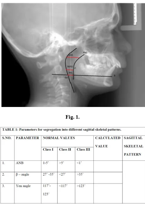
Yen angle (Neela and Mascarenhas, 2004). Fig. 1
31959 Sneh Kalgotra et al.
Orthopaedics, Government Dental
The sample size for this study has been determined
Sample size determination was done using Cohen’s d power table. The following formulae was used to calculate Cohen’s d,
is mean 1 and M2 is mean 2.
, for the power of the study as 80% the sample size of minimum of 30 for each group had to be established. Based on the above calculations, the study would include a total of 60 sample with 30 subjects in Class I skeletal pattern, 30 subjects skeletal pattern. The level of significance was set at 33 years) were included, out of which 25 were male subjects and 35 were female
inclusion criteria
Subjects with history of chronic mouth breathing. Subjects suffering with any medical condition.
standardized cephalometric radiographs, it became important that all the radiographs were taken from the same X-ray machine with the subjects in the natural head position, with teeth in maximum intercuspation and lips at repose. All the ic radiographs were taken by the same operator from the standardized Orthophos XG5 DS CEPH (SIRONA) on a standard Konica Minolta 8 × 10 inch film with an anode to midsubject distance of 5 feet by the same operator. Natural head position was obtained by asking the subject to look straight ahead such that the visual axis was parallel to the floor. Thyroid shield and lead apron were worn by the subject to reduce radiation exposure. All the films were exposed with 64 KVp, 8 mA and an exposure time of 9 seconds.
A sample of 60 lateral cephalograms was taken. Lateral cephalogram were traced upon an A4 size acetate paper with a illuminated viewing screen. The linear measurements were recorded with a ision of 0.5 mm. After going through different studies conducted on the parameters used for posterior discrepancy (Angle, 1907; it was decided to segregate the posterior skeletal patterns on The first parameter used ANB The second parameter used for The thirdparameter used for segregation of radiographs into different skeletal patterns is
[image:2.595.309.558.49.398.2]. Fig. 1
Fig. 1.
Planes and Parameters used
A total of 3 parameters were undertaken in the study .They are as follows:
SAS, superior posterior airway space
behind soft palate along
parallel line to Go-B line)
MAS, middle airway space
line to Go-B line through P)
IAS, inferior airway space
Go-B line).
Landmarks undertaken in the
Eb: Base of the epiglottis P: Tip of the soft palate PNS: Posterior nasal spine Me: Menton
Go: Gonion B: point B
SPSS (Version 16.0) and Microsoft Excel software were to carry out the statistical analysis of data. Data was analyzed with the help of descriptive statistics viz., mean and standard deviation. Student’s independent t
the differences between male and female subjects andin between the groups. A P-value of less than 0.05 was considered statistically significant.
Kalgotra et al. Pharyngeal airway in class i and class ii skeletal patterns
Fig. 1.
A total of 3 parameters were undertaken in the study .They are
SAS, superior posterior airway space (width of airway
MAS, middle airway space(width of airway along parallel B line through P)
IAS, inferior airway space (width of airway space along
the same are as discussed below
Eb: Base of the epiglottis P: Tip of the soft palate PNS: Posterior nasal spine
RESULTS
The observations of the study and the results derived thereof are discussed as follows:
There was no statistically significant difference in superior airway space, middle airway space and inferior airway space dimensions between male and female subjects in skeletal Class I pattern.
There was no statistically significant difference in superior airway space, middle airway space and inferior airway space dimensions between male and female subjects in skeletal Class II pattern.
A statistically significant difference was observed superior posterior, middle and lower airway dimensionsClass I, Class II skeletal patterns.
The P value is statistically significant showing a strong correlation between the two groups.
31960 International Journal of Current Research,
The observations of the study and the results derived thereof
There was no statistically significant difference in superior and inferior airway space dimensions between male and female subjects in skeletal Class
There was no statistically significant difference in superior airway space, middle airway space and inferior airway space female subjects in skeletal Class
A statistically significant difference was observed superior posterior, middle and lower airway dimensionsClass I, Class II
The P value is statistically significant showing a strong
DISCUSSION
The nasopharyngeal dimensions continue to grow rapidly until 13 years of ageand then slow down until adulthood.
this study, the age range was 14
oropharyngeal structures had reached adult size. The reasons for excluding patients with cleft lip and palate was to rule out any syndrome which might affect the skeletal anteroposterior dimensions. Subjects with normal nasal breathing and normal antero-posterior skeletal patterns
The first parameter used to assess antero relationship was ANB angle (Steiner, 1960
considered the most commonly used cephalometric measurement for evaluation of antero
relationship. The validity of this measurement had been investigated by several researchers. Oktay and Ishikawa
et al. reported that ANB angle is one of the most reliable and
accurate measurements of the antero
relationship. Furthermore, Hussels and Nanda reported that the vertical lengths from nasion to point B and from point A to point B are usually affected. Furth
by either growth or orthodontic treatment can also change the ANB reading. Beta angle, does not depend on cranial landmarks or the functional occlusal plane
It uses 3 points located on the jaws
apparent axis of the condyle ‘point C’. In contrast to the ANB angle, the configuration of the Beta angle gives it the advantage to remain relatively stable even when the jaws are rotated. However, precisely tracing the condyle and locating its ce is not always easy. To accurately use that angle, the cephalometric X-rays must be high quality and it still depends upon point A & point B. To overcome all the above mentioned problems, an additional parameter. The Yen angle was considered. It was reported that the Yen angle was not influenced by growth changes and can be easily used in mixed dentition. On comparing 3 parameters within the same skeletal type in males and females it was found that, pharyngeal dimensions were not affected by gender. The
agreement with those reported in the literatureby Al
all, Martin, Handelman CS, which suggest that gender differences in the pharyngeal dimensions were not present (Hanelman et al., 1976; Martin and Muelas, 2006;
1996; Solow et al., 1984; Allhaija and Alkhateeb, 2005
In our study we found that, this difference could be explained by ‘Balter’s philosophy’according to which, Class II
International Journal of Current Research, Vol. 08, Issue, 05, pp.31958-31962, May, 2016
The nasopharyngeal dimensions continue to grow rapidly until 13 years of ageand then slow down until adulthood.[11,12,13] In this study, the age range was 14–33 years to ensure that the had reached adult size. The reasons for excluding patients with cleft lip and palate was to rule out any syndrome which might affect the skeletal anteroposterior dimensions. Subjects with normal nasal breathing and normal posterior skeletal patterns were taken up for this study. The first parameter used to assess antero-posterior skeletal Steiner, 1960). The ANB angle is considered the most commonly used cephalometric measurement for evaluation of antero-posterior jaw relationship. The validity of this measurement had been investigated by several researchers. Oktay and Ishikawa
that ANB angle is one of the most reliable and accurate measurements of the antero-posterior jaw relationship. Furthermore, Hussels and Nanda reported that the vertical lengths from nasion to point B and from point A to point B are usually affected. Furthermore, rotation of the jaws by either growth or orthodontic treatment can also change the ANB reading. Beta angle, does not depend on cranial landmarks or the functional occlusal plane (Baik, 2004).
It uses 3 points located on the jaws- point A, point B and the apparent axis of the condyle ‘point C’. In contrast to the ANB angle, the configuration of the Beta angle gives it the advantage to remain relatively stable even when the jaws are rotated. However, precisely tracing the condyle and locating its center is not always easy. To accurately use that angle, the rays must be high quality and it still depends upon point A & point B. To overcome all the above mentioned problems, an additional parameter. The Yen angle was ported that the Yen angle was not influenced by growth changes and can be easily used in mixed On comparing 3 parameters within the same skeletal type in males and females it was found that, pharyngeal dimensions were not affected by gender. These findings are in agreement with those reported in the literatureby Al- Khateebet
l, Martin, Handelman CS, which suggest that gender differences in the pharyngeal dimensions were not present ., 1976; Martin and Muelas, 2006; Lowe et al.,
., 1984; Allhaija and Alkhateeb, 2005).
malocclusions represent a backward position of tongue, disturbing the cervical region, this impedes respiratory function in the region of larynx and consequently there is faulty deglutition and mouth breathing. In a study conducted by Mergen and Jacobs,they concluded that in nasopharyngeal dimensions were larger in Class I normal subjects than Class II, which is in agreement with Kerr, F.A. Sosa, IffatBatool,who also found the same results. Similarly Kyuny- Min Ohain in a three dimensional analysis reported that retrognathic children had smaller pharyngeal airway as compared to normal children.
Thus, there must be a true relationship between OPA and the cranial base angle (NSBa) and mandibular length (Go-Men). The relationship between Pharyngeal Airway (PA) and mandibular length would appear logical, because as the body of the mandible lengthens, the attachments of the genioglossus and geniohyoid muscles move forward away from the oropharynx, increasing the PA. The facial pattern can be suggested as potential explanation for the discrepancy in the airway as a result of mandibular size and position. Battagel
et al. concluded that mandibular advancement is associated
with an increase in oropharyngeal dimension and subsequent hyoid bone displacement that improves the airway’s permeability (Battagel et al., 1999). Turnbull and Battageldemonstrated a significant decrease in the retrolingual airway dimension after mandibular setback surgery and a significant increase in this dimension after mandibular advancement surgery (Turnbull and Battagel, 2000). Cephalometric radiography has been extensively used as a diagnostic and follow-up technique in the study of craniofacial morphology and the surgical management of craniofacial anomalies. Despite imaging limitations of lateral cephalometry especially in the transverse plane, this technique can provide assessment of the relationship between craniofacial characteristics and nasopharyngeal conditions (Subtelny, 1954). Further, cephalometrics is easy to use, economical, and can provide definitive and quantitative information about the soft palate and naopharynx (Jakhi and Karjodkar, 1990; Wu
et al., 1996).
Serious concerns about Obstructive sleep apnoea (OSA) as well as its investigative procedures are on the rise. OSA is potentially life-threatening disorder caused by repetitive narrowing and occlusion of the upper airway during sleep, and has been associated with loud snoring, excessive daytime sleepiness, intellectual deterioration, hypertension, right heart failure, and cardiac arrhythmias. A narrow upper airway and other predisposing or etiological factors, such as craniofacial deformity, mandibular retrognathia or micrognathia, muscular hypotony, sleep posture, fatty depositions in soft tissue of the upper airway, gender, and age, have been reported. Planning successful treatment for the correction of anatomic abnormalities of upper airway by advancing the mandible in early age with the use of functional appliance or later by surgically advancing mandible depends on extensive knowledge of pharyngeal airway space, and morphology, hyoid bone, and tongue position changes induced by functional appliances and the advancement surgery could help patients with OSA. Although this study was not directly concerned with sleep disorders, there were findings associated with OSAS, the most important being the direct relationship of the OPA to mandibular length because short mandibular length was also a
prime finding in a number of OSAS reports (Valero, 1965; Miles, 1995; Batool, 2010).
In conclusion of this study that was undertaken, following points can be made
No significant difference was found in airway dimensions in male and female subjects in Class I skeletal patterns. No significant difference was found in airway dimensions
in male and female subjects in Class II skeletal patterns. The Superior airway space, Middle airway space & Inferior
airway space was more in Class I subjects when compared to Class II subjects.
Thus we established a strongly positive co-relation between the pharyngeal airway space and different skeletal patterns.
Further studies with a bigger sample size and recent techniques like CBCT are recommended for future research purposes.
REFERENCES
Allhaija, Alkhateeb. Uvulo-glosso-pharyngeal dimensions in different antero- posterior skeletal patterns; Angle Orthod. 2005; 75(6):1012-1018.
Angle EH. Treatment of malocclusion of the teeth. Ed. 7. Philadelphia, PA, SS White Dental manufacturing company;1907.
Baik YC.s A new approach of assessing sagittal discrepancies. The beta angle. Am J OrthodDentofacOrthop. 2004; 126:1005-1012.
Baik. Cephalometric& obstruction sites in OSA. Angle Orthod. 2002; 72:124-134.
Batool I. Comparison of upper and lower pharyngeal airway space in Class II high angle and low angle cases. Pak
dental journal. 2010; 65:25-34.
Battagel JM, Johal A, L’Estrange PR, Croft CB, Kotecha B. Changes in airway and hyoid position in response to mandibular protrusion in subjects with obstructive sleep apnea (OSA). Eur J Orthod. 1999; 21:363–375.
Hanelman CS, Growth of nasopharynx& adenoid development from one to eighteen years. Angle Orthod. 1976; 46:243-259.
Harvold EP, EP. Expansion on the developing dentition. Am J
OrthodDentofac Orthop.1972; 74-77.
Jakhi SA, Karjodkar FR. Use of cephalometry in diagnosing resonance disorders. Am J OrthodDentofacOrthop. 1990; 98:323– 332.
Jeans WD, Fernando DCJ, Maw AR, Leighton BC. A longitudinal study of the growth of the nasopharynx and its contents in normal children. Br J Radiol. 1981; 54:117– 121.
Linder – Aronson S. Effects of adenoidectomy on mode of breathing, size of adenoid & nasal airflow. Orl J
Otorhinolaryngeal Rel. Space. 1973; 35: 283-301.
Lowe AA, Ono T, Ferguson KA, Pae EK, Ryan CF, Fleetham JA. Cephalometric comparisons of craniofacial and upper airway structure by skeletal subtype and gender in patients with obstructive sleep apnea. Am J OrthodDentofacOrthop.
1996; 110:653-664.
Martin, Muelas. Nasopharyngeal cephalometric study of ideal occlusion. Am J OrthodDentofacOrthop. 2006:436 -436.
Miles PG. Craniofacial morphology and obstructive sleep apnea syndrome – a qualitative analysis and met-analysis of the literature. Am J OrthodDentofac Orthop.1995; 107:459-472.
Neela PK, Mascarenhas R. A new sagittal dysplasia indicator. The Yen angle. World J Orthod. 2004; 32; 37-45.
Solow B, Siersbæk-Nielsen S, Greve E. Airway adequacy, head posture, and craniofacial morphology. Am J Orthod. 1984; 86:214–223.
Steiner CC. Use of cephalometrics as an aid to planning and assessing orthodontic treatment. Am J Orthod. 1960; 46: 721-735.
Subtelny JD. The significance of adenoid tissue in Orthodontia. Angle Orthod. 1954; 36:62-69.
Subtelny JD. The significance of adenoid tissue in Orthodontia. Angle Orthod. 1980; 50: 147.
Tome CS. On the developmental origin of V-shaped contracted maxilla. Monthly Rev. Dent. Surg. 1872; 32:1.2-5
Toume LP. Growth of pharynx & its physiologic implications.
Am J OrthodDentofacOrthop. 1991; 99:129-139.
Turnbull NR, Battagel JM. The effects of orthognathic surgery on pharyngeal airway dimensions and quality of sleep. J
Orthod. 2000; 27:235-47.
Valero A, Alory G. hypoventilation in acquired micrognthia.
Archives of Internal Medicine.1965; 85: 714-719.
Wu JT, Huang GF, Huang CS, Noordhoff MS. Nasopharyngoscopic evaluation and cephalometric analysis of velopharynx in normal and cleft palate patients. Ann
Plast Surg. 1996; 36:117– 122.