THE IMPACT OF NOTTINGHEM PROGNOSTIC INDEX (NPI) ON THE OCCURRENCE OF RELAPSES
IN HER
*
,1DzengisJasar,
2Snezhana Smichkoska,
1
Clinical Hospital ACIBADEM/SISTINA, Skopje, Skupi street No. 5a, Republic of MACEDONIA
2University Clinic of Radiotherapy and Oncology, Medical
3
Institute of Pathology, Medical Faculty Skopje, University “Ss. Cyril and Methodius”, Republic of MACEDONIA
ARTICLE INFO ABSTRACT
Introduction:
the evaluation of breast cancer. According to data in the literature this marker, which applies as unfavorable prognos
who will benefit from adequate treatment with Herceptin. Aim of th histopathological and immunohistochemical parameters and NPI of relapses in HER
Material and Methods: treated
clinical parameters
HER-2 / neu status, the Ki67 proliferative index gene p53. During the follow
Results:
in 99 (57%) patients. The tumor was poorly differentiated in 70 patients (40%). I of HER
30 of the 33 HER2 / neu of HER
differentiation, l proliferative index Ki67 ( Conclusion:
lymphonodal
the course of the disease in the HER
Copyright © 2015DzengisJasar et al. This is an open access article distributed under the Creative Commons Att distribution, and reproduction in any medium, provided the original work is properly cited.
INTRODUCTION
The incidence of breast cancer, in the world and in our country is the most common cancer in women (Ferlay
Receptor of the Human Epidermal growth factor (HER or c-erbB2) is identified by the chemical carcinogen ethyl nitroso-urea, which was induced in the rat neuroblastomas (Fehm et al., 2004). Human homologue of HER
present in the 17th chromosome and encodes a 185 kd transmembrane glycoprotein. In breast cancer, amplification of the HER-2 / neu and expression of HER-2 / neu protein is
*Corresponding author: DzengisJasar,
Clinical Hospital ACIBADEM/SISTINA, Skopje, Skupi street No. 5a, Republic of MACEDONIA
ISSN: 0975-833X
Vol.
Article History:
Received 21st August, 2015
Received in revised form 30th September, 2015 Accepted 16th October, 2015
Published online 30th November,2015
Key words:
Breast cancer, HER2 / neu, NPI, Relapse.
Citation: DzengisJasar, Snezhana Smichkoska, Katerina Kubelka
nottinghem prognostic index (NPI) on the occurrence of relapses in her
Research, 7, (11), 22566-22570.
RESEARCH ARTICLE
THE IMPACT OF NOTTINGHEM PROGNOSTIC INDEX (NPI) ON THE OCCURRENCE OF RELAPSES
IN HER-2 / NEU POSITIVE BREAST CANCER
Snezhana Smichkoska,
1Katerina Kubelka-Sabit,
1VanjaFilipovski and
3Gordana Petrushevska
Clinical Hospital ACIBADEM/SISTINA, Skopje, Skupi street No. 5a, Republic of MACEDONIA
University Clinic of Radiotherapy and Oncology, Medical Faculty Skopje, University “Ss. Cyril and Methodius”
Republic of MACEDONIA
Institute of Pathology, Medical Faculty Skopje, University “Ss. Cyril and Methodius”, Republic of MACEDONIA
ABSTRACT
Introduction: Immunohistochemical detection of Human epidermal growth factor (HER
the evaluation of breast cancer. According to data in the literature, 10-30% of breast cancers show expression of this marker, which applies as unfavorable prognostic factor. Therefore, it is important in the selection of patients who will benefit from adequate treatment with Herceptin. Aim of this study is to determine the impact of clinical, histopathological and immunohistochemical parameters and NPI- Nottingham
of relapses in HER-2 / neu positive breast cancer.
Material and Methods: In this retrospective study, 174 patients were included from primary breast cancer, and were analyzed in histopatholog
clinical Hospital in the period from June 2007 to June 2010. Beside the analysis of clinical and histopathological parameters as well as the NPI, additional immunohistochemicaltests were evalu
2 / neu status, the Ki67 proliferative index, and the expression of the protein gene p53. During the follow-up period (42 to 80 months) relapses were observed Results: The age of patients ranged from 28-83 (average 55.48, +10.0) years. in 99 (57%) patients. The tumor was poorly differentiated in 70 patients (40%). I of HER-2 / neu was observed in 33 patients (19%) and relapses of the disease
30 of the 33 HER2 / neu-positive patients (90%), NPI was higher than 3.4. Despite this association of HER-2 / neu was correlated with tumor diameter, lymph node status
differentiation, lymph-vascular invasion, stage of the disease and the expression of p53 protein product and proliferative index Ki67 (p<0.05).
Conclusion: The score of theNottingham prognostic index higher than 3.4 which included lymphonodal status and degree of histological differentiation of the tumor
the course of the disease in the HER-2 / neu positive breast cancers compared to the occurrence of relapses.
is an open access article distributed under the Creative Commons Attribution License, which distribution, and reproduction in any medium, provided the original work is properly cited.
reast cancer, in the world and in our country,
Ferlay et al., 2013). Receptor of the Human Epidermal growth factor (HER-2 / neu erbB2) is identified by the chemical carcinogen
ethyl-urea, which was induced in the rat neuroblastomas . Human homologue of HER-2 / neu is present in the 17th chromosome and encodes a 185 kd transmembrane glycoprotein. In breast cancer, amplification
2 / neu protein is
Clinical Hospital ACIBADEM/SISTINA, Skopje, Skupi street No. 5a,
associated with a poor prognosis with an average survival of 3 years (Fehm et al., 2004; Chen
6-7 year survival in HER-2 / neu negative cases.
(Herceptin®) is a human monoclonal antibody that selectively binds to the extracellular part of the 185 kd HER
protein (Miles, 2001). It is the first "target" or gene t
breast cancer which showed a good response and improved survival. Testing the status of HER
introduced into routine practice in the world and is performed by immunohistochemical analysis, which gives good results considering the standardized procedures and protocols established by the American Association of Clinical Oncologists (ASCO) (Wolff et
subjective assessment of evaluation
International Journal of Current Research
Vol. 7, Issue, 11, pp.22566-22570, November, 2015
INTERNATIONAL
DzengisJasar, Snezhana Smichkoska, Katerina Kubelka-Sabit,VanjaFilipovski and Gordana Petrushevska
nottinghem prognostic index (NPI) on the occurrence of relapses in her-2 / NEU positive breast cancer”,
THE IMPACT OF NOTTINGHEM PROGNOSTIC INDEX (NPI) ON THE OCCURRENCE OF RELAPSES
VanjaFilipovski and
Clinical Hospital ACIBADEM/SISTINA, Skopje, Skupi street No. 5a, Republic of MACEDONIA
Faculty Skopje, University “Ss. Cyril and Methodius”
Institute of Pathology, Medical Faculty Skopje, University “Ss. Cyril and Methodius”, Republic of MACEDONIA
Human epidermal growth factor (HER-2 / neu) is necessary in 30% of breast cancers show expression of tic factor. Therefore, it is important in the selection of patients study is to determine the impact of clinical, Nottingham prognostic index on the occurrence
included. All of the patients were previously analyzed in histopathological laboratory in Acibadem / Sistina the analysis of clinical and histopathological were evaluated in order to determine the and the expression of the protein product of the tumor suppressor
observed in 38 patients (21.8%).
10.0) years. Lymphnode metastases were found in 99 (57%) patients. The tumor was poorly differentiated in 70 patients (40%). Immunohistochemical expression observed in 33 patients (19%) and relapses of the disease were present in 13 of them (40%). In positive patients (90%), NPI was higher than 3.4. Despite this association, the expression status, mitotic index, degree of histological invasion, stage of the disease and the expression of p53 protein product and
than 3.4 which included the tumor size, nd degree of histological differentiation of the tumor, is an excellent parameter in evaluating
east cancers compared to the occurrence of relapses.
ribution License, which permits unrestricted use,
associated with a poor prognosis with an average survival of 3
Chen et al., 2004) compared with the 2 / neu negative cases. Trastuzumab (Herceptin®) is a human monoclonal antibody that selectively binds to the extracellular part of the 185 kd HER-2 / neu . It is the first "target" or gene therapy for breast cancer which showed a good response and improved Testing the status of HER-2 / neu oncogene is introduced into routine practice in the world and is performed immunohistochemical analysis, which gives good results the standardized procedures and protocols established by the American Association of Clinical
et al., 2007), which reduces the
evaluation (Wolff et al., 2013).
INTERNATIONAL JOURNAL OF CURRENT RESEARCH
VanjaFilipovski and Gordana Petrushevska, 2015. “The impact of
Fluorescent in situ hybridization (FISH) and other hybridization methods (conventional - CISH, hybridization with silver – SISH, etc.) represent additional sensitive and accurate methods for the quantitative evaluation of HER-2 / neu gene status, but are more expensive and require special laboratory conditions and equipment as well as more time for evaluation. Therefore, hybridization techniques are not recommended in routine practice in evaluation of HER-2 / neu status and are reserved for selected patients in which the immunohistochemicalexpression is incomplete according to the criteria of ASCO (Wolff et al., 2013).
The aim of this study is to determine the impact of Nottingham prognostic index on the occurrence of relapses in HER-2 / neu positive breast cancers and to distinguish important prognostic clinicopathological parameters (age, tumor diameter, lymphnode status, degree of histological differentiation, nuclear grade, mitotic index, lymph-vascular invasion, stage of disease) and immunohistochemical parameters (expression of the protein product of the tumor suppressor gene p53 and proliferative index Ki67), in this group of patients.
MATERIAL AND METHODS
An audit of 174 histopathological findings of breast cancers paatients was performed. All the patients were diagnosed in the histopathological laboratory of the Acibadem / Sistina clinical Hospital, from June 2007 to June 2010. From this group of patients,according to immunohistochemical analysis, 33 (19%) of the patients belong to the category of HER-2 / neu positive tumors. Routine sections were first analyzed with standard staining for hematoxylin and eosin. In routine breast cancer diagnostic procedures, classical histological parameters were evaluated: tumor diameter - T, lymphnode status – N, and degree of histological differentiation - G. The degree of histological differentiation is evaluated according to the modified Nottingham scoring system of Bloom-Richardson (Bloom and Richardson, 1957). Of those three parameters of the Nottingham scoring system, nuclear grade and mitotic index were evaluated separately.
These histological parameters are incorporated in performing the Nottingham prognostic index - NPI which is calculated according to the equation (0,2 x T) + N + G (Galea et al., 1992). In this equation the values for N are 1 = without metastatic deposits, 2 = 1-3 positive nodes and 3 = more than three positive nodes, and the values of G, 1 = well differentiated, 2 = moderately differentiated and 3 = poorly differentiated tumors. The obtained results are categorized into three prognostic groups - low-risk group with the value up to 3.4; with high-risk score from 3.4 to 5.4 and a very high risk to score over 5.4 (Galea et al., 1992). In our study intermediate-risk and high-intermediate-risk group are allocated in one category (high-risk) in terms of low-risk group for convenience of statistical analysis. With the other routine analyzes, the age of the patients is incorporated, as well as the stage of the disease, according to a postoperative histopathological classification of malignant neoplasms of the breast of the American Joint Cancer Committee - AJCC, from 2010 (Edge et al., 2010). Representative samples of the tumor were selected and further processed for immunohistochemical analysis. Freshly cut
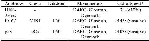
[image:2.595.307.556.501.571.2]sections with a thickness of 2.5 microns, were placed in special pretreated slides (Poly-l-lysine) and dried overnight at a temperature of 600С. They were dewaxed in xylene and dehydrated in alcohol with different concentrations (100%, 96%, 80%). Epitope retrieval of HER2 / neu receptor was used in water bath at a temperature of 960С, and forimmunohistochemical studies of proliferative index Ki67, and protein products of the p53 tumor suppressor gene citrate buffer was used in a microwave oven of 700W, according to the requirements of the manufacturer. Imunohistohemical analysis was performed by avidin-biotin complex technique. Evaluation of HER-2 / neu gene was performed by ready-to-use, HercepTest kit (DAKO, Glostrup, Denmark), and the for the immunohistochemical analysis of Ki67 proliferative index and the protein product of the tumor suppressor gene p53 prepared monoclonal murine antibody (DAKO, Glostrup, Denmark) and visualizing system (Dako REAL ™ EnVision ™ Detection System, Peroxidase / DAB +, Rabbit / Mouse) have been used with the dilution of 1:50. At the slides with a HER-2 / neu positive and negative controls were added for accurate evaluation and control staining. After the performed steps of immunohistochemical staining, slides were counterstained with hematoxylin. Evaluation of HER-2 / neu consists in detecting strong membrane staining in at least 10% of malignant cells in the invasive front of the tumor, which was considered as positive result and indicated with 3+ (Wolff et al., 2007). Evaluation of the nuclear signal for Ki67 proliferative index and the protein product of the tumor suppressor gene p53 was performed according to the rules of Bhargava (2009) and Reed (2000) by counting at least 10 high power fields (HPF, x40) which were considered as positive on 14% of nuclei of malignant cells for Ki67 and over 10% of nuclei of cancer cells for p53. The type of the antibody, clone, dilution, the manufacturer and the cut-off point of a positive signal for the tested antibodies are shown in Table 1.
Table 1. Type of the antibody, clone, dilution, manufacturer and the cut-off point of positive/negative signal
Antibody Clone Dilution Manufacturer Cut-offpoint*
HER-2/neu
- DАКО, Glostrup,
Denmark
3+ (>10%)
Ki-67 MIB1 1:50 DАКО, Glostrup, Denmark
>14% (positive)
p53 DO7 1:50 DАКО, Glostrup, Denmark
>10% (positive)
* Evaluation of Herceps test was performed according the criteria of the American Society of Clinical Oncology (ASCO). For the other two antibodies, modified proposals of Bhargava (2009) and Reed (2000) were accounted.
The categorization of the analyzed parameters were represented by numbers (percentages), and their association in respect of expression of HER-2 / neu were expressed by Pearson-'s χ2 and Fisher-'s exact test. Statistical significance was determined by the values of p<0.05.
RESULTS
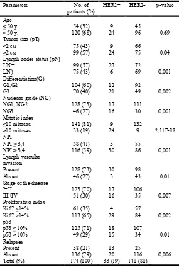
entire group was over 50 years old. Ninety-nine (57%) of these 174 patients, at the time of diagnosis had a tumor diameter greater than 2 cm with positive lymph node status. In 116 patients (59%) NPI was higher than 3.4, that belonged to the unfavorable prognostic group of patients. High proliferative activity of the primary tumor determined by Ki67 antibody, was observed in 113 patients (65%), and in this study relapses were identified in 38 patients (21%).
[image:3.595.37.289.376.753.2]The expression of HER-2 / neu oncogene was identified in 33 patients (19%). There was a positive correlation between expression of HER-2 / neu oncogene with the parameters of the postoperative histopathological classification of cancer, namely, a tumor diameter - pT (p = 0.04) and lymph node status - pN (p = 0.001) as well as and the stage of the disease (p = 0.007). There was also a strong association with the parameters of Bloom-Richardson’s modified Nottingham scoring system that determine the degree of histological differentiation - G (p = 0.002), or nuclear grade - NG (p = 0,001) and mitotic index (p <0,01). All these parameters were reflected along the NPI which shows that it is more pronounced with HER-2 / neu positive breast cancers that are disadvantageous prognostic group (p = 0,001) compared with other patients. Lymph-vascular invasion was present in 30 of 33 (90%) HER-2 / neu positive breast cancers (p = 0.01).
Table 2. Clinical and histological parameters in the group of 174 patients with breast cancer related to the expression of HER-2 / neu oncogene
Parameters No. of
patients (%)
HER2+ HER2- p-value
Age < 50 y. > 50 y.
54 (32) 120 (68)
9 24
45
96 0,69
Tumor size (рТ) <2 см >2 см
75 (43) 99 (57)
9 24
66
75 0,04
Lymph nodes status (pN) LN +
LN )
99 (57) 75 (43)
27 6
72
69 0,001 Differentiation(G)
G1,G2 G3
104 (60) 70 (40)
12 21
92
49 0,002 Nucleaar grade (NG)
NG1, NG2 NG3
128 (73) 46 (27)
17 16
111
30 0,001 Mitotic index
<10 mitoses >10 mitoses
141 (81) 33 (19)
9 24
132
9 2,11Е-18 NPI
NPI < 3,4 NPI > 3,4
58 (41) 116 (59)
3 30
55
86 0,001 Lymph-vascular
invasion Present Absent
128 (73) 46 (27)
30 3
98
43 0,01
Stage of the disease I+II
III+IV
123 (70) 51 (30)
17 16
106
35 0,007 Proliferative index
Ki67 <14% Ki67 >14%
61 (35) 113 (65)
4 29
57
84 0,002 p53
p53 < 10% p53 > 10%
125 (71) 49 (29)
18 15
107
34 0,01
Relapses Present Absent
38 (21) 136 (79)
13 20
25
116 0,006 Total (%) 174 (100) 33 (19) 141 (81)
In terms of immunohistochemical parameters, the level of proliferative activity of the tumor cells determined by Ki67 antibody was more pronounced in the HER-2 / neu positive tumors (p = 0.002) as well as and the expression of the protein product of tumor suppressor gene p53 (p = 0.01). The analyzed parameters and their ratios are presented in Table 2. Regarding the present relapses, identified in 38 patients, 13 of them belongto the HER-2 / neu-positive patients (34%). Of these 13 patients, loco-regional metastases were found in 9 (70%), bone metastases in 2 (15%), and metastasis in visceral organs in 2 (15%) patients.
Legend: HER2 - human epidermal growth factorreceptor, NPI - Nottingham prognostic index; p-value refers to the parameters categorized as: the age group of the patients equal or up to 50 years, compared to the group over 50 years old, tumor status (size of the primary tumor) up to 2 cm (pT1) and more than 2 cm (pT2, pT3, pT4 ), LN + positive and LN- negative lymph nodes, well and intermediate differentiated tumors (G1 / G2) in relation to poorly differentiated (G3), low and intermediate nuclear differentiation (NG1 / NG2) compared the high degree of nuclear differentiation (NG3) and disease stages I and II compared to more advanced stages of the disease III and IV.
DISCUSSION
In our study, the age of the patients as a clinical parameter, did not show any association with the expression of HER-2 / neu oncogene that were confirmed in studies of Partridge (2013)
and Arvold (2011). In these studies,with a large series of patients (1703 patients in the first and 1434 in the other study),the age of the HER-2 / neu positive patients is an independent prognostic factor compared with other types of breast cancer.
The size of the primary tumor in our study was proved to be an important parameter in correlation with HER-2 / neu status. As in other studies (Korkolis et al., 2004; Cortesi et al., 2013), our results showed that the HER-2 / neu positive breast cancers have larger tumor diameter, but not to the extent that was found in other molecular subgroups, such as the "triple" negative cancers cancer (Kim et al., 2006). Similar results were reported in a study by Michaelson and coworkers (Michaelson
et al., 2002).
The lymph-node status is a strong prognostic parameter in all types of breast cancers and in HER-2 / neu positive breast cancers it was associated with lymph-vascular invasion as shown in our as well as in some other published studies (Slamon et al., 1987; Millar et al., 2009). Cited data in the report of Cardoso et al. (2001), showed thateven negative finding of the ipsilateral axillary lymph nodes, increases the risk of the recurrent disease in HER-2 / neu positive primary tumors.
parameters are analyzed, there is a strong association between them and the expression of HER-2 / neu oncogene was found (21,22).
Aaltomaa and colleagues (Ménard et al., 2001) in 1991 published a study that included 688 patients with breast cancer in which emphasize the importance of nuclear grade and mitotic index as important prognostic parameters in the evaluation of the course and outcome of breast cancer. With the introduction of new immunohistochemical and molecular biological methods of evaluation Weigel and Dowsett in 2010 suggest that these classical parameters have not lost their importance, but rather only confirm their role in the classification of favorable and unfavorable groups of patients with breast cancer (Weigel and Dowsett, 2008). In all studies that describe the association of HER-2 / neu with the occurrence of relapses (Bhargava et al., 2009; Slamon et al., 1987; Millar et al., 2009) mitotic index and nuclear grade incorporated in the degree of histological differentiation are important factors in the prediction of the course of the disease as was outlined in our study.
The Nottingham Prognostic Index (NPI) unites classic parameters of postoperative histopathological classification of breast - the tumor diameter, lymph-node status, and thedegree of histological differentiation of the tumor which through the Nottingham scoring system incorporate two important elements - mitotic index and the nuclear grade (Lee and Ellis, 2008). Taking into account all previously described parameters that are strongly correlated with the expression of HER-2 / neu, as it was expected, and this parameter reflects the importance of prediction of recurrent disease in HER-2 / neu positive breast cancer as it is described in our and in other studies (Ménard
et al., 2001; Miller et al., 2004; Kollias et al., 1999). In our series, 30 out of 33 patients with HER-2 / neu positive breast cancers are in unfavorable prognostic group (90%). This result is similar to the results of Tovey (2009) that reported 68% and
Chia (2008) with 73% in a series of patients that have a negative lymph-node status, up to 87% of the patients reported in the series of Sidoni and coworkers (Sidoni et al., 2004). The parameters that are incorporated in the postoperative histopathological classification which determine the stage of the disease, showed that they correlate with the expression of Her-2 / neu thus, the stage of the disease, is an important element in determining the course and outcome of the disease.
Lymph-vascular invasion is a phenomenon which is more prevalent in certain molecular subtypes of breast cancers and is associated with lymph-node status (Pinder et al., 1993; Simpson and Page, 1994) and the expression of HER-2 / neu, as it is published in some studies (Kim et al., 2006; Millar
et al., 2009) and as shown results in our series.
Immunohistochemical parameters reported in our series showed a positive correlation with the expression of HER-2 / neu. According to the results, as it was expected, the high mitotic index reflects the strong expression of proliferative index determined by monoclonal antibody Ki67 in this particular group of patients, especially that in most studies this parameter is treated as an independent prognostic factor regardless of the type of breast cancer (Bhargava et al., 2009; Reed et al., 2000; Millar et al., 2009). Expression of the protein
product of the tumor suppressor gene p53, according to recent studies (Melhem-Bertrandt et al., 2012) is a powerful parameter which is associated with positive expression of HER-2 / neu, as published in the study of Reed and coworkers (Reed et al., 2000) where in a series of 613 patients was singled out as an independent prognostic parameter.
Conclusion
According to the results of our study, the classic histopathological parameters incorporated in postoperative histopathology classification of breast, along with the stage of the disease, and the parameters involved in determining the degree of histological differentiation of primary breast cancer are relevant factors that reflect the values of Nottingham Prognostic Index (NPI). Compared with the HER-2 / neu negative breast cancers, NPI was proved as excellent predictor in classifying HER-2 / neu positive breast cancer that in our series have a high rate of recurrent or metastatic disease. On the other hand, in addition of determining the prognostic groups of HER-2 / neu positive breast cancer, other immunohistochemical parameters must be taken into account that will imply adequate treatment of these patients.
REFERENCES
Aaltomaa S, Lipponen P, Eskelinen M, Alhava E, Syrjänen K. Nuclear morphometry and mitotic indexes as prognostic factors in breast cancer. Eur J Surg., 1991;157:319–324 Arvold ND, Taghian AG, Niemierko A, et al. Age, breast
cancer subtype approximation, and local recurrence after breast-conserving therapy. J ClinOncol., 29(29):3885-91, 2011.
Bhargava R, Striebel J, Beriwal S, Flickinger JC, Onisko A, Ahrendt G, et al. Prevalence, morphologic features and proliferation indices of breast carcinoma molecular classes using immunohistochemical surrogate markers.
IntClinExpPathol., 2:444–455, 2009
Bloom HJG, Richardson WW. Histological grading and prognosis in breast cancer. A study of 1409 cases of which 359 have been followed for 15 years. Br J Cancer, 1957;9:359–377.
Cardoso F, Di Leo A, Larsimont D, et al. Evaluation of HER2, p53, bcl-2, topoisomerase II-α, heat shock proteins 27 and 70 in primary breast cancer and metastatic ipsilateral axillary lymph nodes. Annals of oncology, 2001; 12(5):615-620.
Chen CH, Lin YS, Lin CC, Yang YH, Ho YP, Tsai CC. Elevated serum levels of a c-erbB2 oncogene product in oral squamous cell carcinoma patients. Journal of Oral
Pathology and Medicine, 2004; 33(10):589-94.
Chia S, Norris B, Speers C, Cheang M, Gilks B, Gown AM, Huntsman D, Olivotto IA, Nielsen TO, Gelmon K. Human epidermal growth factor receptor 2 overexpression as a prognostic factor in a large tissue microarray series of node-negative breast cancers. J ClinOncol., 2008;26:5697–5704. Cortesi, L., Marcheselli, L., Guarneri, V., Cirilli, C., Braghiroli,
B., Toss, A., Sant, M., Ficarra, G., Conte, P. F. and Federico, M. Tumor size, node status, grading, HER2 and estrogen receptor status still retain a strong value in patients with operable breast cancer diagnosed in recent years. Int.
Edge, S.B., Byrd, D.R., Compton, C.C. et al. AJCC Cancer
Staging Manual. 7th ed. Springer, New York; 2010
Fehm T, Jäger W, Krämer S, Sohn C, Solomayer E, Wallwiener D et al. Prognostic significance of serum HER-2 and CA 15-3 at the time of diagnosis of metastatic breast cancer. Anticancer Research, 2004; 24(3b):1987-92. Ferlay J, Shin HR, Bray F, Mathers C and Parkin DM.
GLOBOCAN 2012, Cancer Incidence and Mortality worldwide: IARC Cancer Base No. 10 (Internet). Lyon, France: International Agency for Research on Cancer, 2013. Galea MH, Blamey RW, Elston CE, Ellis IO: The Nottingham PrognosticIndex in primary breast cancer. Breast Cancer
Res Treat, 1992,22(3):207-219.
Kim MJ, Ro JY, Ahn SH, Kim HH, Kim SB, Gong G. Clinicopathologic significance of the basal-like subtype of breast cancer: a comparison with hormone receptor and Her2/neu-overexpressing phenotypes. Hum Pathol.,
37(9):1217-26, 2006.
Kollias J, Murphy CA, Elston CW, Ellis IO, Robertson JF, Blamey RW: Theprognosis of small primary breast cancers.
Eur J Cancer, 1999,35(6):908-912.
Korkolis DP, Tsoli E, Fouskakis D, et al. Tumor histology and stage but not p53, Her2-neu or cathepsin-D expression are independent prognostic factors in breast cancer patients.
Anticancer Res, 2004;24:2061–2068,
Lee AH, Ellis IO: The Nottingham prognostic index for invasive carcinoma of the breast. Pathol Oncol Res., 2008, 14(2):113-115.
Melhem-Bertrandt A, Bojadzieva J, Ready KJ, Obeid E, Liu DD, Gutierrez-Barrera AM, Litton JK, Olopade OI, Hortobagyi GN, Strong LC, Arun BK.Early onset HER2-positive breast cancer is associated with germline TP53 mutations. Cancer, 2012 Feb 15;118(4):908-13. MénardS, Fortis S, Castiglioni F, Agresti R, Balsari. A HER2
as a prognostic factor in breast cancer. Oncology, 2001;61Suppl 2:67-72.
Michaelson JS, Silverstein M, Wyatt J, et al. Predicting the survival ofpatients with breast carcinoma using tumor size.
Cancer, 2002;95:713–723.
Miles DW. Update on HER-2 as a target for cancer therapy: Herceptin in the clinical setting. Breast Cancer Research, (online). 2001 Oct 11; 3(6):380-4.
Millar EK, Graham PH, O'Toole SA, et al. Prediction of local recurrence, distant metastases, and death after breast-conserving therapy in early-stage invasive breast cancer using a five-biomarker panel. J ClinOncol., 27(28):4701-8, 2009.
Miller DV, Leontovich AA, Lingle WL, Suman VJ, Mertens ML, Lillie J,Ingalls KA, Perez EA, Ingle JN, Couch FJ, et
al: Utilizing NottinghamPrognostic Index in microarray
gene expression profiling of breastcarcinomas. Mod
Pathol., 2004, 17(7):756-764.
Partridge AH1, Gelber S, Piccart-Gebhart MJ, Focant F, Scullion M, Holmes E, Winer EP, Gelber RD. Effect of age on breast cancer outcomes in women with human
epidermal growth factor receptor 2-positive breast cancer: results from a herceptin adjuvant trial. J ClinOncol., 2013 Jul 20;31(21):2692-8.
Pinder SE, Ellis IO, Galea M, et al. Pathological prognostic factors inbreast cancer. III. Vascular invasion: Relationship with recurrence andsurvival in a large study with long-term follow-up. Histopathology,1993;24:41–47.
Prati R, Apple SK, He J, Gornbein JA, Chang HR. Histopathologic characteristics predicting HER-2/neu amplification in breast cancer. Breast Journal, 2005; 11(6):433-9.
Rakha EA, et al.: Breast cancer prognostic classification in the molecular era: the role of histological grade. Breast Cancer
Research, 2010, 12:207.
Reed W, Hannisdal E, Boehler PJ, Gundersen S, Host H, Marthin J. The prognostic value of p53 and c-erb B-2 immunostaining is overrated for patients with lymph node negative breast carcinoma: a multivariate analysis of prognostic factors in 613 patients with a follow-up of 14–30 years. Cancer, 88:804–813, 2000.
Sidoni A, Bellezza G, Cavaliere R, et al. Prognostic indexes in breast cancer: Comparison of the Nottingham and Adelaide indexes. Breast, 2004;13:23–27.
Simpson JF, Page DL. Status of breast cancer prognostication based on histopathologic data. Am J ClinPathol., 1994;102(Suppl 1):53–58.
Slamon DJ, Clark GM, Wong SG, et al. Human breast cancer:Correlation of relapse and survival with amplification of the HER-2/neu oncogene. Science, 1987;235:177–182.
ToveySM, Brown S, Doughty JC, Mallon EA, Cooke TG, Edwards J. Poor survival outcomes in HER2-positive breast cancer patients with low-grade, node-negative tumours. Br
J Cancer, 2009 March 10; 100(5): 680–683.
Weigel MT and Dowsett M: Biomarkers in breast cancer.
Endocrine-Related Cancer, 2010; 17: R245–R262
Wolff AC, Hammond ME, Hicks DG, Dowsett M, McShane L M, Allison KH, et al. Recommendations for human epidermal growth factor receptor 2 testing in breast cancer: American society of clinical oncology/college of American pathologists clinical practice guideline update. J
ClinOncol., 2013;31:3997–4013.
Wolff AC, Hammond ME, Schwartz JN, Hagerty KL, Allred D C, Cote RJ, et al. American society of clinical oncology/college of American pathologists guideline recommendations for human epidermal growth factor receptor 2 testing in breast cancer. ArchPathol Lab
Med., 2007;131:18–43.
Yuan P, Xu BH, Chu DT. Correlation between serum HER-2 oncoprotein and patients with breast cancer. Chinese
Medical Science Journal, 2004 ; 19(3):212-5.