ISSN Online: 2164-3067 ISSN Print: 2164-3059
DOI: 10.4236/ojts.2018.83010 Sep. 14, 2018 57 Open Journal of Thoracic Surgery
Successful Extracorporeal Membranous
Oxygenation with Possible Transfusion-Related
Acute Lung Injury after Pulmonary
Endarterectomy
Kayo Sugiyama
*, Hirotaka Watanuki, Masaho Okada, Yasuhiro Futamura,
Atomu Tajima, Rokuki Kiyosawa, Katsuhiko Matsuyama
Department of Cardiac Surgery, Aichi Medical University Hospital, Nagakute, Japan
Abstract
Transfusion-related acute lung injury (TRALI) is characterized by acute se-vere hypoxemia with bilateral noncardiogenic pulmonary edema after trans-fusion of a plasma-containing blood component. In patients undergoing car-diac surgery, the incidence of TRALI is high; however, the detailed clinical course is unknown. Here, we report a case of life-threatening TRALI follow-ing pulmonary thrombectomy, which was successfully treated with extracor-poreal membranous oxygenation (ECMO).
Keywords
Transfusion-Related Acute Lung Injury, Extracorporeal Membrane Oxygenation, Acute Pulmonary Emboli
1. Introduction
Transfusion-related acute lung injury (TRALI) develops during or within 6 hours of transfusion and it is a potentially fatal complication. However, its me-chanism and risk factors are still unknown. The term “possible TRALI” was de-veloped because of indistinguishability between TRALI and acute respiratory distress syndrome. Moreover, although cardiac surgery is a risk factor of TRALI, there have been few reports on TRALI after cardiac surgery. Although the op-timal management of TRALI with severe deteriorated lung condition is contro-versial, the use of extracorporeal membranous oxygenation (ECMO) can be an effective treatment. Especially, veno-arterial (V-A) ECMO has the advantage of How to cite this paper: Sugiyama, K.,
Watanuki, H., Okada, M., Futamura, Y., Tajima, A., Kiyosawa, R. and Matsuyama, K. (2018) Successful Extracorporeal Mem-branous Oxygenation with Possible Trans-fusion-Related Acute Lung Injury after Pulmonary Endarterectomy. Open Journal of Thoracic Surgery, 8, 57-62.
https://doi.org/10.4236/ojts.2018.83010
Received: July 30, 2018 Accepted: September 11, 2018 Published: September 14, 2018
Copyright © 2018 by authors and Scientific Research Publishing Inc. This work is licensed under the Creative Commons Attribution International License (CC BY 4.0).
DOI: 10.4236/ojts.2018.83010 58 Open Journal of Thoracic Surgery reducing right ventricular volume and enables safe tracheal suction. In this re-port, we present the case of a patient with life-threatening TRALI following pulmonary thrombectomy, which was successfully treated with ECMO. Moreo-ver, we observed rebound hypercoagulability after abrupt cessation of direct oral anticoagulant (DOAC) administration.
2. Case Report
A 55-year-old woman with acute pulmonary emboli was treated with DOAC administration. The patient was recovering without any symptoms or pulmo-nary hypertension. However, a week before admission, she experienced severe melena due to hemorrhoids and DOAC administration was ceased. She was then transferred to our institute because of acute progressive hypoxia.
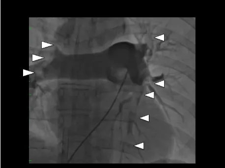
The patient’s hemodynamics had already collapsed on admission and emer-gency ECMO was initiated to stabilize her hemodynamics. Because pulmonary angiography showed multiple defects in the bilateral pulmonary arteries (Figure 1) with significant pulmonary hypertension (mean pulmonary artery pressure, 62 mmHg), the patient was transferred to the operating room for emergency surgery. After establishment of cardiopulmonary bypass with ascending aortic cannulation and bilateral venous drainage through the median sternotomy, fresh thrombus was successfully removed from the bilateral pulmonary arteries on the beating heart.
[image:2.595.258.487.520.691.2]However, at the time of weaning from the cardiopulmonary bypass, fresh blood flowed out of the tracheal tube because of pulmonary bleeding, and trans-fusion was needed to control the bleeding. Consequently, the patient received 24 units of red blood cell concentrates, 30 units of fresh frozen plasma, and 55 units of platelet concentrates over a 2-hour period. Although pulmonary bleeding was partially controlled prior to the completion of transfusion, clear and frothy fluid flowed out of the tracheal tube. Despite pure oxygen inhalation and high positive end-expiratory pressure, the partial pressure of the arterial oxygen gradually
Figure 1. Preoperative pulmonary angiography showing multiple defects (white arrow
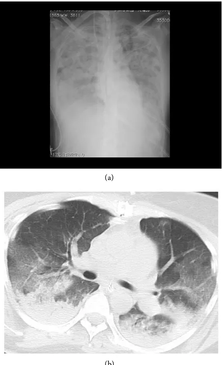
DOI: 10.4236/ojts.2018.83010 59 Open Journal of Thoracic Surgery decreased. Finally, V-A ECMO was started through the femoral artery cannula-tion and the superior vena caval drainage. The transesophageal echocardiogra-phy showed almost normal cardiac function and the urine output was sufficient. Chest radiography conducted in the operating room revealed diffuse bilateral pulmonary infiltrates (Figure 2(a)). Chest computed tomography showed bila-teral gravitational atelectasis and peribronchial infiltrates with no pleural effu-sion (Figure 2(b)). In the intensive care unit, the patient received meticulous treatment including steroid administration and nitric oxide inhalation. Frequent bronchoscopic suction was performed for lung recruitment. The impaired oxy-genation gradually improved and ECMO was weaned on the fourth day. Al-though tracheostomy was temporally performed, the patient was discharged on postoperative day 80 without any complications after sufficient rehabilitation. The pulmonary hypertension improved to almost normal range. The patient is currently recovering well without recurrent thromboembolic events and respi-ratory failure at the 14-month follow up.
(a)
[image:3.595.261.487.306.677.2](b)
Figure 2. (a) Chest radiography performed in the operating room showing diffuse
DOI: 10.4236/ojts.2018.83010 60 Open Journal of Thoracic Surgery The patient provided consent to publish the details and results of the case. The identity of the patient has been protected.
3. Discussion
TRALI is a potentially fatal complication of blood transfusion that is characte-rized by dyspnea, hypotension, and hypoxemia and usually develops during or within six hours after transfusion [1]. Although this definition appears straightforward, the characteristics of TRALI are indistinguishable from those of acute lung injury due to other causes, such as sepsis or lung contusion. To diffe-rentiate this condition, the term “possible TRALI” was introduced [2], which al-lows for the presence of another risk factor for acute lung injury. The typical findings of TRALI are diffuse bilateral pulmonary infiltrates with the absence of cardiac dysfunction and without pleural effusion on chest radiography and computed tomography.
A two-hit hypothesis has been proposed for TRALI [2]. The first hit concerns patient-related factors: adherence of neutrophils to the pulmonary endothelium. The second hit concerns discharge of mediators that activate the endothelial cells and pulmonary neutrophils, resulting in capillary leakage and subsequent pul-monary edema.
The incidence of TRALI is estimated to be between 0.08% and 15% in patients receiving blood transfusion. Furthermore, the incidence of TRALI is 50 - 100 times higher in critically ill patients than in the general hospital population [2]. In general, TRALI has a good prognosis and mortality is considered to be low at approximately 5% - 10% [2]. However, TRALI mortality in surgical patients was higher than in transfused controls [2].
Although this definition appears straightforward, the characteristics of TRALI are sometimes indistinguishable from those of acute respiratory distress syn-drome because of deteriorated patient status. To identify such cases, the term “possible TRALI” was developed [2], which allows the presence of another risk factor for acute lung injury.
Cardiac surgery is a risk factor of TRALI [3]. During cardiac surgery, the lungs are deflated and remain nonventilated for several hours, which may cause injury to the pulmonary endothelium (the first hit). In addition, the use of cardiopulmonary bypass can cause pulmonary edema (the second hit) [3]. Besides these possible risk factors, patients often receive blood transfusion. The patient in the present case received considerable blood transfusion. Therefore, the present case is identified as possible TRALI based on the use of cardiopulmonary bypass, highly invasive procedures, and absence of cardiac dysfunction.
con-DOI: 10.4236/ojts.2018.83010 61 Open Journal of Thoracic Surgery troversial because of high incidence of morbidities, early use of ECMO enables us to reduce the patient’s fraction of inspiratory oxygen and peak inspiratory pressure and can prevent ventilator-induced lung injury [5]. V-A ECMO has the advantage of reducing right sided heart volume including of the pulmonary arte-ries, allowing for the recovery of ventricular function and optimizing oxygen transport by improving cardiac output and oxygen content.
Kuroda et al. reported successful management of TRALI with V-V ECMO [4]. V-V ECMO is associated with a lower complication rate than V-A ECMO [7]. In V-V ECMO, there are fewer bleeding complications [7] and greater supply of oxygenated blood in the coronary circulation. However, hypotension is a com-mon sign in TRALI and usually does not respond to IV fluid infusion. V-A ECMO was selected for the treatment of patients who appeared to be at near cardiac arrest, although respiratory failure usually requires only V-V ECMO [8]. Furthermore, as V-A ECMO decreases the transpulmonary blood flow, it may help control both airway bleeding from the pulmonary circulation and pulmo-nary edema secondary reperfusion [9]. Therefore, V-A ECMO enables sufficient and safe bronchoscopic tracheal suction. In the present case, V-A ECMO as-sisted the impaired respiratory system and enabled us to perform bronchoscopic suction safely.
During V-A ECMO, the patient received meticulous treatment including ad-ministration of methylprednisolone and nitric oxide inhalation. Inhalation of nitric oxide reduces pulmonary vascular resistance in patients with pulmonary hypertension [10]. Perioperative corticosteroid administration can reduce com-plement activation and release of inflammatory cytokines associated with cardi-opulmonary bypass. However, the clinical benefits of this treatment are
uncer-tain [11].
4. Conclusion
V-A ECMO placement plays an important role in the treatment of patients with acute severe hypoxemia due to possible TRALI after cardiac surgery. In particu-lar, it can prevent ventilator-induced lung injury and enable safe bronchoscopic tracheal suction.
Acknowledgements
We thank the Honyaku Center Inc. for reviewing and editing the manuscript. We also thank our colleagues for their helpful comments.
Conflicts of Interest
There are no conflicts of interest to declare. We did not receive any financial support for this study.
References
Considera-DOI: 10.4236/ojts.2018.83010 62 Open Journal of Thoracic Surgery tions in Transfusion-Related Acute Lung Injury. Transfusion, 25, 573-577. https://doi.org/10.1046/j.1537-2995.1985.25686071434.x
[2] Vlaar, A.P.J. and Juffermans, N.P. (2013) Transfusion-Related Acute Lung Injury: A Clinical Review. The Lancet, 382, 984-994.
https://doi.org/10.1016/S0140-6736(12)62197-7
[3] Vlaar, A.P.J., Hofstra, J.J., Determann, R.M., et al. (2011) The Incidence, Risk Fac-tors, and Outcome of Transfusion-Related Acute Lung Injury in a Cohort of Car-diac Surgery Patients: A Prospective Nested Case-Control Study. Blood, 117, 4218-4225. https://doi.org/10.1182/blood-2010-10-313973
[4] Kuroda, H., Masuda, Y., Imaizumi, H., et al. (2009) Successful Extracorporeal Membranous Oxygenation for a Patient with Life-Threatening Transfusion-Related Acute Lung Injury. Journal of Anesthesia, 23, 424-426.
https://doi.org/10.1007/s00540-009-0760-5
[5] Lindem, V., Palmer, K., Reinhard, J., et al. (2000) High Survival in Adult Patients with Acute Respiratory Distress Syndrome Treated by Extracorporeal Membrane Oxygenation, Minimal Sedation, and Pressure Supported Ventilation. Intensive Care Medicine, 26, 1630-1637. https://doi.org/10.1007/s001340000697
[6] Lewandowski, K., Rossaint, R., Pappert, D., et al. (1997) High Survival Rate in 122 ARDS Patients Managed According to Clinical Algorithm Including Extracorporeal Membrane Oxygenation. Intensive Care Medicine, 23, 819-835.
https://doi.org/10.1007/s001340050418
[7] Makdisi, G and Wang, I. (2015) Extra Corporeal Membrane Oxygenation (ECMO) Review of a Lifesaving Technology. Journal of Thoracic Disease, 7, E166-E176. [8] Lee, A.J., Koyyalamudi, P.L. and Martinez-Ruiz, R. (2008) Severe
Transfu-sion-Related Acute Lung Injury Mamaged with Extracorporeal Membrane Oxyge-nation (ECMO) in an Obstetric Patient. Journal of Clinical Anesthesia, 20, 549-552. https://doi.org/10.1016/j.jclinane.2008.05.019
[9] Boulate, D., Mercier, O., Mussot, S., et al. (2016) Extracorporeal Life Support after Pulmonary Endarterectomy as a Bridge to Recovery or Transplantation: Lessons from 31 Consecutive Patients. The Annals of Thoracic Surgery, 102, 260-268. https://doi.org/10.1016/j.athoracsur.2016.01.103
[10] Giaid, A. and Saleh, D. (1995) Reduced Expression of Endothelial Nitric Oxide Synthase in the Lungs of Patients with Pulmonary Hypertension. The New England Journal of Medicine, 333, 214-221. https://doi.org/10.1056/NEJM199507273330403 [11] Kerr, K.M., Auger, W.R., Marsh, J.J., et al. (2012) Efficacy of Methylprednisolone in
Preventing Lung Injury Following Pulmonary Thromboendarterectomy. Chest, 141, 27-35. https://doi.org/10.1378/chest.10-2639
Abbreviations
TRALI: Transfusion-related acute lung injury ECMO: Extracorporeal membrane oxygenation V-A: Veno-Arterial