ISSN Print: 2152-2197
DOI: 10.4236/jep.2018.910067 Sep. 7, 2018 1082 Journal of Environmental Protection
Toxicity Evaluation of Acrylamide on the Early
Life Stages of the Zebrafish Embryos
(
Danio rerio
)
Hattie Spencer
1, Joseph Wahome
1, Mary Haasch
21Department of Natural Sciences and Environmental Health, Mississippi Valley State University, Itta Bena, Mississippi, USA
2National Center for Natural Products Research, Environmental Toxicology Research Program, University of Mississippi, Oxford, Mississippi, USA
Abstract
Acrylamide is a chemical used mainly in industrial applications and the treatment of drinking and wastewater, making it easy to enter aquatic ecosys-tems. There are few studies known about the toxicity of acrylamide to aquatic organisms which have shown evidence of a number of histopathological ef-fects. To assess the effects of acrylamide to freshwater fish, Zebrafish (Danio rerio) embryos were exposed to serial concentrations of acrylamide (0, 100, 300, and 500 mg/L) to investigate the acute toxicity effects on teleost em-bryogenesis. Embryos less than 24 hrs old were exposed under static non-renewal conditions for ten days or until hatching. The toxic endpoints evaluated include: egg/embryo viability, hatchability, and morphologi-cal/developmental anomalies during organogenesis. The acute toxicity test resulted in a 48 h-LC50 of 585 mg/L for egg viability. Exposure of embryos significantly reduced hatchability and larval survival, in a concentration de-pendent manner. Dimethyl sulfoxide (DMSO) was used as a solvent carrier to permeate the uptake of acrylamide through the chorion membrane. No sig-nificant damages or complications were observed in embryos exposed to DMSO. At 500 mg/L, the highest test concentration, the survival of embryos was greatly reduced within 24 hrs of exposure. The lower test, 100 mg/L, produced a significant number of developmental anomalies to the Zebrafish that included dorsal tail flexure, severe pericardial edema, facial and cranial defects and decreased heartrate (40 bpm). Premature hatching of embryos and developmental arrest was observed in all concentrations. The severity of these anomalies was concentration-dependent and resulted in low survival rate and high frequency of malformations. These results indicate that acryla-mide is teratogenic and provide support for sub-lethal toxicity testing using Zebrafish embryos.
How to cite this paper: Spencer, H., Wa-home, J. and Haasch, M. (2018) Toxicity Evaluation of Acrylamide on the Early Life Stages of the Zebrafish Embryos (Danio rerio). Journal of Environmental Protec-tion, 9, 1082-1091.
https://doi.org/10.4236/jep.2018.910067
Received: June 20, 2018 Accepted: September 4, 2018 Published: September 7, 2018
Copyright © 2018 by authors and Scientific Research Publishing Inc. This work is licensed under the Creative Commons Attribution International License (CC BY 4.0).
DOI: 10.4236/jep.2018.910067 1083 Journal of Environmental Protection
Keywords
Acrylamide, Toxicity, Zebrafish, Malformation, Embryonic Development
1. Introduction
Acrylamide is a chemical intermediate used in the production and synthesis of polyacrylamide [1] [2]. It is a synthetic chemical compound commonly used in many branches of industry. The largest use for polyacrylamide is treating mu-nicipal drinking water and wastewater to remove suspended solids. The polymer is used to remove suspended solids from industrial wastewater before discharge, reuse, or disposal. Recent discoveries have shown that people are exposed to small amounts of acrylamide through its presence in some starchy foods cooked at high temperatures [3]. Effects of acrylamide on human health and the envi-ronment depend on how much acrylamide is present and the length and fre-quency of exposure [4]. The most important environmental contamination re-sults from the use of acrylamide in soil grouting and drinking-water contamina-tion by the use of polyacrylamide flocculants that contain residual acrylamide monomer [5] [6] and from acrylamide-based sewer grouting and wastepaper re-cycling [7] [8].
The EPA’s Toxics Release Inventory reported environmental releases of 8,797,482 lb of acrylamide from 42 facilities in the US, 99.9% of which was re-leased to underground injection wells [9]. Levels detected of 400 mg/l in well-water in Japan had been contaminated from a grouting operation and resi-dual acrylamide concentrations for water-treatment plants ranged from 0.5 to 600 ppm [10] [11]. Acrylamide monomers may not be removed in many water treatment processes and remain stable for more than two months in tap water and can become a potential source of pollution.
Laboratory animals exposed to acrylamide exhibited a decrease in glutathione level and in the activity of glutathione-S-transferase on the brain and liver. The inhibition of glutathione-S-transferase by acrylamide, which catalyzed conjuga-tion with glutathione, may lead to the accumulaconjuga-tion of the monomer and it en-hanced neurotoxicity [12]. In addition, laboratory studies in animals have also shown that exposure to acrylamide can induce cancer, genetic damage and ad-verse effects on reproduction and development. Several studies have shown acrylamide to have a moderate acute toxicity effect to aquatic organisms [13]. At present, little is known about the toxic effects of acrylamide in fishes. Acryla-mide toxicity study conducted in two species of aquatic macroinvertebrates and three species of fish demonstrated that acrylamide was moderately toxic aquatic organisms [14]. There was also histopathological changes and correlation with genotoxicity and metabolic alterations when Carassius auratus hepatopancreas
(goldfish) was exposed to acrylamide [15].
un-DOI: 10.4236/jep.2018.910067 1084 Journal of Environmental Protection
derstand and evaluate its impact on the early life stage development of aquatic organisms. Fish is an important component of the ecosystem and the early life stages of fish have been found to be more sensitive to pollutants than adult [16]. Proliferating embryonic tissue can be particularly sensitive to damage from reac-tive metabolites generated by xenobiotic metabolizing enzymes [17].
In this study the zebrafish, (Danio rerio) was the model used to assess the de-velopmental processes and toxicological effects of acrylamide on developing embryo. The zebrafish is idea due to ease of maintenance, translucent and non-sticky eggs, and a short reproduction cycle [18]. A non-static renewal bio-assay was used to assess hatchability, and development. Furthermore, the study will determine if acrylamide is teratogenic to the developing embryo.
2. Materials and Experimental Methods
2.1. Test Material
Acrylamide (C3H5NO)-Lot No. 148571-25G, Purity 97%, and Dimethyl
Sulfox-ide,CAS No. 67-68-5, was both purchased from Sigma-Aldrich Chemical Com-pany (St. Louis, MO). Dimethyl Sulfoxide (DMSO) was used as a solvent carrier. Instant Ocean Sea Salt was used for preparing embryo rearing medium (egg wa-ter) and was obtained from Aquarium Systems (Vernon, CT). The embryos were obtained from a continuous culture at the University of Mississippi, (Oxford, MS) Biology department. The water temperature was set at 27˚C with a cycle of 14 hours light: 10 hours dark. Eggs were collected within 5 hours of spawning and examined microscopically for fertilization and stage development. Embryos were transferred to a clean 20 ml vials containing 15 ml of rearing medium (60 µg/ml), 15 ul of DMSO and appropriate concentration of test chemical. The rearing medium used in this study consisted of the following: 10 g NaCl, 0.3 g KCl, 0.4 g CaCl2, and 1.63 g MgSO4 in 1000 mL of deionized water. Normal
hatching period for untreated zebrafish embryo was 56 hours after fertilization. Embryos, both treated and untreated, completing the hatching process were recorded daily. Hatched larvae were not fed due to the duration of the test. Ferti-lized eggs were examined daily to observe embryonic development, time point of lesion and abnormal development, developmental arrest and hatching success.
2.2. Acute Exposure
The study was conducted in a static non-renewal system. The rearing medium consisted of Instant Ocean Sea Salts (40 g) added to 1liter distilled water. The stock solution was diluted with rearing medium to the desired acrylamide con-centrations of 100, 300 and 500 mg/L. These concon-centrations are based on the high levels of acrylamide released in the effluents from treatment plants into the aquatic environments.
DOI: 10.4236/jep.2018.910067 1085 Journal of Environmental Protection
signs of early life stage toxicity. There were two control groups, a positive (rear-ing medium only) and vehicle control (DMSO plus rear(rear-ing medium). DMSO made the embryo more sensitive to the uptake of acrylamide to reach sufficient concentrations needed for the exposure.
Dead and hatched embryos were counted and removed daily. Embryo deve-lopmental stage, time point of lesion and mortality was determined by micro-scopic examination. Egg mortality was based on the number of embryos that died prior to hatching and that was opaque in appearance. Embryos were ex-posed to concentrations until hatched.
2.3. Statistical Analysis
The EPA Probit analysis method was used to calculate the LC50 value and its 95%
confidence intervals, from the 96-h acute toxicity data. For the sub-acute expe-riments, descriptive statistics were applied to determine the mean values of each experimental data set. An analysis of variance (ANOVA, F-test) was performed to determine whether there were significant differences among these means. The Student t-test was used to compare each of the treated measurements with the control measurements, which included the DMSO vehicle control. The level of significance was set at p = 0.05 (95% confidence level).
3. Results and Discussion
3.1. Time Hatching and Egg Mortality
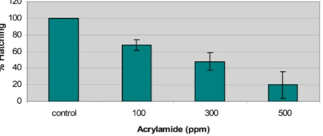
[image:4.595.214.539.559.696.2]The hatching success of non-exposed (control) and exposed zebrafish embryos are presented in Figure 1. The acute exposure of zebrafish embryos to acryla-mide caused a significant decrease in hatching that correlated with increasing acrylamide concentrations. Embryos were exposed to acrylamide after fertiliza-tion at concentrafertiliza-tions of 100, 300, and 500 mg/L. Hatching for zebrafish em-bryos began at 56 hours where 100% of emem-bryos hatched in the control group and 95% fry hatched in the vehicle control. No death was observed in the DMSO exposed embryos during the first 24 hours, however, the mean percentage of egg mortality for DMSO embryos were 5% for the remainder of development.
DOI: 10.4236/jep.2018.910067 1086 Journal of Environmental Protection
DMSO did not appear to have a negative effect on the development of the zebra-fish during the hatching period. The non-exposed embryos showed normal signs of teleostean development based on the developmental series for the zebrafish embryo [19]. Hatching and egg mortality disparities were observed in all con-centrations as well as delayed hatching and a reduction in the number of em-bryos that completed the hatching process. Egg mortality was shown to be centration dependent. Death in exposed was observed within 24 hours and con-tinued to increase throughout embryos development. Statistical analysis using the F-test (ANOVA) shows highly significant differences (p = 0.01) in mean percentages of hatching among experimental groups. Therefore, it could be con-cluded that hatching was inversely correlated with acrylamide concentrations. Statistical analysis using the F test (ANOVA) shows highly significant difference among hatching experimental groups.
The diffusion of the acrylamide in the zebrafish embryo was extremely slow in which slow infusion of a chemical is a protective mechanism of the outer cho-rion and membrane systems that protect the embryo from chemical insult [20]. DMSO made the embryo more sensitive to the uptake of acrylamide to reach sufficient concentrations needed for the exposure.
3.2. Teratogenic Effects of Acrylamide
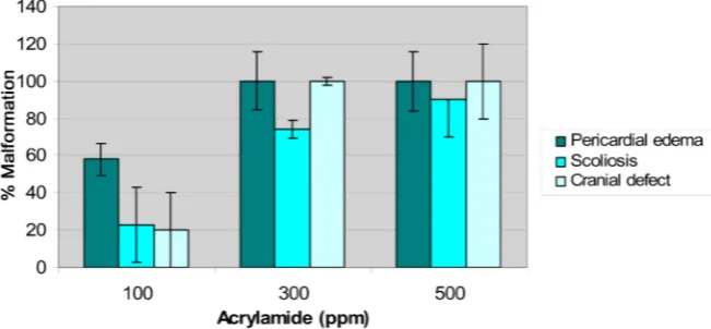
[image:5.595.211.537.531.682.2]Characteristics signs of acrylamide early life stage toxicity in embryo are pre-sented in Figure 2. The embryonic abnormalities were observed within 32 hours of exposure, indicating that acrylamide may be teratogenic. Major signs of acry-lamide toxicity observed were severe pericardial and yolk sac edema, scoliosis (dorsal tail flexure), and cranial defects (sloping forehead). As illustrated, there was a concentration-response relationship with respect to the induction of each of these developmental abnormalities by acrylamide. The earliest adverse physi-ological effect produced by induced acrylamide toxicity on the developing zebra-fish embryo was pericardial and yolk sac edema at a concentration of 100 ppm.
DOI: 10.4236/jep.2018.910067 1087 Journal of Environmental Protection
For instance, 0.0, 23, 74 and 90 percent of scoliosis malformations were observed in embryos exposed to 0 (Control), 100, 300 and 500 ppm of acrylamide, respec-tively.
3.3. Effects of Acrylamide on Heartrate and Blood Flow in
Zebrafish Embryo
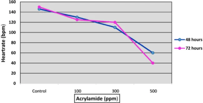
When exposed to acrylamide shortly after fertilization, zebrafish embryos exhi-bit reduced heartrate and blood flow by 32 hours post fertilization (hpf). The ef-fects of acrylamide on the heartrate of the zebrafish embryo are shown in Figure 3. Data illustrated indicates a dose-response relationship related to the toxicity of acrylamide. All control embryos exhibit a normal heartrate throughout hatching. The average heart rate for control at 32 hr was 142 beats per minutes (bpm). Within 32 hours, embryos began to show sign of stress. All treated embryos, be-gan to show oscillation blood flow pattern associated with a decreased blood flow and heartrate in all concentrations after 32 and 48 hours of exposure, but more profound at 48 hrs. Upon 48 hours of exposure, the average percentage heartrate for embryos in each concentration were 146, 83, 70, 40 bpm at concen-trations of 0, 100, 300 and 500 ppm, respectively. Oscillation blood flow pattern usually occurred in conjunction with slow blood flow. Similar patterns of blood flow were also observed in the yolk sac in the vitelline vein of the embryo. After 48 hours of exposure, the vitelline vein became a broad band in the treated em-bryos. With an increase in concentration, the number of lesions (blood clots, hemorrhage, and cessation of blood flow) increased in treated embryos.
3.4. Effects of Increasing Concentrations of Acrylamide on
Malformation Development in Zebrafish Embryo
[image:6.595.211.536.504.669.2]The primary site of action of acrylamide in the zebrafish embryos was the pro-found effects on the circulatory and nervous systems. The earliest sign of acrylamide
DOI: 10.4236/jep.2018.910067 1088 Journal of Environmental Protection
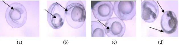
induced toxicity after post-fertilization was the cardiovascular and nervous sys-tem. An increase in concentration resulted in an increase in the percentage ab-normalities occurring in the zebrafish embryos. A normal control zebrafish embryo and fry shown in Figure 4(a) illustrates normal zebrafish embryonic and larvae development. A treated embryo and fry from (100 ppm) is shown in
Figure 4(b) with moderate pericardial and yolk-sac edema, sloping forehead and
scoliosis after induction of acrylamide toxicity, indicating that organogenesis may be the most sensitive stage of embryonic development. A severe form of pe-ricardial edema and scoliosis was also observed at 300 ppm (Figure 4(c)). The same effects were notable but more severe than observed in the 100 ppm treated embryo. The embryo shown in Figure 4(d) reflects a greater degree of severity of the lesion cause by acrylamide exposure. After 72 hours, most embryos in this concentration were not hatched.
Acrylamide produce more severe forms of pericardial edema, cranial defect and scoliosis at 500 ppm than seen at any other concentrations. The severity of these malformations also produced a high death rate in the embryo which indi-cates that acrylamide becomes increasingly fatal to embryos at higher concentra-tions. Pericardial edema gives rise to the collapse of the yolk sphere which pre-vents the heart from undergoing normal chamber formation [21].
Scoliosis of the spine was commonly observed in all concentrations. Scoliosis in fish is most likely caused by agents that act on the central nervous system, or the neuromuscular junctions [22]. Similar lesion observed in zebrafish exposed has been attributed to calcium depletion [23].
4. Conclusion
This study illustrated that acrylamide was acutely toxic to the development of the zebrafish embryo. The stage of embryonic development through the larval stage of a fish life is most sensitive to environmental contaminants. The expo-sure to sublethal concentrations of acrylamide resulted in a significant reduction in hatching, as well as the development of a number of morphological and physio-logical abnormalities in the zebrafish. Acrylamide exposure induces developmental
[image:7.595.217.532.552.631.2](a) (b) (c) (d)
DOI: 10.4236/jep.2018.910067 1089 Journal of Environmental Protection
abnormalities in the zebrafish embryos, which leads to developmental arrest and delayed hatching, especially in the highest concentration. The lowest tested con-centration produced a significant number of developmental effects to the zebra-fish embryo, including pericardial edema, dorsal tail flexure, cranial defect, and defects in heart morphology. The severity of these malformations also produced a high death rate in the embryo. These results indicated that acrylamide becomes increasingly fatal to embryos, reducing survival at higher concentrations when exposed during early development. In addition, the zebrafish is a good test mod-el for assessing and screening the wide range of effect following exposure to acrylamide and the potential for risks to human health at low levels.
Acknowledgements
We thank Dr. Michael Smith for his expertise and assistant with this research and Dr. Ali Ishaque and Dr. Frank von Hippel for critically reading the manu-script. This research was financially supported by a grant from the National In-stitute of Child Health and Human Development (NICHD) from the National Institute of Health (NIH) grant number: GII HD 37065-05 and MBRS grant.
Conflicts of Interest
The authors declare no conflicts of interest regarding the publication of this pa-per.
References
[1] IARC, International Agency for Research on Cancer (1986) IARC Monographs on the Evaluation of the Carcinogenic Risk of Chemicals to Humans. Some Chemicals used in Plastics and Elastomers, 39, 403.
http://citeseerx.ist.psu.edu/viewdoc/download?doi=10.1.1.173.3428&rep=rep1&type =pdf
[2] IARC, International Agency for Research on Cancer (1994) IARC Monographs on the Evaluation of Carcinogenic Risks to Humans. Some Industrial Chemicals, 60, 560.
http://citeseerx.ist.psu.edu/viewdoc/download?doi=10.1.1.173.3428&rep=rep1&type =pdf
[3] National Institute of Environmental Health Sciences (2004) NTP-CERHR Mono-graph on the Potential Human Reproductive and Developmental Effects of Acryla-mide. Center for the Evaluation of Risks to Human Reproduction (CERHR).
https://ntp.niehs.nih.gov/ntp/ohat/acrylamide/acrylamide_monograph.pdf
[4] U.S.EPA. (1994) U.S. Environmental Protection Agency. Office of Pollution Pre-vention and Toxics. Chemical in the Environment: Acrylamide. EPA 79—06-1, Washington DC.
[5] NSF (1988) Drinking Water Treatment Chemicals—Health Effects. Ann Arbor, MI, National Sanitation Foundation (Standard 60).
http://www.who.int/water_sanitation_health/dwq/chemicals/acrylamide.pdf [6] WHO (1985) Acrylamide. Environmental Health Criteria 49. World Health
Organ-ization, Geneva. http://www.iprev.it/uploads/media/ACRYLAMIDE.pdf
DOI: 10.4236/jep.2018.910067 1090 Journal of Environmental Protection the Degradation of Acrylamide Monomer. Water Research, 14, 775-778.
http://www.iprev.it/uploads/media/ACRYLAMIDE.pdf
[8] Brown, L., Rhead, M.M., Hill, D. and Bancroft, K.C.C. (1982) Rapid Screening Technique Utilizing High-Performance Liquid Chromatography for Assessing Acrylamide Contamination in Effluents. Analyst, 107, 749-754.
http://www.iprev.it/uploads/media/ACRYLAMIDE.pdf
[9] TRI (2009) TRI Explorer Chemical Report. U.S. Environmental Protection Agency.
http://www.epa.gov/triexplorer
[10] Igisu, H., Goto, I., Kawamura, Y., Kato, M., Izumi, D. and Kuroiwa, Y. (1975) Acry-lamide Encephaloneuropathy Due to Well Water Pollution. Journal of Neurology, Neurosurgery, and Psychiatry, 38, 581-584.
[11] Howard, P.H. (1989) Acrylamide. In: Handbook of Environmental Fate and Expo-sure Data for Organic Chemicals, Vol 1, Large Production and Priority Pollutants, Lewis Publishers, Chelsea, MI, 13-19.
[12] Shanker, R. and Seth, P.K. (1986) Toxic Effects of Acrylamide in a Freshwater Fish,
Heteropneustes fossilis. The Bulletin of Environmental Contamination and Toxi-cology, 37, 274-280.
[13] Kegley, S.E., Hill, B.R., Orme, S. and Choi, A.H. (2016) PAN Pesticide Database, Pesticide Action Network, North America, (Oakland, CA).
http://www.pesticideinfo.org
[14] Krautter, G.R., Mast, R.W., Alexander, H.C., Wolf, C.H., Friedman, M.A., Koschier, F.J. and Thompson, C.M. (2009) Acute Aquatic Toxicity Tests with Acrylamide monomer and Macroinvertebrates and Fish. Environmental Toxicology and Che-mistry, 5, 373-377. https://doi.org/10.1002/etc.5620050406
[15] Larguinho, M., Costa, P.M., Sousa, G., Costa, M.H., Diniz, M.S. and Baptista, P.V. (2013) Histopathological Findings on Carassius auratus Hepatopancreas upon Ex-posure to Acrylamide: Correlation with Genotoxicity and Metabolic Alterations.
Journal of Applied Toxicology, 34, 12.
[16] McKim, J.M. (1977) Evaluation of Tests with Early Life Stages of Fish for Predicting Long-Term Toxicity. Journal of the Fisheries Research Board of Canada, 34, 1148-1154. https://doi.org/10.1139/f77-172
[17] Harbison, R.D. (1975) Parathion-Induced Toxicity and Phenobarbital-Induced Protection against Paratihion during Prenatal Development. Toxicology and Ap-plied Pharmacology, 32, 482-493. https://doi.org/10.1016/0041-008X(75)90113-1 [18] Oberemm, A. (2000) The Use of a Refined Zebrafish Embryo Bioassay for the
As-sessment of Aquatic Toxicity. Laboratory Animal, 29, 32-40.
[19] Westerfield, M. (1995) The Zebrafish Book. Guide for the Laboratory Use of Zebra-fish (Danio rerio). University of Oregon Press, Eugene.
[20] Jung, M.H., Halter, C., Friesenhengst, A., Walzer, J. and Czerny, T. (2013) Diffusion of Small Molecules into Medaka Embryos Improved by Electroporation. BMC Bio-technology, 13, 53.https://doi.org/10.1186/1472-6750-13-53
[21] Wisk, J.D. and Cooper, K.R. (1990) The Stage Specific Toxicity of 2,3,7,8, Tetrach-lorodibenzo-p-Dioxin in the Embryos of the Japanese Medaka (Oryzias latipes).
Environmental Toxicology and Chemistry, 9, 1159-1169. https://doi.org/10.1897/1552-8618(1990)9[1159:TSSTOT]2.0.CO;2
[22] Couch, J.A., Winstead, J.T. and Goodman, L.R. (1977) Kepone-Induced Scoliosis and Its Histological Consequences in Fish. Science, 197, 585-587.