ISSN Online: 2164-3016 ISSN Print: 2164-3008
Advantages of Ilizarov External Fixation in an
Elderly Patient with Pilon Fracture with Severe
Soft Tissue Injury and Severe Osteoporosis:
A Case Report
Koji Nozaka
*, Naohisa Miyakoshi, Hidetomo Saito, Shuichi Chida, Hiroyuki Tsuchie,
Yusuke Yuasa, Motoki Mita, Yoichi Shimada
Department of Orthopedic Surgery, Akita University Graduate School of Medicine, Hondo, Akita, Japan
Abstract
Introduction: Pilon fracture in elderly individuals is characterized by senile skin atrophy, poor dermal extensibility, and thin subcutaneous tissue. The use of bulky internal fixation material can thus cause the swelling that accompa-nies the fracture to induce secondary injury to skin tissue. In addition, initia-tion of postoperative weight-bearing is delayed due to bone fragility and dif-ficulties with partial weight-bearing, causing a tendency toward prolonged hospitalization. Mean duration of hospitalization after pilon fracture for el-derly patients in our department was 79.2 days. Case Presentation: An 80-year-old woman with pilon fracture with soft tissue injury and severe os-teoporosis was transferred to our department. The fracture was treated using Ilizarov external fixation. Fourteen days postoperatively, walking with full weight-bearing was permitted. The hospital stay was 28 days. The external fixator of the ankle was removed 87 days postoperatively, at which time the patient was anatomically and functionally recovered and able to walk unaided. Conclusion: Ilizarov external fixation may represent a useful option in elderly patients with pilon fracture showing severe soft tissue injury and severe os-teoporosis. The present case provides evidence that this procedure can be suc-cessfully applied to the management of such pilon fractures in elderly patients.
Keywords
Ilizarov External Fixation, Elderly Patient, Pilon Fracture, Soft Tissue Injury, Osteoporosis
How to cite this paper: Nozaka, K., Miya-koshi, N., Saito, H., Chida, S., Tsuchie, H., Yuasa, Y., Mita, M. and Shimada, Y. (2019) Advantages of Ilizarov External Fixation in an Elderly Patient with Pilon Fracture with Severe Soft Tissue Injury and Severe Os-teoporosis: A Case Report. Open Journal of Orthopedics, 9, 14-22.
https://doi.org/10.4236/ojo.2019.91002
Received: November 25, 2018 Accepted: January 25, 2019 Published: January 28, 2019
Copyright © 2019 by author(s) and Scientific Research Publishing Inc. This work is licensed under the Creative Commons Attribution International License (CC BY 4.0).
http://creativecommons.org/licenses/by/4.0/ Open Access
The mean duration of hospitalization after pilon fracture in our department is 79.2 days. The length of hospital stay after pilon fracture in Japan is much longer than in Western countries, largely because there are extremely few nursing homes and rehabilitation hospitals in Japan [1]. Ilizarov external fixation for soft tissue injury and bones with osteoporosis, even in pilon fractures where the cor-tical bone is thin and use of screws for internal fixation is difficult, allows suita-ble fixation using multiple Ilizarov wires [2] [3]. Ilizarov external fixation is to prevent soft tissue complications by avoiding unnecessary internal fixation of fragile skin. The other is to enable early weight bearing, early ambulation, early independence, and early hospital discharge. We have been performing osteo-synthesis using an Ilizarov external fixator and early weight-bearing in an elderly patient who sustained a pilon fracture with severe soft tissue injury and osteo-porosis [4].
2. Case Presentation
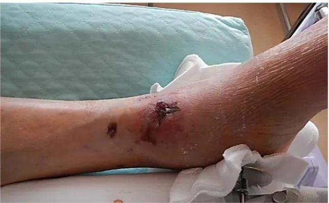
An 80-year-old woman was injured when she fell on a snowy road. She was transported by ambulance to another hospital. Since comminuted fracture was observed, she was treated with skeletal traction. However, repositioning the fractured bone proved very difficult. Because of her low weight (35 kg), it was very difficult to apply skeletal traction with a heavy weight. Severe osteoporosis with cortical thinning at the fracture site was evident (Figure 1). Since skin con-dition was also not improving, she was transferred to our department, where left pilon fracture was diagnosed (OTA [Orthopaedic Trauma Association’s Fracture and Dislocation Compendium] 43C3) (Figure 2). The bone mineral density of the lumbar spine (L2–4, 0.549 g/cm2, T-score: −3.78 S.D.) and proximal femur (0.622 g/cm2, T-score: −2.57 S.D.) confirmed a diagnosis of osteoporosis. Skin necrosis was identified at the anterior fracture site (Figure 3). Given these find-ings, the risk of skin disorders with the use of bulky internal fixation materials appeared high. The decision was therefore made to use closed indirect reduction techniques with an Ilizarov ring fixator for rigid fixation [5]. One day after ad-mission to our institute, Ilizarov ring fixator surgery was performed with the pa-tient under general anesthesia in a supine position with trans-calcaneal traction. We used 4 rings for the Ilizarov fixator. The amount of reduction achieved by ligamentotaxis was checked using intensification. The foot was incorporated by olive wires inserted into the calcaneus and fixed to a foot ring connected to the
Figure 1. Preoperative radiographs on initial consultation showing pilon fracture.
Figure 2. Computed tomography (CT) and 3-dimensional CT on initial consultation at our hospital show pilon fracture (OTA (Orthopaedic Trauma Association’s Fracture and Dislocation Compendium) type 43C3).
Figure 3. Skin condition at the time of transfer to our department. Necrosis is evident at the anterior fracture site.
[image:3.595.213.537.494.694.2]An olive wire was also introduced through the tibial malleolus, first to reduce the fracture tilted in varus and anteriorly and then to compress the fracture site
(Figure 3). After restoration of articular congruity, a 1.8 mm Ilizarov wire was
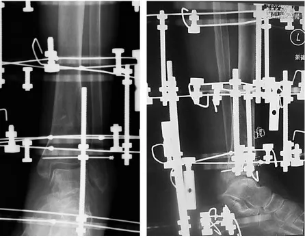
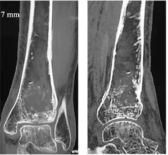
[image:4.595.262.485.521.695.2]passed parallel to the articular surface in the anteroposterior view on X-ray, ap-proximately 15 mm from the joint line in the tibial epiphysis. Additional 1.8 mm Ilizarov wires were inserted in safe corridors to improve alignment and increase stability. Three wires were inserted onto the distal tibial ring. The wires were fixed to the rings of the fixator and tensioned. The foot was fixed in neutral posi-tion to avoid supinaposi-tion and equinus posiposi-tion. Olive wires were used in an op-posing configuration, allowing variable degrees of interfragmentary compression to be achieved. This was of particular benefit in putting transverse compressive forces on a spiral metaphyseal fracture (Figure 4). We treated the patient with an ankle-hinge Ilizarov external fixator to allow early implementation of range-of-motion exercises (Figure 5) [7]. Fourteen days postoperatively, walk-ing with full weight-bearwalk-ing was permitted. The duration of hospitalization was 28 days. The external fixator of the ankle was removed at 87 days after surgery. Radiographs and computed tomography (CT) showed healing of the fracture at 87 days postoperatively (Figure 6 and Figure 7). At follow-up 3 years after sur-gery, the patient was satisfied with the procedure and was able to walk unaided. Clinical outcomes were measured using postoperative American Orthopaedic Foot & Ankle Society scale ankle/hindfoot scale (AOFAS) score, Short Form-36
Figure 4. Anteroposterior- and lateral-view plain radiograph after surgery, showing ana-tomical reduction of the articular surface.
Figure 5. Ankle-hinge Ilizarov external fixation. (a) Neutral position; (b) Extended position.
Figure 6. Radiograph after removal of the external fixator, showing bone union.
Figure 7. Anteroposterior- and lateral-view CT after removal of the external fixator, showing anatomical reduction of the articular surface.
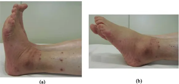
(SF-36), and Visual Analogue Scale (VAS) pain questionnaires. AOFAS score was 97 after surgery. Postoperative SF-36 subscores were 40.2 for physical com-ponent summary after surgery, and 60.9 for mental comcom-ponent summary. Visual analogue scale score was 0 after surgery. As of the last visit, the degree of dor-sal/plantar flexion was 0˚/35˚, range of motion of the ankle on the operated kle had returned to almost matching the range of motion in the unoperated an-kle (Figure 8).
[image:5.595.289.460.418.576.2]Figure 8. Final function of the ankle. (a) Extended position; (b) Flexed position.
3. Discussion
OTA type C pilon fractures are among the most difficult orthopedic trauma in-juries to treat in the elderly [8]. Pilon fracture is characterized by thinning of the subcutaneous tissue, poor dermal extensibility, and, particularly in elderly indi-viduals, senile skin atrophy [9]. Cutaneous aging manifests as a progressive re-duction in maximum function and reserve capacity of skin tissue. Collagen atrophy is a major factor in skin aging, which is associated with a progressive increase in extensibility and reduced elasticity. With increasing age, the skin also becomes more fragile and susceptible to trauma, leading to more lacerations and bruising [10]. Furthermore, wound healing is impaired in the elderly. In recent years, lower profile metallic implants have significantly reduced soft-tissue com-plications, but cases of implant-related soft-tissue problems are still encountered [8]. For these reasons, treatment of type C pilon fracture remains challenging and controversial. External fixation techniques preserve soft tissues and the pe-riosteum, yet provide stable reduction for OTA type C pilon fractures. Soft-tissue complications and deep infections are infrequent when external fixa-tion is combined with minimally invasive surgery, with a frequency of 5% re-ported by Wyrsch, 1 of 17 cases by Tornetta, and 3 of 37 cases by Barbieri. Lovi-setti et al. reported no cases of pseudoarthrosis or deep infection [6] [11] [12]. They attributed the 100% union rate to meticulous preservation of soft tissues in the fracture zone, made possible by the treatment strategy. Despite the quality of reduction achieved and lack of complications observed, clinical outcomes in their series have been less favorable than described by others. Treatment of pilon fractures by circular external fixation allows for less soft tissue dissection and represents a reliable method for achieving stabilization and healing of distal tibi-al intra-articular fractures with fewer soft tissue complications in the elderly.
Use of the Ilizarov external fixator is a safe method for pilon fracture in the elderly, with the advantage of immediate mobilization at full weight-bearing. The ankle-hinged Ilizarov external fixator and early joint movement allows ef-fective management of tibial pilon fractures, usually accompanied by soft-tissue injuries [7]. Many reports have emphasized the negative impacts of non-weight- bearing, including an approximately 50% reduction in tibial cancellous bone
mass in just 1 week, and the necessity for weight-bearing lasting at least twice as long for bone mass recovery even once weight-bearing has been restarted [13]
[14] [15] [16]. The drop in physical fitness with aging is an unavoidable fact.
Crutches require patients to maintain balance while weight is shifted in a swing-ing motion to move forward. Crutches are unstable for the elderly and represent a potential for further injury. Otherwise, body strength and bone strength dete-riorate rapidly when the patient is confined to a wheelchair. Buildings and hous-es are very small in Japan, compared to the United Stathous-es. This means it is diffi-cult for a patient to move in a room with a Roll-A-Bout [17]. We do not have the Roll-A-Bout in Japan. Rehabilitation function is very weak at hospitals in Japan. Numbers of staff and nurses at hospitals in Japan are very low. In the present case, the patient made satisfactory progress to the point where discharge was possible at 28 days postoperatively. Rigid fixation and closed reduction of the Ilizarov external fixation enabled early weight-bearing and early discharge in the present case with severe soft tissue injury and severe osteoporosis.
4. Conclusion
In conclusion, Ilizarov external fixation for soft-tissue injury and bones with os-teoporosis, even in pilon fractures where the cortical bone is thin and the use of screws for internal fixation is difficult, allows suitable fixation and closed reduc-tion to be achieved using multiple Ilizarov wires. We have been performing os-teosynthesis using an Ilizarov external fixator and early weight-bearing in elderly patients with pilon fracture with severe soft-tissue injury and osteoporosis.
Acknowledgements
There is no substantial direct or indirect commercial financial incentive associ-ated with publishing this article.
Consent
Written informed consent was obtained from the patient for publication of this Case Report and any accompanying images. A copy of the written consent is available for review by the Editor-in-Chief of this journal.
Conflicts of Interest
The authors declare that they have no competing interests. There is no substan-tial direct or indirect commercial financial incentive associated with publishing this article.
Each author certifies that he or she has no commercial associations (e.g., con-sultancies, stock ownership, equity interest, patent/licensing arrangements, etc.) that might pose a conflict of interest in connection with the submitted article.
Ethical Review Committee Statement
It does not need to approve for a case report at Medical Ethics Review Committee
Orthopedics, 2013, Article ID: 653146.
[3] Cavusoglu, A.T., Ozsoy, M.H., Dincel, V.E., Sakaogullari, A., Basarir, K. and Ugur-lu, M. (2009) The Use of a Low-Profile Ilizarov External Fixator in the Treatment of Complex Fractures and Non-Unions of the Distal Femur. Acta Orthopaedica Belgi-ca, 75, 209-218.
[4] Black, J.D., Bhavikatti, M., Al-Hadithy, N., Hakmi, A. and Kitson, J. (2013) Early Weight-Bearing in Operatively Fixed Ankle Fractures: A Systematic Review. The Foot, 23, 78-85. https://doi.org/10.1016/j.foot.2013.05.002
[5] Bozkurt, M., Ocguder, D.A., Ugurlu, M. and Kalkan, T. (2008) Tibial Pilon Fracture Repair Using Ilizarov External Fixation, Capsuloligamentotaxis, and Early Rehabil-itation of the Ankle. The Journal of Foot and Ankle Surgery, 47, 302-306.
https://doi.org/10.1053/j.jfas.2008.02.013
[6] Lovisetti, G., Agus, M.A., Pace, F., Capitani, D. and Sala, F. (2009) Management of Distal Tibial Intra-Articular Fractures with Circular External Fixation. Strategies in Trauma and Limb Reconstruction, 4, 1-6.
https://doi.org/10.1007/s11751-009-0050-7
[7] Fırat, A., Tecimel, O., Işık, C., Ozdemir, M., Oçgüder, A. and Bozkurt, M. (2013) Ilizarov External Fixator in the Management of Tibial Pilon Fractures: Ankle Hinged vs Ankle Fixed Frame. Eklem Hastalıkları ve Cerrahisi, 24, 133-138. https://doi.org/10.5606/ehc.2013.30
[8] Bacon, S., Smith, W.R., Morgan, S.J., Hasenboehler, E., Philips, G., Williams, A., Ziran, B.H. and Stahel, P.F. (2008) A Retrospective Analysis of Comminuted In-tra-Articular Fractures of the Tibial Plafond: Open Reduction and Internal Fixation versus External Ilizarov Fixation. Injury, 39, 196-202.
https://doi.org/10.1016/j.injury.2007.09.003
[9] Crist, B.D., Khazzam, M., Murtha, Y.M. and Della Rocca, G.J. (2011) Pilon Frac-tures: Advances in Surgical Management. Journal of the American Academy of Or-thopaedic Surgeons, 19, 612-622.
https://doi.org/10.5435/00124635-201110000-00005
[10] Calleja-Agius, J., Muscat-Baron, Y. and Brincat, M.P. (2007) Skin Ageing. Meno-pause International, 13, 60-64. https://doi.org/10.1258/175404507780796325 [11] Wyrsch, B., McFerran, M.A., McAndrew, M., Limbird, T.J., Harper, M.C., Johnson,
K.D. and Schwartz, H.S. (1996) Operative Treatment of Fractures of the Tibial Pla-fond. A Randomized Prospective Study. The Journal of Bone and Joint Surgery, 78, 1646-1657.https://doi.org/10.2106/00004623-199611000-00003
[12] Tornetta, P., Weinwer, L., Bergman, M., Watnik, N., Steuer, J., Kelley, M. and Yang, E. (1993) Pilon Fractures: Treatment with Combined Internal and External Fixa-tion. Journal of Orthopaedic Trauma, 7, 489-496.
https://doi.org/10.1097/00005131-199312000-00001
[13] Sakai, A., Nakamura, T., Tsurukami, H., Okazaki, R., Nishida, S., Tanaka, Y.,
rimura, T. and Suzuki, K. (1996) Bone Marrow Capacity for Bone Cells and Trabe-cular Bone Turnover in Immobilized Tibia after Sciatic Neurectomy in Mice. Bone, 18, 479-486. https://doi.org/10.1016/8756-3282(96)00042-7
[14] Sakai, A., Sakata, T., Ikeda, S., Uchida, S., Okazaki, R., Norimura, T., Hori, M. and Nakamura, T. (1999) Intermittent Administration of Human Parathyroid Hormone (1-34) Prevents Immobilization-Related Bone Loss by Regulating Bone Marrow Capacity for Bone Cells in ddY Mice. Journal of Bone and Mineral Research, 14, 1691-1699.https://doi.org/10.1359/jbmr.1999.14.10.1691
[15] Sakata, T., Sakai, A., Tsurukami, H., Okimoto, N., Okazaki, Y., Ikeda, S., Norimura, T. and Nakamura, T. (1999) Trabecular Bone Turnover and Bone Marrow Cell De-velopment in Tail-Suspended Mice. Journal of Bone and Mineral Research, 14, 1596-1604.https://doi.org/10.1359/jbmr.1999.14.9.1596
[16] Sakai, A. and Nakamura, T. (2001) Changes in Trabecular Bone Turnover and Bone Marrow Cell Development in Tail-Suspended Mice. Journal of Musculoskeletal & Neuronal Interactions, 14, 387-392.
[17] Goldman, S.M.and Chang, T. (1999) Roll-A-Bout: Complete Off-Loading Made Easy. The Journal of Foot and Ankle Surgery,38, 427.
https://doi.org/10.1016/S1067-2516(99)80045-3