ISSN Print: 2156-8553
DOI: 10.4236/as.2018.98075 Aug. 31, 2018 1085 Agricultural Sciences
Cloning and Expression Analysis of RrGT1
Gene Related to Anthocyanin Biosynthesis in
Rosa rugosa
Xiaoming Sui
1*, Pengyuan Zhang
2*, Yu Wang
1, Mingyuan Zhao
1, Xu Han
1, Lanyong Zhao
1#,
Zongda Xu
1#1College of Forestry, Shandong Agricultural University, Taian, China
2Forestry Seedlings and Flower Department, Forestry Bureau of Shandong, Jinan, China
Abstract
Glycosylation modification fulfills an important role in increasing the stabili-ty and solubilistabili-ty of anthocyanin in plants. In this study, based on the tran-scriptional database of R. rugosa, a gene with full length cDNA of 1161 bp, encoding 386 amino acids, designated as RrGT1, was isolated from flowers of
R. rugosa ‘Zizhi’ and then functionally characterized. According to online software prediction, the molecular formula of the protein encoded by the
RrGT1 gene is C1879H2964N494O556S14, the relative molecular mass is 41,820.02 Da, and the theoretical isoelectric point is pI = 5.03. The result of the RrGT1
protein 3D model construction showed that it had the highest homology with the UDP-glucose: anthocyanidin 3-O-glucosyltransferase protein model in the database (47.01%). Sequence alignments with the NCBI database showed that the RrGT1 protein is a member of the GTB superfamily. Homology analysis revealed that the coding regions of RrGT1 was highly specific among different species, but still had typical conserved amino acid residues called PSPG that are crucial for RrGT1 enzyme activity. RrGT1 transcripts were de-tected in five flowering stages and seven tissues of R. rugosa ‘Zizhi’, R. rugosa ‘Fenzizhi’ and R. rugosa ‘Baizizhi’, and their expression patterns corres-ponded with the accumulation of anthocyanins. Therefore, we speculated that glycosylation of RrGT1 plays a crucial role in anthocyanin biosynthesis in R.
rugosa.
Keywords
Rosarugosa, RrGT1 Gene, Clone, Gene Expression, Anthocyanin
*These authors contribute equally. #Corresponding authors.
How to cite this paper: Sui, X.M., Zhang, P.Y., Wang, Y., Zhao, M.Y., Han, X., Zhao, L.Y. and Xu, Z.D. (2018) Cloning and Ex-pression Analysis of RrGT1 Gene Related to Anthocyanin Biosynthesis in Rosa rugo-sa. Agricultural Sciences, 9, 1085-1096.
https://doi.org/10.4236/as.2018.98075
Received: August 8, 2018 Accepted: August 28, 2018 Published: August 31, 2018
Copyright © 2018 by authors and Scientific Research Publishing Inc. This work is licensed under the Creative Commons Attribution International License (CC BY 4.0).
http://creativecommons.org/licenses/by/4.0/
DOI: 10.4236/as.2018.98075 1086 Agricultural Sciences
1. Introduction
Rosarugosa is an important ornamental plant which belongs to the genus Rosa
in the family Rosaceae. It is native to China and is widely distributed in the world. Because of its unique fragrance, color, cold resistance and drought resis-tance, it has great development potential in garden application [1]. There are many varieties of roses, but most of them are traditional colors such as pink, purple, etc. A few varieties are white, lacking yellow, bright red, orange and compound color, etc. [2]. Therefore, how to innovate rose color has become the main goal of breeders. The analysis of the pigment composition of rose and the study of the expression characteristics of the key enzymes encoding genes that catalyze the synthesis of rose pigment are the important prerequisite for rose color oriented molecular breeding [3]. Anthocyanin determines the color of higher plant organs. Its biosynthesis pathway related structural genes (CHS,
CHI, F3H, F3’H, DFR, ANS, 3GT etc.) and regulatory genes (MYB, mostly R2R3 MYB, BHLH and WD40 classes) have been cloned, sequenced and protein func-tion studies in many plants, such as petunia, maize, snapdragon and so on. But less research has been done on R. rugosa.
Anthocyanins, derived from the anthocyanin biosynthesis pathway, are the largest group of water-soluble plant flavonoids found in organs of plants and crops [4] [5]. Anthocyanins are unstable in plants, mainly in the form of glyco-sides in the vacuole [6]. Anthocyanins play an important role in insect pollina-tion, auxin transport, protection of leaves from ultraviolet radiapollina-tion, inhibition of diseases and insect pests, etc. [7]. In addition, as a safe, non-toxic natural food pigment, anthocyanins also have anti-oxidation, anti-cancer and anti-arteriosclerosis functions [8].
Anthocyanin biosynthesis pathway is one of the secondary metabolic path-ways in plants. At present, the research on it has been clearer. Firstly, naringin was formed by the catalysis of CHS and CHI with coumaryl coenzyme A and malonyl Co A. Then flavonols were formed under the action of F3H. The next step is to form colored anthocyanins under the action of DFR and ANS. Finally, through glycosylation, methylation, acylation, hydroxylation and other modifi-cations to form a variety of anthocyanins with a stable structure [9]. Flavonoid 3-0-glycosyltransferase (3GT) gene is a downstream gene in anthocyanin syn-thesis pathway. It can catalyze the glycosylation of UDP glucose to replace the 3 hydroxyl groups of anthocyanin and make anthocyanin glycosylation to produce colored and stable anthocyanins. And move the maximum absorption spectrum to the ultraviolet end, thus increasing the blue tone of anthocyanins [10]. Glyco-sylation can change the hydrophilicity, biochemical activity and subcellular loca-lization of anthocyanins, which is beneficial to the transport and storage of an-thocyanins in cells and organisms [11].
Some studies have shown that the anthocyanin content of plants lacking 3GT
DOI: 10.4236/as.2018.98075 1087 Agricultural Sciences amino acids at its C-terminal, known as plant secondary product glycosyltrans-ferase (PSPG) box. At present, 3GT has been cloned and analyzed in many plants such as Zeamays [13], Vitis vinifera [14], Gentianatrflora [15], Petunia hybrida [16] and so on, which has laid a foundation for understanding the me-tabolic regulation of anthocyanin synthesis pathway.
At present, the studies on R. rugosa are mainly focused on morphological classification, geographical distribution, essential oil extraction and food quality evaluation, and there are few reports on the anthocyanin biosynthesis mechan-ism, so we don’t know exactly how it works. In this study, based on the R. rugosa
transcriptome data, we cloned and identified RrGT1 gene from the petals of R.
rugosa ‘Zizhi’ for the first time. We carried out detailed bioinformatics analysis, homology analysis and the temporal and spatial expression pattern analysis of the RrGT1 gene in order to provide some useful informations for the subsequent color improvement project in R. rugosa.
2. Materials and Methods
2.1. Plant Materials
For R. rugosa, three varieties (R. rugosa ‘Zizhi’, R. rugosa ‘Fenzizhi’ and R. ru-gosa ‘Baizizhi’) cultivated in Rose germplasm nursery of Shandong Agricultural University was used as the test material. We collected the petals at the budding stage, initial opening stage, half opening stage, full opening stage and wilting stage in the forenoon on sunny days from 20 April to 10 May 2017. Seven tissues (roots, stems, leaves, petals at the budding stage, sepals, stamens and pistil) of R.
rugosa ‘Zizhi’ were collected at the same time. After quick freezing of liquid ni-trogen, all samples collected with three replicates were put into −80˚C refrigera-tor for srefrigera-torage.
2.2. Extraction of Total RNA and Synthesis of the First-Strand
cDNA
The extraction of total RNA is based on the specification of EASY spin plant RNA rapid extraction kit (Aidlab Biotech, Beijing, China). The integrity of RNA was detected by gel electrophoresis with 1.0% nondenatured agarose, the purity and concentration of RNA were detected by Nanodrop2000C ultramicro spec-trophotometer (Thermo Fisher Scientific, Wilmington, Delaware, USA), and the qualified RNA was preserved at −80˚C. The first-strand cDNA was synthesized by referring to the steps of 5× All-In-One RT MasterMix reverse transcription kit (ABM Company, Vancouver, Canada) and synthesized according to the re-quirements of RT-PCR and qRT-PCR.
2.3. Full-Length CDS Cloning of the
RrGT1
Gene
se-DOI: 10.4236/as.2018.98075 1088 Agricultural Sciences quence in transcriptome data and the 3' terminal sequence obtained by RACE technique were spliced with DNAMAN software to obtain the full-length cDNA sequence of the RrGT1 gene. According to the sequence obtained by splicing, the upstream primer RrGT1-F containing the starting codon of the RrGT1 gene and the downstream primer RrGT1-R containing the terminating codon were de-signed and amplified. The estimated length of amplification was 1161 bp.
2.4. Bioinformatics Analysis of the
RrGT1
Gene
Bioinformatics analysis softwares and tools were used to predict the physico-chemical properties and structural functions of the protein encoded by the
RrGT1 gene, which provided a reference for the future research and application of the gene. The basic physical and chemical properties of the protein encoded by the RrGT1 gene were analyzed by using Prot-Param tools in ExPasy
(http://web.expasy.org/protparam/). The CD-Search function
(https://www.ncbi.nlm.nih.gov/Structure/cdd/wrpsb.cgi) on NCBI was used to
predict the conserved domain in the protein encoded by the RrGT1 gene. The 3D structure of the RrGT1 protein was predicted by online tool SWISS-MODEL. DNAMAN software and Blast tool in NCBI were used to analyze the amino acid sequence of the RrGT1 gene. The phylogenetic tree is constructed by using MEGA5.0 software.
2.5. qRT-PCR Detection
We analyzed the gene expression by qRT-PCR on a Bio-Rad CFX96TM Real-Time PCR instrument (Bio-Rad, Inc., USA). The qRT-PCR mixture (20 μL total vo-lume) contained 10 μL of SYBR® Premix Ex TaqTM (TaKaRa, Inc., Japan), 8.2 μL of ddH2O, 0.4 μL of each primer and 1 μL of cDNA. The PCR program was car-ried out with an initial step of 95˚C for 30 s; 40 cycles of 95˚C for 5 s, 60˚C for 30 s; and then, 95˚C for 10 s, 65˚C for 5 s and 95˚C for 5 s for the dissociation stage. Each gene was assessed with three biological replications. The relative expression levels of the genes were calculated by the 2−ΔΔCt method [17].
3. Results
3.1. Cloning of
RrGT1
and Sequence Analysis
We obtained the RrGT1 gene 3' terminal sequence of 277 bp length by using nested PCR method (Figure 1(a)). The full-length sequence (Figure 1(b)) of the
RrGT1 gene was cloned by full-length primers (Table 1) and confirmed by se-quencing. The RrGT1 gene has a complete open reading frame from the starting codon ATG to the termination codon TAA, encodes a 386-amino acids protein and has a polyA tail. Sequence alignments with the NCBI database showed the
DOI: 10.4236/as.2018.98075 1089 Agricultural Sciences
Figure 1. The results of 3' RACE amplification and full-length CDS amplification of the
RrGT1 gene. (a) 3' RACE amplification product of the RrGT1 gene; (b) Full-length CDS amplification product of the RrGT1 gene.
Table 1. Primers used in the present study.
Primer Name Sequence (5' - 3') Description
3' RrGT1-F-outer ATGTCTTGGTCCTCGCATTTC 3' RACE for RrGT1
3' RrGT1-F-inner GAGGGGAAGAAGATGAGAGGTAAAG
B26 GACTCGAGTCGACATCGATTTTTTTTTTTTTTTTT
RrGT1-F ATGTCAGGAAATCCACTGGATGC cDNA for RrGT1 Full-length
RrGT1-R TTAAGGCGAAGAAATCAAATCTACC
RrGAPDH-F TTCTGCCTGCTCTCAATG qRT-PCR for RrGAPDH
RrGAPDH-R TGCCTTCTTCTCAAGTCTG
qRrGT1-F GTATTTGCCAACACACTGAGTAA qRT-PCR for RrGT1
qRrGT1-R CTGCAGTGGTAATGAGAGGGAG
molecular formula of the RrGT1 protein is C1879H2964N494O556S14, the relative mo-lecular mass is 41,820.02 Da, and the theoretical isoelectric point is pI = 5.03.
3.2. Protein 3D Model Construction
During the construction of the RrGT1 protein 3D model, templates search with BLAST and HHBlits has been performed against the SWISS-MODEL template library. It was found that it had the highest homology with the
UDP-glucose:anthocyanidin 3-O-glucosyltransferase protein model in the data-base (47.01%), so the RrGT1 protein 3D model (Figure 2) was constructed on the basis of its model.
3.3. Homology Analysis
[image:5.595.211.537.323.505.2]DOI: 10.4236/as.2018.98075 1090 Agricultural Sciences
Figure 2. The protein modelling results and quality estimation of RrGT1. (a) Visualizable 3D model of RrGT1; (b) Local quality
estimation of the protein; (c) As a statistical value, Z-score can be used to represent the matching degree between template pro-teins and propro-teins to be tested. QMEAN: A comprehensive scoring function for model quality assessment. All Atom: Normalized all-atom potential energy of the residue calculated from the short-range statistical potentials. CBeta: Normalized cbeta potential energy of the residue calculated from the short-range statistical potentials. Solvation: Normalized solvation potential energy of the residue calculated from the short-range statistical potentials. Torsion: Torsion energy of the residue exposed relative solvent ac-cessibility, calculated by dividing the maximally accessible surface area (ASA) of a residue by the observed value; (d) Comparison with Non-redundant Set of PDB (Protein Data Bank) structures; (e) Alignment between the model and the template.
Figure 3. Amino acid sequences homologous analysis of the RrGT1 gene and other
[image:6.595.212.539.545.650.2]DOI: 10.4236/as.2018.98075 1091 Agricultural Sciences
RrGT1 protein has strong species specificity in the N-terminal region and PSPG conserved domain in the C-terminal region. Using MEGA5.0 software, phylo-genetic tree (Figure 4) was constructed from 21 plant amino acids sequences in-cluding R. rugosaRrGT1. The results showed that the RrGT1 gene had the clos-est genetic relationship with the EsUF3GT gene, and its homology reached 59%, which indicated that GTs gene is highly specific among different species.
3.4. Temporal and Spatial Expression Patterns of the
RrGT1
Gene
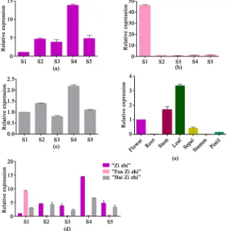
The expression levels of the RrGT1 gene, which significantly differed, were as-sessed during five flowering stages. For R. rugosa ‘Zizhi’ (Figure 5(a)), the high-est expression level was observed during the full opening stage, and the lowhigh-est was observed during the budding stage. For R. rugosa ‘Fenzizhi’ (Figure 5(b)), the highest expression level was observed during the budding stage, and the ex-pression levels of other four stages were relatively low. And for R. rugosa ‘Bai-zizhi’ (Figure 5(c)), the expression level was also highest during the full opening stage but lowest during the half opening stage. The expression patterns of the
[image:7.595.238.509.342.658.2]RrGT1 gene in R. rugosa ‘Zizhi’ and R. rugosa ‘Baizizhi’ showed the same trend.
Figure 4. Phylogenetic tree of RrGT1 and GT members from other plant species. The tree
DOI: 10.4236/as.2018.98075 1092 Agricultural Sciences
Figure 5. The temporal and spatial expression patterns of RrGT1. (a) The relative
expres-sions of the RrGT1 gene in five flowering stages of R. rugosa ‘Zizhi’; (b) R. rugosa ‘Fen-zizhi’; (c) R. rugosa ‘Baizizhi’; (d) Comparison of relative expressions of RrGT1 in five flowering stages of three varieties above; (e) The relative expression of RrGT1 in seven different tissues of R. rugosa ‘Zizhi’. RrGAPDH was used as the internal control. Error bars represent the SDs of triplicate reactions. The experiment was repeated three times with similar results.
Then the expression levels of the RrGT1 gene in three varieties was compared in five flowering stages (Figure 5(d)). In the budding stage, R. rugosa ‘Fenziz-hi’ > R. rugosa ‘Baizizhi’ > R. rugosa ‘Zizhi’, while in the initial opening , half opening ,full opening and wilting stage, the trends of gene expression in the three varieties were consistent: R. rugosa ‘Zizhi’ > R. rugosa ‘Baizizhi’ > R. rugo-sa ‘Fenzizhi’.
The expression levels of the RrGT1 gene, which also significantly differed, were assessed in seven different tissue types of R. rugosa ‘Zizhi’ (Figure 5(e)). The expression level in the leaves, stems and flower buds was relatively high but was relatively low in the other tissues.
4. Discussion
DOI: 10.4236/as.2018.98075 1093 Agricultural Sciences color, there are few reports of downstream structural genes such as GTs. The fi-nal formation of anthocyanins depends on the glycosylation of GTs, so it is very important to elucidate the function and influence of the RrGT1 gene in R. rugo-sa color formation. In this study, we successfully cloned RrGT1 gene with full length cDNA of 1161bp, encoding 386 amino acids from the petals of R. rugosa
‘Zizhi’. It was predicted that the molecular formula of the protein encoded by the
RrGT1 gene is C1879H2964N494O556S14, the relative molecular mass is 41820.02 Da, the theoretical isoelectric point pI = 5.03, which belongs to GTB superfamily. During the construction of the RrGT1 protein 3D model, it was found that the
RrGT1 protein had the highest homology with the existing UDP-glucose: an-thocyanidin 3-O-glucosyltransferase protein model (47.01%), which suggested that the protein encoded by RrGT1 gene was related to the glycosylation of an-thocyanin.
The evolutionary analysis of flavonoids GTs by Sawada etal. [18] showed that GTs, which catalyze the glycosylation of flavonoids in different positions (3-O, 5-O, 7-O), were formed into different evolutionary branches (F3Gly T, F5Gly T, F7Gly T) without restriction of species. It suggested that the region-specific of flavonoids GTs to glycosyl receptors (catalytic site specificity) was formed before species differentiation. In the course of evolution, the ability to utilize UDP-sugar (UDP-glucose, UDP-rhamnose, UDP-galactose) was obtained. Phylogenetic tree analysis showed that RrGT1 was linked to EsUF3GT of 3-O-glycosylation of flavonoids, suggesting that RrGT1 may be involved in the glycosylation process of 3-O positions of flavonoids.
The alignment of amino acid sequences between RrGT1 and glycosyltransfe-rases from the other 21 species indicated that RrGT1 possessed a common PSPG motif of the glycosyltransferases superfamily (Figure 3). Previous studies have shown that the conserved region of PSPG is related to substrate recognition and catalytic activity of enzyme proteins [19] [20] [21] [22]. If the 44 amino acids of the PSPG domain are numbered, the amino acids at position 22, 23 and 44 play an important role in the selection of enzyme proteoglycan donors. The twen-ty-second position of tryptophan (Trp, W) can correctly locate UDP-glucose, while arginine (Arg, R) can make UDP-glucuronic acid locate correctly; the twenty-third position of serine (Ser, S) is highly conserved in UDP-glucurono- syltransferase [23] [24] [25]; the forty-fourth glutamine (Gln, Q) and histidine (His, H) have strong conservatism in glucosyltransferase and galactotransferase respectively [22]. In the PSPG domain of the RrGT1 gene, the amino acids at position 22, 23 and 44 are cysteine (Cys, C), asparagine (Asn, N) and histidine (His, H), respectively. Therefore, we speculated that the RrGT1 gene is using UDP-glucose or galactose as the main glycosyl donor, but has no glucuronyl-transferase activity.
The expression levels of the RrGT1 gene during flower development and in different tissues were investigated. It was found that the expression of the RrGT1
ru-DOI: 10.4236/as.2018.98075 1094 Agricultural Sciences
gosa varieties, indicating that the expression of the RrGT1 gene was develop-mentally regulated in the process of anthocyanin biosynthesis. Studies have shown that the accumulation of anthocyanin in red skinned sand pear, strawber-ries and litchi is positively correlated with the activity of UF3GT. Boss etal. [26] also detected the expression of UF3GT in the peels of red grape which accumu-lated anthocyanin, but not in other tissues of red grape and white grape without anthocyanin accumulation. Gong et al.’s [27] studies showed that the partial structural genes of Perillafrutescens anthocyanin metabolic pathway were only expressed in the leaves of red varieties, but not in green varieties or the expres-sion in the green leaves was very low. About the tissue-specific expresexpres-sion in R.
rugosa ‘Zizhi’, besides the high expression level in flowers, it is worth mention-ing that the stems of R. rugosa ‘Zizhi’ are purple, which is consistent with the high expression level of the RrGT1 gene. In addition, RrGT1 was also highly ex-pressed in the leaves, so we infer that RrGT1 is also involved in the glycosylation of secondary metabolites in leaves and plays an important role.
In conclusion, the cloning and expression analysis of the RrGT1 gene was beneficial to analyzing the molecular synthesis and regulation mechanism of anthocyanins, and also provided some important informations for the im-provement of R. rugosa flower color in the future.
Acknowledgements
This project was supported by the Agricultural Seed Project of Shandong Prov-ince ([2014] No. 96).
Conflicts of Interest
The authors declare that they have no conflicts of interest.
References
[1] Li, M. (2006) Survey and Quality Evaluation of Shandong Rose Varieties. Shandong University of Traditional Chinese Medicine, Jinan.
[2] Feng, L.G., Shao, D.W., Sheng, L.X., etal. (2009) Study on Investigation and Mor-phological Variation of Wild Rosarugosa in China. JournalofShandong Agricul-turalUniversity, 40, 484-488.
[3] Chen, S.-M., Zhu, X.-R., Chen, F.D., etal. (2010) Expression Characteristics of An-thocyanin Structural Genes in Different Flower Color Chrysanthemum Cultivars.
JournalofNorthwestPlants, No. 3, 453-458.
[4] Kim, S.H., Lee, J.R., Hong, S.T., etal. (2003) Molecular Cloning and Analysis of Anthocyanin Biosynthesis Genes Preferentially Expressed in Apple Skin. Plant Science, 165, 403-413. https://doi.org/10.1016/S0168-9452(03)00201-2
[5] Martens, S., Preuß, A. and Matern, U. (2010) Multifunctional Flavonoid Dioxyge-nases: Flavonol and Anthocyanin Biosynthesis in Arabidopsis thaliana L. Phyto-chemistry, 71, 1040-1049. https://doi.org/10.1016/j.phytochem.2010.04.016
DOI: 10.4236/as.2018.98075 1095 Agricultural Sciences
[7] Forkmann, G. (1991) Flavonoids as Flower Pigments: The Formation of the Natural Spectrum and Its Extension by Genetic Engineering. PlantBreeding, 106, 1-26.
https://doi.org/10.1111/j.1439-0523.1991.tb00474.x
[8] Springob, K., Nakajima, J.I., Yamazaki, M., et al. (2003) Recent Advances in the Biosynthesis and Accumutation of Anthocyanins. Natural Product Reports, 20, 288-303. https://doi.org/10.1039/b109542k
[9] Yonekura-Sakakibara, K., Nakayama, T., Yamazaki, M., etal. (2009) Modification and Stabilization of Anthocyanins. Springer, New York, 169-190.
[10] Zheng, Z.L. (1994) Flower Color Gene Engineering of Flower Crops. Northern Hor-ticulture, 3, 37-38.
[11] Vogt, T. and Jones, P. (2000) Glycosyltransferases in Plant Natural Product Synthe-sis: Characterization of a Supergene Family. TrendsinPlantScience, 5, 380-386.
https://doi.org/10.1016/S1360-1385(00)01720-9
[12] Tohge, T., Nishiyama, Y., Hirai, M.Y., etal. (2005) Functional Genomics by Inte-grated Analysis of Metabolome and Transcriptome of Arabidopsis Plants Over-Ex- pressing an MYB Transcription Factor. ThePlantJournal, 42, 218-235.
https://doi.org/10.1111/j.1365-313X.2005.02371.x
[13] Goto, T., Kondo, T., Tamura, H., etal. (1982) Structure of Gentiodelphin, an Acy-lated Anthocyanin IsoAcy-lated from Gentiana makinoi, That Is Stable in Dilute Aqueous Solution. TetrahedronLetters, 23, 3695-3698.
https://doi.org/10.1016/S0040-4039(00)88660-8
[14] Hall, D., Yuan, X.X., Murata, J., etal. (2012) Molecular Cloning and Biochemical Characterization of the UDP-Glucose: Flavonoid 3-0-Glucosyltransferase from Concord Grape (Vitislabrusca). Phytochemistry, 74, 90-99.
https://doi.org/10.1016/j.phytochem.2011.10.007
[15] Tanaka, Y., Yoshikazu, K., Fukuchi-Mlizutani, M., etal. (1996) Molecular and Bio-chemical Characterization of Three Anthocyanin Synthetic Enzymes from Gentiana triflora. PlantandCellPhysiology, 37, 711-716.
https://doi.org/10.1093/oxfordjournals.pcp.a029004
[16] Yamazaki, M., Yamagishi, E., Gong, Z.Z., etal. (2002) Two Flavonoid Glucosyl-transferases from Petuniahybrida: Molecular Cloning, Biochemical Properties and Developmentally Regulated Expression. PlantMolecularBiology, 48, 401-411.
https://doi.org/10.1023/A:1014043214943
[17] Schmittgen, T.D. and Livak, K.J. (2008) Analyzing Real-Time PCR Data by the Comparative C (T) Method. NatureProtocols, 3, 1101-1108.
https://doi.org/10.1038/nprot.2008.73
[18] Sawada, S., Suzuki, H., Ichimaida, F., etal. (2005) UDP-Glucuronic Acid: Antho-cyanin Glucuronosyltransferase from Red Daisy (Bellis perennis) Flowers. The JournalofBiologicalChemistry, 280, 899-906.
https://doi.org/10.1074/jbc.M410537200
[19] Gachon, C.M., Meurinne, M.L. and Saindrenan, P. (2005) Plant Secondary Meta-bolism Glycosyltransferases: The Emerging Functional Analysis. Trends in Plant Science, 10, 542-549. https://doi.org/10.1016/j.tplants.2005.09.007
[20] Sakakibara, K.Y. and Hanada, K. (2011) An Evolutionary View of Functional Diver-sity in Family 1 Glycosyltransferases. ThePlantJournal, 66, 182-193.
https://doi.org/10.1111/j.1365-313X.2011.04493.x
DOI: 10.4236/as.2018.98075 1096 Agricultural Sciences https://doi.org/10.1016/j.febslet.2009.09.042
[22] Kubo, A., Arai, Y., Nagashima, S., etal. (2004) Alteration of Sugar Donor Specifici-ties of Plant Glycosyltransferases by a Single Point Mutation. Archivesof Biochemi-stryandBiophysics, 429, 198-203. https://doi.org/10.1016/j.abb.2004.06.021
[23] Shao, H., He, X.Z., Achnine, L., etal. (2005) Crystal Structures of a Multifunctional Triterpene/Flavonoid Glycosyltransferase from Medicago Truncatula. Plant Cell, 17, 3141-3154. https://doi.org/10.1105/tpc.105.035055
[24] Li, L.N., Modolo, L.V., Trevino, L.E., etal. (2007) Crystal Structure of Medicago truncatula UGT85H2—Insights into the Trusctural Basis of a Multifunctional (Iso) Flavonoid Glycosyltransferase. JournalofMolecularBiology, 370, 951-963.
https://doi.org/10.1016/j.jmb.2007.05.036
[25] Modolo, L.V., Li, L.N., Pan, H.Y., etal. (2009) Crystal Structures of Glycosyltrans-ferase UGT/8G1 Reveal the Molecular Basis for Glycosylation and Deglycosylation of (Iso) Flavonoids. JournalofMolecularBiology, 392, 1292-1302.
https://doi.org/10.1016/j.jmb.2009.08.017
[26] Boss, P.K., Davies, C. and Robinson, S.P. (1996) Expression of Anthocyanin Bio-synthesis Pathway Genes in Red and White Grapes. PlantMolecular Biology, 32, 565-569. https://doi.org/10.1007/BF00019111