Printed in Great Britain © The Company of Biologists Limited 1992
CALCIUM AND POTASSIUM CURRENTS IN LEG
MOTONEURONS DURING POSTEMBRYONIC DEVELOPMENT
IN THE HAWKMOTH MANDUCA SEXTA
BY JON H. HAYASHI1 AND RICHARD B. LEVINE1 2
1
Division of Neurobiology and 2Department of Physiology, University of Arizona, Tucson, AZ 85721, USA
Accepted 30 June 1992
Summary
During insect metamorphosis the nervous system is reorganized to accommo-date changes in behavior. In the hawkmoth, Manducasexta, many identified larval motoneurons persist to innervate new adult muscles, while undergoing changes in dendritic morphology and synaptic connections. The thoracic leg motoneurons, for example, innervate different sets of muscles in the larva and adult and participate in distinct types of locomotor behavior in the two stages of life. To determine whether changes in the biophysical properties of these motoneurons accompany the structural and functional modifications that have been described, we used the whole-cell voltage-clamp technique to compare the Ca2+ and K+ currents expressed by leg motoneurons isolated from the larval, pupal and adult stages of Manduca. After 24h in culture, the somata of leg motoneurons isolated from all three stages expressed voltage-sensitive Ca2+ currents that could be blocked by Cd2+, Co2 + or Ni2 +. The currents were larger with Ba2+ as the charge carrier. The Ca2+ current density was significantly lower in these motoneurons during the early pupal stage than in either the larva or adult. Similar experiments revealed both transient and sustained K+ currents in the leg motoneurons that could be blocked with Cs+. There was a significant decrease in the density of the transient, inactivating outward current in leg motoneurons isolated from the early pupal stage. Thus, the levels of some types of ionic currents are modulated during metamorphosis. These changes may be important for the developmental or behavioral changes that accompany metamorphosis.
Introduction
Levine, 19886).
The dendritic structures and synaptic interactions of the thoracic leg motoneur-ons change markedly during metamorphosis, as might be expected given the dramatic differences between leg movement and structure in the larva and adult (Kent and Levine, 1988a,£>). Little is known, however, about the intrinsic functional properties of these motoneurons. The roles of different membrane currents in the production of patterned motor activity during locomotion have never been investigated, nor is it known whether the types of ion channels expressed by the leg motoneurons change during metamorphosis as functional requirements are modified.
Changes in ion channel expression during development have been documented in several systems (McCobb et al. 1989; O'Dowd et al. 1988; Salkoff and Wyman, 1981a) where different types of channels appear in a stereotyped sequence. This sequential appearance, in addition to reflecting the orderly differentiation of mature characteristics, may be important for certain developmental processes. For example, the expression of Ca2+ currents may be modulated during development to regulate neurite outgrowth or synapse formation (Mattson and Kater, 1987; Holliday and Spitzer, 1990). Since similar developmental processes are associated with metamorphosis, changes in the expression of ion channels may be important during this time.
ion channels in Drosophila (Atkinson et al. 1991; Kamb et al. 1987; Papazian et al. 1987) opens the possibility of examining regulation at the molecular level.
To investigate the types of voltage-sensitive currents expressed by the thoracic leg motoneurons in Manduca, we have applied the whole-cell patch-clamp technique to neurons maintained in vitro. This was accomplished using techniques that were adapted from those devised for the maintenance of dissociated interneurons from the antennal lobes of Manduca (Hayashi and Hildebrand, 1990). The leg motoneurons were labelled with a vital dye prior to dissociation to facilitate their identification after dissociation of ganglia from animals of different stages (Prugh et al. 1992). We find that the leg motoneurons express a variety of voltage-sensitive currents and that the levels of these currents change during metamorphosis. Some of these results have been reported previously in abstract form (Levine and Hayashi, 1990).
Materials and methods
Animals
Manduca sexta (L.) (Lepidoptera: Sphingidae) were reared from eggs on
artificial diet (modified from Bell and Joachim, 1976) on a long-day photoperiod regimen (17 h light/7 h dark) at 26°C and 50-60% relative humidity. Following hatching, larvae feed continually and pass through five larval instars. Near the end of the last (fifth) larval instar, in response to changes in the ecdysteroid and juvenile hormone titers, animals begin metamorphosis. Adult development begins soon after the molt of the larva into the pupa and proceeds through 18 stages, corresponding roughly to days 0-18 (P0-P18). Pupae were staged as previously described (Sanes and Hildebrand, 1976; Tolbert et al. 1983).
Preparation of cultures
The thoracic ganglia were dissociated and maintained in culture using a modification of techniques that were originally developed for central antennal neurons in Manduca (Hayashi and Hildebrand, 1990). To obtain neurons from different stages of development, animals were dissected at stage LI (day 2 of the last larval instar), stage P0 (the first day of the pupal stage) or stage P18 (at the end of adult development, but before emergence). For most experiments the three thoracic ganglia were dissected from two animals in sterile insect saline (see below) and pinned in a Sylgard-lined Petri dish. One surface of each ganglion was treated for 1 min with a 3 % solution of collagenase/dispase (Boehringer Mann-heim), then the ganglia were desheathed and separated from the interganglionic connectives and peripheral nerves. The individual desheathed ganglia were transferred to a test tube containing supplemented saline (see below; Hayashi and Hildebrand, 1990).
allow 100/il for each culture dish (usually six dishes were used for six thoracic ganglia). The cells were allowed to settle and adhere to the coated surface of the culture dish (see below) for 2h, after which the dish was filled with 2 ml of medium. The cultures were maintained at 26°C in a humidified incubator. Normally this procedure resulted in 5-10 motoneurons per dish.
Cells were grown in miniwells made by cutting an 8 mm hole in the bottom of a plastic 35 mm culture dish. Glass coverslips were sealed to the bottom of the dishes with Sylgard (Dow Corning). After ultraviolet sterilization, the glass was coated by exposure to a solution of 200 ^g ml"1 concanavalin A and 2 fig ml"1 laminin for 2h at 37 CC. The dishes were then rinsed with sterile distilled water and air-dried in a sterile hood.
The contents of the solutions used were derived from Hayashi and Hildebrand (1990).
Insect saline. NaCl, lOOmmoll"1; KC1, 4 mmol P1; CaCl2, e m m o l P1; Hepes, 10 mmol P1, pH7; and glucose, SmmolP1; adjusted to 360mosmolP1 with mannitol.
Supplemented saline. NaCl, 149.9 mmol I"1; KC1, SmmolP1; CaCl2, SmmolP1; MgCl2, 0.5 mmol I"1; Tes 10 mmol I"1; D-glucose, 11 mmol P1; lactal-bumin hydrolysate 6.5 g P1; TC Yeastolate (Difco) 5 g P:; 10% foetal bovine serum (FBS); penicillin lOOunitsmP1; streptomycin lOO^igmP1; pH7; 360mos-moll"1.
Culture medium. To 500ml of Leibovitz's L15 (Gibco) were added:
alpha-ketoglutaric acid 185 mg, fructose 200 mg, glucose 350 mg, malic acid 335 mg, succinic acid 30 mg, TC yeastolate 1.4 gm, lactalbumin hydrolysate 1.4 gm, niacin 0.01 mg, imidazole 30 mg, streptomycin 100/UgmP1, penicillin 100 units ml"1, 20-hydroxyecdysone 1/igmP1 (Sigma) and stable vitamin mix 2.5 ml. A 5 ml stock solution of vitamin mix consists of: aspartic acid 15 mg, cystine 15 mg, beta-alanine 5mg, biotin 0.02 mg, vitamin B12 2mg, inositol 10 mg, choline chloride 10 mg, lipoic acid 0.05 mg, p-aminobenzoic acid 5mg, fumaric acid 25 mg, coenzyme A 0.4 mg, glutamic acid 15 mg, Phenol Red 0.5 mg. The medium was adjusted to pH7, and 360mosmolP1. Both the supplemented saline and the medium were filter-sterilized prior to use.
Identification of cells in culture
these remained labelled for the duration of adult development (Kent and Levine, 19886, 1992). Cells labeled in this manner could be identified under fluorescent illumination in cultures derived from ganglia dissociated at any stage of metamor-phic adult development. The labelled cells remained viable in culture for up to 4 weeks (Griffin and Levine, 1989; Prugh et al. 1992). Normally, Dil was injected into the region of many of the leg muscles in all six legs of the animal so that several (10-20) motoneurons were labelled in each ganglion. In the experiments that follow, therefore, we did not necessarily record from the same leg moto-neuron each time. All thoracic leg motomoto-neurons share the same fate during metamorphosis (Kent and Levine, 19886).
Biophysical techniques
The data reported in this paper are based on records from 58 motoneurons in 42 experiments. Records were obtained from the neurons 18-24 h after plating, before the cells had grown significant processes (Fig. 1). Membrane currents were recorded using the whole-cell configuration of the patch-clamp technique (Hamill
etal. 1981; Fenwick et al. 1982) using an Axopatch 1-B or 1-D amplifier (Axon
Instruments). Patch-clamp electrodes with resistances between 1 and 3mQ were fabricated from borosilicate glass with a Narishige PP-83 puller. Gigaohm seals between the electrode and the cell membrane were easily formed after the culture medium had been replaced by a recording medium that lacked fetal bovine serum (see below). Junction potentials were nulled prior to seal formation. Intracellular access was achieved by rupturing the membrane beneath the electrode with a combination of suction and a brief high-voltage pulse.
Data were gathered and analyzed using pClamp software (Axon Instruments, version 5.1) run on an IBM-compatible '386' computer. Membrane currents were routinely filtered at 10 kHz with a four-pole Bessel filter and sampled at intervals of 100 [JS. Three runs were averaged for each voltage step. Linear leakage currents were subtracted electronically from all records. The pipette resistances were low (1-3 mQ; see above), and upper estimates of the series resistance (as calculated from the capacitive charging transient) were always less than 5 MQ. Nevertheless, significant discrepancies between actual and measured values of the membrane voltage may have arisen during the measurement of the largest peak currents, which approached 8nA in some cells. When possible, the series resistance was compensated electronically at 70-90%. The current/voltage (I/V) plots and records shown in most figures were obtained under these conditions, suggesting a maximum voltage error (with a peak current of 7 nA) of 3-10 mV. The accuracy of the measurements was evaluated by comparing the voltage sensitivity of inward current activation in cells with large currents to that under conditions in which the current was reduced by decreasing the Ba2+ concentration to one-twelfth of the normal level (see below), by comparing Ca2+ to Ba2+ currents, or by investigating neurons with small inward currents. In these cases the voltage-sensitivity of activation was similar to that of cells with large inward currents.
electroni-Fig. 1. Photographs of leg motoneurons in culture. (A) Leg motoneuron from a thoracic ganglion of a stage PO pupa; 24 h in vitro. The cell body is viewed at 400x with Hoffmann differential interference contrast optics. Note the absence of extensive processes. (B) Same cell as A viewed under fluorescence illumination to reveal punctate Dil labelling. The larval legs of this animal had been injected with Dil. After dissociation of the thoracic ganglia several days later at stage PO, the leg motoneurons could be identified by virtue of the Dil labelling. (C) Another leg motoneuron like that shown in A, but viewed at 200x under phase optics. Note the halo of processes. (D) A leg motoneuron after 1 week in culture. Scale bars, 50jum.
cally were removed from the records digitally by subtracting from the total current the linear current scaled up from that induced by a 10-20 mV step in a non-active region of the membrane potential. Estimates of the whole-cell capacitance for the calculation of current density were obtained from the time constant of the membrane charging transient during a voltage step from — 70 mV to — 90 mV.
[image:6.595.126.494.115.464.2]10~8moir1; Sigma) was added to block Na+ channels, and 5xlO~4moll~1 CdCl2 was added to abolish the Ca2+ currents. Alternatively, Ca2+ currents were blocked by replacing Ca2+ with equimolar Ni2 +. The osmolarity was set at 360mosmol I"1 with mannitol. The pipette solution consisted of potassium aspartate or KC1, 1 5 0 m m o i rl; MgCl2, 2mmoH~1; NaCl, lOmmoll"1; CaCl2, lmmoll"1; and EGTA l l m m o i r1. The pH was adjusted to 7 with Hepes (SmmolP1) and the osmolarity was set at 321mosmoll~1 with mannitol.
To record currents through Ca2+ channels, the cells were superfused with modified saline in which CaCl2 was replaced by BaCl2 (6mmoll~1), and TTX (10~8mol~1) and tetraethylammonium chloride (TEA+, SOmmoll"1; Sigma) were added to block Na+ and some K+ currents, respectively. The intracellular solution in these experiments resembled the solution used to record K+ currents, except that CsCl (lSOmmolP1) was used in place of potassium salts. In some experiments the Ba2+ was partially replaced (O.Smmoll""1 BaC^/S.Smmoll"1 MgCl2) to reduce the inward current magnitude.
Intracellular recordings from the cultured motoneurons were obtained by impaling the cell body with microelectrodes fashioned with a Brown and Flaming puller (Sutter Instruments) from fiber-filled glass microcapillary tubes (Fred Haer). When filled with 2moll"1 potassium acetate the electrodes had resistances between 75-100 MQ. Records were obtained using an Axoclamp 2A microelec-trode amplifier (Axon Instruments) and recorded on video tape (Vetter Instru-ments).
Results
Intracellular recording
The leg motoneurons could be identified in vitro by virtue of the fluorescent label (Fig. 1). The motoneurons could also be recognized by their large somata. After a day in culture, these cells have formed a halo of neurites but lack the extensive arborizations that develop over 3-4 weeks in vitro (Fig. 1; Prugh et al. 1992). Leg motoneurons isolated from all three stages and examined after 18-24 h in culture had resting membrane potentials of - 6 0 to - 7 0 mV and generated action potentials in response to the injection of depolarizing current through an intracellular microelectrode (Fig. 2, stages P0 and P18; stage LI not shown). These resting potentials are similar to those normally observed in vivo (Kent and Levine, 1988a). Ion substitution experiments suggested that both Na+ and Ca2+ currents contribute to the rising phase of the action potential. Action potentials that were evoked from a depolarized membrane potential (-35 mV) were increased in width compared to those evoked from more hyperpolarized mem-brane potentials (-60mV), especially in motoneurons from stage P18 (Fig. 2B). Thus, inactivating outward currents may contribute to the repolarizing phase of the action potential (McCobb et al. 1990). These currents are considered in more detail below.
P18
25 mV 100 ms
Depolarized
25mV| 20 ms
[image:8.595.204.403.136.436.2]Hyperpolarized
Fig. 2. Intracellular recordings from cultured leg motoneurons. (A) Intracellular recordings from leg motoneurons isolated from stage PO and P18 animals and maintained for 24h in culture. (B) Leg motoneuron from stage P18. In the top trace the membrane potential has been depolarized from —60 to —35 mV by passing positive current through the intracellular electrode. Note the increased width of the action potential.
not generate overshooting action potentials in vivo (Kent and Levine, 1988a). Following treatment with TEA+ in vivo, however, large plateau-like potentials
observed in previous studies of acutely isolated insect neuron somata (Lees et al. 1987).
Calcium currents
To isolate voltage-activated inward currents, outward currents were blocked by replacing K+ with Cs+ in the internal solution and adding TEA+ (SOmmoir1) to the external solution (see Materials and methods). Immediately after establishing the whole-cell clamp configuration, large outward currents were observed in response to depolarizing voltage commands, but these disappeared after 1-2 min. Our interpretation is that TEA4"-resistant outward currents are present and that these are blocked as the Cs+-containing pipette solution replaces the cytoplasm of the cell.
After blockade of the outward currents, depolarizing voltage commands activated inward currents with both transient and sustained components. Current traces from a whole-cell patch-clamp recording made from a leg motoneuron after 1 day in culture are presented in Fig. 3A. This example was selected because the sustained inward current was particularly small in this cell. A fast, transient inward current with a threshold of about — 45 mV was activated by depolarizing commands (Fig. 3B). Motoneurons from all the stages examined expressed the fast, transient inward current when recorded after 18h in culture. Although not considered in detail in the present study, this transient current was blocked by TTX (10~8mol~x) and may represent a Na+ current that contributes to the action potentials recorded in vitro. The TTX block was irreversible after up to l h of washing in TTX-free saline.
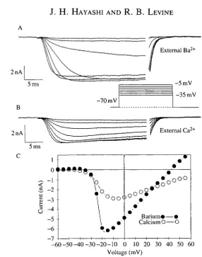
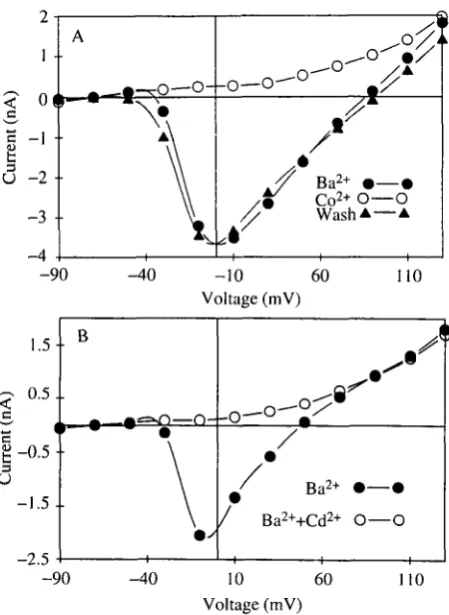
To isolate the sustained inward current, the fast, transient inward current was blocked with TTX and the outward currents were blocked as described. In addition, Ba2+ replaced Ca2+ as the divalent cation. Under these conditions, a prolonged inward current was revealed that activated in response to voltage commands more depolarized than - 4 0 m V and peaked at about - l O m V (Fig. 4). With Ca2+ as the divalent cation in the external solution rather than Ba2 +, the peak amplitude of the inward current was smaller, and there was a faster decline of the current during the voltage step (Fig. 5A,B). The voltage-sensitivity of activation, however, was similar to that in Ba2+ (Fig. 5C). The Ca2+ or Ba2+ currents could be blocked reversibly by substituting equimolar Co2 +, Ni2+ or Mg2+ in the external solution (Fig. 6A). Adding Cd2+ ( 5 x l O "4m o i r1) to the Ba2+ solution blocked the inward current (Fig. 6B). The block was irreversible (not shown). A small outward current remained in the presence of Cd2+, TEA+ and internal Cs+, as has been observed in other systems (Lewis and Hudspeth, 1983). Taken together, these results suggest that the leg motoneurons express voltage-sensitive Ca2+ channels. The voltage-sensitivity of the Ca2+ current in these motoneurons is similar to that reported in other insect systems (Byerly and Leung, 1988).
inA
-15mV
-55 mV
3 . 5 •
2 . 5 •
1.5 •
"c 0.5
-I -°-
5
'
5-1.5-- 2 . 5 •
3.5 -
-4.5-\ •
1 1 1 1 1
. *
i i i i i -60 -50 -40 -30 -20 -10 0 10 20 30 40 50 60
[image:10.595.125.459.105.496.2]Voltage (mV)
Fig. 3. The fast, transient inward current. This motoneuron was isolated from a stage P0 animal and maintained overnight in culture. Whole-cell voltage-clamp records are shown, with normal saline externally and ISOmmoll"1 CsCl internally. (A) In the absence of TTX, depolarizing voltage commands evoke a fast inward current. This was followed by a small, sustained inward current. (B) Voltage-dependence of the peak inward current.
25
2nA 5 ms
-70 raV
-5mV -40 mV
<
c
Curren
t
1-0< - 1 •
2 3 4 5
-• •
••••'
•
•
• • •
1 1 1 1 1 1 - 5 0 - 4 0 - 3 0 - 2 0 - 1 0 0 10 20 30 40 50 60 70
Voltage (mV)
Fig. 4. The Ba2+ current. Whole-cell voltage-clamp records obtained with TTX
8 l + l 2 1 2
(10-8moirl), TEA+ (30mmoirl) and Ba2 1
replacing Ca2+ in the )
external solution, and ISOmmoll"1 CsCl in the internal solution. (A) Depolarizing voltage commands evoke a sustained inward current. (B) Voltage-dependence of the peak inward current.
charge carrier. Following return to a holding potential of — 70 mV, the large Ba2+ current recovered from inactivation, but rarely returned to its original level. By contrast, with Ca2+ as the charge carrier the recovery was complete. Thus, prolonged Ba2+ influx may alter ion channel function or cause a build-up of internal Ba2+ that the cell is unable to regulate.
Changes in the level of Ca2+ currents during metamorphosis
2nA
2nA External Ca
2+
5 ms
0 < > • » # § • $
-a
2
--4 5 --6 -7
,8*
-o-oBarium*—• CalciumO—O
[image:12.595.139.426.110.474.2]_60-50-40-30-20-10 0 10 20 30 40 50 60 Voltage (mV)
Fig. 5. Comparison of the sustained inward current with Ca2+ and Ba2+ as the charge carriers. (A) Whole-cell voltage-clamp records obtained as in Fig. 4. (B) The same motoneuron as in A, but after changing the external solution to one containing 6mmol I"1 Ca2+ rather than Ba2+. Note that the inward current is smaller and decays more rapidly. (C) Voltage-dependence of the peak inward currents with Ca2+ and Ba2+ as the charge carriers.
animals. Both components of the Ba2+ current were present in all stages and displayed similar voltage sensitivities, with the inactivating component always being larger than the sustained component.
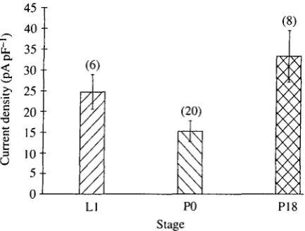
The calculated whole-cell capacitance values increased somewhat during metamorphosis (Ll = 136±27pF; P0=185±27pF; P18=207±44pF; mean±
S.E.M.), as expected given the small increase in cell size. It should be noted,
Ba2+ • — < Co2+ O — O Wash A — •
-90 -40 -10 60 Voltage (mV)
1 . 5 ••
0 . 5 . .
t - ° -5 • • O
- 1 . 5
J--2.5
B
—O--Q
J Ba2+ • — «
/ Ba2++Cd2+ O—C
-90 -40 10 60 Voltage (mV)
[image:13.595.181.406.145.453.2]110
Fig. 6. Blockage of the Ba2+ current in leg motoneurons. (A) Current/voltage relationship of the peak inward current obtained with the whole-cell voltage-clamp technique as in Fig. 4. Changing from an external solution containing Ba2+ (6mmoll~') to one containing Co2+ (6mmolP1) blocks the inward current reversibly. (B) In a similar experiment, adding Cd2+ (500^moir') to the Ba2+-containing external solution blocked the inward current irreversibly.
Potassium currents
To isolate K+ currents, Na+ currents were blocked with TTX and Ca2+ currents were blocked either with Cd2+ or by substituting Ni2 + for Ca2 +. No differences in the outward currents between the two blocking techniques were noted. Outward currents were similar whether the internal solution contained potassium aspartate or KC1. Under these conditions, the response to depolarizing voltage commands differed depending upon the stage from which the cells were dissociated (see below). The following description applies to motoneurons cultured from fifth-instar larvae (LI).
7 0 -•
60 -•
50 -•
4 0 -•
30 -•
2 0 -•
10 -•
0 * -90
'pulse/'max •
/test//max O O
O O ()
-80 -70 -60 -50 -40 -30 - 2 0 - 1 0 0 Voltage (pulse or pre-pulse)
—I—
[image:14.595.149.423.112.325.2]10 20
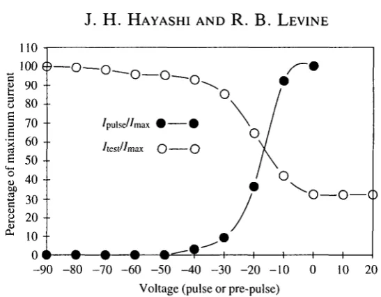
Fig. 7. Steady-state inactivation of the Ca2+ current. Whole-cell voltage-clamp of a leg motoneuron with Ca2+, TTX and TEA+ in the external solution and 150mmoll~L CsCl in the internal solution. Open circles, the cell was held at —70 mV and stepped to -lOmV test pulses following 120 ms pre-pulses to the membrane potentials shown on the x-axis. The peak inward current during the test pulse (/test) is plotted as the
percentage of the maximum current evoked (/max; pre-pulse of -90mV). Filled circles,
the peak inward current evoked with pulses to the membrane potentials shown on the x-axis (/pUise) is plotted as a percentage of the current evoked by a step to 0 mV (/max).
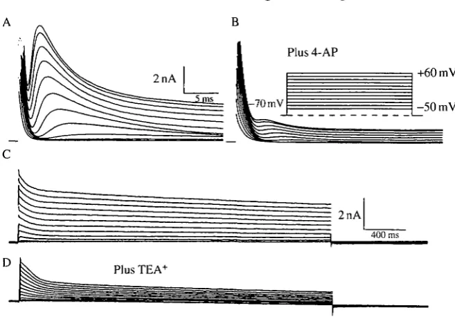
current and compared to that of the sustained component (measured at the end of 30ms depolarizing steps). The transient component activated at about - 3 0 m V (Fig. 11B), whereas the sustained current had a higher threshold for activation (about OmV; Fig. 11B), although the threshold of the sustained current was difficult to measure precisely because of contamination from the falling phase of the transient component. The transient component of the outward current was reduced by external 4-AP (4-aminopyridine, lOmmoll"1; Fig. 12A,B), which also caused a small decrease in the sustained component. TEA+ (30 mmol I"1) reduced both the transient and sustained outward currents (Fig. 12C,D). Both blockers took 2-4min for full effect and were reversible.
-70 mV holding potential
2nA
-70 mV
-20 mV holding potential
2nA
5 ms -20 mV-0.5
O • • »
- 0 . 5 -•
- 1 . 5 -• | 2 . 5
-¥ -3.5
4-- 5 . 5 4--•
- 6 . 5 -•
-7.5
-O O r
Vh=-70mV • — •
Vh=-20mV O — O
-60 -50 -40 -30 -20 -10 0 Voltage (mV)
Fig. 8. Current/voltage relationships of the Ba2+ current with holding potentials of —70 mV and —20 mV. (A) Whole-cell voltage-clamp records of a leg motoneuron with Ba2+, TTX and TEA+ in the external solution and CsCl in the internal solution. The cell was held at -70 mV and stepped to the depolarized potentials shown. (B) Same cell as A, but now with a holding potential of — 20 mV. Note that a smaller, but non-inactivating, inward current is evoked by depolarizing commands. (C) Voltage-sensitivity of the peak inward current with holding potentials of -70 mV and -20 mV.
2nA
D 1
-0« - 1 • 2 -- 3 •
- 5 •
- 6 i •
fc A A A A A
• ^A
1 1 1 1 • •
A' .A'
- 1 1
; • ' •
A> Ll •
/ PO O -P18A~
1 1 1-n-O-t
••••
- •
- O
1 ——f—
I
u
[image:16.595.148.424.167.614.2]- 6 0 - 5 0 - 4 0 - 3 0 - 2 0 - 1 0 0 10 20 30 40 50 Voltage (mV)
31
Q4 a. >-, densi t §4 5
4 0
- 35- 30- 25- 20- 15- 10- 5-
0-(!
(6) T 1l
y/
//
(20) T \J\SN
v \
X X \ 5) X X 0 X X X0
0
$81
AA /\X y yLI PO P18
[image:17.595.187.404.153.317.2]Stage
Fig. 10. Comparison of the peak Ba2+ current (mean±s.E.M.) in leg motoneurons isolated from early last instar larvae (LI), early pupae (P0) and the fully developed adult (P18). The current density was calculated by dividing the peak inward current (holding potential — 70 mV) by the whole-cell capacitance of each neuron. Numbers in parentheses refer to the number of neurons examined in each stage. The level of this current declines significantly in stage P0 (P<0.05; Student's Mest), before increasing during adult development (P<0.05).
Changes in potassium currents during metamorphosis
The levels of the transient and sustained outward currents in leg motoneurons changed during metamorphosis. Unlike the motoneurons from stage LI and P18, those from stage P0 did not have an obvious transient component of the total outward current (Fig. 14). The relative contributions of the two components of the outward current described above were estimated by measuring the peak transient and the late outward currents for voltage steps to +55 mV. The peak transient current density decreased significantly in the early pupal stage (P0), then increased during adult development (P18; Figs 14 and 15). The sustained outward current did not decrease significantly at the onset of metamorphosis, but increased significantly during adult development (Figs 14 and 15). A reduction in the level of the inactivating outward current during stage P0 was also suggested by comparing the outward currents evoked by steps to +55 mV from holding potentials of - 7 0 and —20 mV. A large, transient component of the outward current was eliminated at holding potentials of - 2 0 mV in stage P18 and LI motoneurons. By contrast, cells isolated from early pupal stage animals (P0) displayed little inactivating outward current (not shown).
Discussion
Relevance of these findings to the normal function of motoneurons in vivo
-60 mV
lnA
<
c
3 3
2 1 0
Transient • — • Sustained O—O
•o-o-o
v Ok
,o-o•o-o•O
[image:18.595.135.432.101.556.2]- 6 0 - 5 0 - 4 0 - 3 0 - 2 0 - 1 0 0 10 20 30 40 50 Voltage (mV)
Fig. 11. Potassium currents in a leg motoneuron. (A) Whole-cell voltage-clamp record of a leg motoneuron isolated from stage LI. The external solution contained Ba2+ ^mmoU"1), Cd2+ (SOOjumolP1) and TTX (HP8moll"1); the internal solution contained ISOmmolP1 potassium aspartate. The cell was held at -70 mV and stepped to the membrane potentials indicated in the inset. (B) Voltage-sensitivities of the peak outward current (transient) and of the current at the end of a 30 ms step (sustained).
A B
Plus 4-AP
1
2nA
I §
[image:19.595.133.462.133.362.2]-70 mV
Fig. 12. Effect of potassium channel blockers on the outward currents. (A) A leg motoneuron isolated from a stage LI animal was held at — 70 mV and given voltage commands as indicated in the inset. Internal and external solutions as in Fig. 11. (B) Same cell as in A, but with 10 mmol I"1 4-AP added to the external solution. (C) A leg motoneuron from stage P18; experimental protocol as in A, but longer voltage commands were given. The transient outward current is present as the brief peak at the far left of the records. (D) Same cell as C but with 30 mmol T1 TEA+ added to the external solution.
neurons that contribute to their activity patterns at different stages of life. Changes in the intrinsic biophysical properties of the motoneurons may contribute to the behavioral changes that accompany metamorphosis.
a
oI
6 .
4-2
0 1 1 1— 1 1 1 1—
- 2 0 - 1 0 0 10 20 30
Voltage (mV)
40 50 -10 0 10 20 30 40 50
Voltage (mV)
'pulse/' max # # /test//max O O
vV
c
100 $90 80 70 60 50 40 30 20 10
0
[image:20.595.70.509.110.486.2]-90 -80 -70 -60 -50 -40 -30 -20 -10 0 10 20 Voltage (pulse or pre-pulse)
Fig. 13. Characteristics of outward currents. (A) Voltage-dependence of the rate of activation. A leg motoneuron isolated from stage LI was held at -70 mV and stepped to the membrane potentials indicated; internal and external solutions as in Fig. 11. The time-to-peak of the outward current decreases with more depolarizing commands. (B) Voltage-dependence of the inactivation time constant (T). Same neuron as in A. Inactivation of the peak outward current is faster with more depolarizing commands. (C) Voltage-dependence of inactivation. Open circles, a leg motoneuron from stage LI (external and internal solutions as in Fig. 11) was held at — 70 mV and stepped to — lOmV following 120 ms pre-pulses to the membrane potentials indicated on the x-axis. The peak outward current (Aest) w as plotted as a percentage of the maximum outward current (Imax; with a pre-pulse of — 90 mV). Filled circles, the leg motoneuron
was held at — 70 mV and stepped to the membrane potentials indicated on the x-axis. The peak outward current (/puise) is plotted as a percentage of the outward current
evoked by a step to +20 mV (/max).
35
[image:21.595.210.395.148.329.2]Ll
Fig. 14. Comparison of the outward currents at different stages. (A,B,C) Leg motoneurons isolated from stages Ll, PO and P18, respectively, were voltage-clamped as in Fig. 11 and stepped to +55 mV from a holding potential of -70 mV. Note that the transient outward current is much larger in cells from stages Ll and P18 than in the cell from stage PO.
axonal processes will require a more direct voltage-clamp analysis of the distal processes of the neurons in culture. Preliminary studies, in which patch electrodes were used to voltage-clamp the distal processes of motoneurons cultured for 1-2 weeks, revealed Ca2+ currents similar to those described in this report (J. H. Hayashi, M. D. Withers and R. B. Levirie, unpublished observation). In addition, it will be important to examine in more detail the biophysical properties of the leg motoneurons in vivo.
One important variable that needs to be addressed more fully is the identity of the leg motoneuron. Although the present study was restricted to leg motoneur-ons, we did not attempt to distinguish between different identified cells. The specific biophysical properties of the cells may vary among motoneurons that innervate different leg muscles, or between slow and fast motoneurons to the same muscle.
Calcium currents
u
[image:22.595.181.393.159.303.2]P18
Fig. 15. Comparison of the potassium currents at different stages of metamorphosis. The open bars indicate the peak value of the transient outward current; the filled bars indicate the level of the sustained outward current at the end of a 30 ms step (mean±s.E.M.). Current densities were calculated as in Fig. 10. Numbers in paren-theses indicate the number of leg motoneurons examined at each stage. Note that the level of the transient current declines significantly at the beginning of the pupal stage (P0) (F<0.05; Student's Mest), then increases again during adult development (P<0.05). The sustained outward current does not decline significantly at the onset of pupation, but increases significantly during adult development (P<0.05).
Ca2+-dependent K+ current, since such currents should be blocked by the use of internal Cs+. The small component of the Ba2+ or Ca2+ current that persists when the cells are maintained for several seconds at depolarized holding potentials may represent an independent class of Ca2+ channels or may reflect a small fraction of the channels in a single class that remain available for activation even after prolonged depolarizing steps. The former possibility is suggested by the change in the voltage threshold and peak of the I/V relationship when the holding potential is changed from —70 to — 20mV. Our ability to distinguish between these possibilities awaits single-channel measurements or a selective blocker that could discriminate between channel types. Neurons cultured from embryonic
Dros-ophila also display Ca2+ currents with inactivating and sustained components (Byerly and Leung, 1988; Leung etal. 1989; Leung and Byerly, 1991). The non-inactivating component is blocked preferentially by a venom fraction from the spider, Hololena curta (Leung et al. 1989), suggesting the presence of at least two channel types.
antennal-lobe interneurons of Manduca also revealed similar properties (Hayashi and Hildebrand, 1990). The Ca2+ currents in Manduca neurons do not correspond clearly to any of the three types of channels described for vertebrate neurons (Nowycky et al. 1985; Fox et al. 1987a,b) in terms of their profiles of threshold, inactivation kinetics and sensitivity to inorganic blockers. In addition, the Ca2+ currents in Manduca leg motoneurons were insensitive to organic blockers, such as conotoxin and dihydropyridines, that have helped to distinguish between the channel types found in vertebrate neurons (J. H. Hayashi and R. B. Levine, unpublished observation).
Potassium currents
Manduca leg motoneurons express voltage-sensitive outward currents that are
blocked by extracellular TEA+ and 4-AP and by substituting Cs+ for K+ in the internal solution. Outward currents were similar with KC1 or potassium aspartate in the intracellular solution. Based on these observations, we conclude that these outward currents are carried by K+. We have divided these K+ currents into transient (rapidly inactivating) and late (slowly or non-inactivating) components. Similar studies of cultured neurons from embryonic (Saito and Wu, 1991), larval (Sole and Aldrich, 1988) and pupal (Baker and Salkoff, 1990) Drosophila, and single-electrode voltage-clamp studies of non-spiking interneurons in the locust (Laurent, 1991), have revealed K+ currents with comparable properties. The two components were sensitive to 4-AP and TEA+ as reported in other systems, but it is not clear that the blockers can be used to separate the two components reliably. The small reduction in the late outward current that was caused by 4-AP may reflect an effect on the falling phase of the transient current, although the same reduction in the late current was observed during longer voltage steps.
The rapidly inactivating K+ current (or A current) observed in cultured
Drosophila neurons (Sole and Aldrich, 1988) is generally distinguished from that
in muscle (Zagotta et al. 1988; Salkoff and Wyman, 1983; Singh and Wu, 1989) on the basis of several characteristics. The A current measured from the somata of cultured neurons (A2 channels) has an activation threshold of about - 5 0 mV, whereas that of the A current in muscle (Aj channels) is 20 mV more depolarized. In addition, the muscle A current has a more depolarized steady-state inactivation curve and a faster inactivation rate and its rate of inactivation is voltage-dependent, unlike that of neurons. At channels from muscle are dependent upon the shaker locus (Salkoff and Wyman, 19816; Wu and Haugland, 1985; Timpe and Jan, 1987), whereas A2 channels in neurons are s/ia£:er-independent (Sole et al. 1987; Baker and Salkoff, 1990). Interestingly, the shaker mutation causes hyperexcitability of presynaptic terminals (Jan et al. 1977; Ganetzky and Wu, 1982) and broadening of giant fiber action potentials (Tanouye et al. 1981), leading to the suggestion that At channels may be the predominant contributors to A currents in neuronal processes, while A2 channels predominate on the soma (Sole and Aldrich, 1988).
One possibility is that after 1 day in culture the motoneuron somata are expressing A currents that would normally be destined for the processes. The rather positive values at which the leg motoneuron A current inactivates suggest that it might normally be important for action potential repolarization (McCobb etal. 1990). This is consistent with the finding that the action potential width was increased when it was evoked from motoneurons at depolarized membrane potentials.
A second component of the K+ current in Manduca leg motoneurons did not inactivate fully during prolonged steps to depolarized membrane potentials and was less sensitive to 4-AP. This current was also partially blocked by SOmmolP1 T E A+. It thus displays many characteristics of the delayed rectifier K+ currents described in Drosophila (Sole and Aldrich, 1988; Singh and Wu, 1989) and
Manduca (Zufall etal. 1991).
Our separation of outward currents into two components is probably an over-simplification. Multiple species of K+ channels are derived from alternative splicing of the shaker locus (Schwartz et al. 1988; Pongs et al. 1988; Kamb et al. 1988) and from members of the extended shaker gene family in Drosophila (Wei
et al. 1990). Voltage-clamp studies have revealed multiple variants of the rapidly
inactivating K+ currents in neurons (Baker and Salkoff, 1990), many of which do not depend upon the shaker locus. Similarly, Sole and Aldrich (1988) distinguish between slowly inactivating (KD) and non-inactivating (K{) K+ channels in
Drosophila neurons, which may have been considered together in our studies. We
also did not examine the possible expression of Ca2+- or Na+-dependent K+ currents in the leg motoneurons. Cation-dependent K+ currents, which contribute significantly to the outward current in other insect neurons (Thomas, 1984; Nightingale and Pitman, 1989), would not have been observed under the conditions used in the present experiments. A complete analysis of the channel species expressed by the leg motoneurons also awaits measurements of single-channel currents.
Changes in currents during metamorphosis
A comparison of the voltage-gated currents in leg motoneurons at different
stages of metamorphosis reveals that the levels of Ca2+ and inactivating K+ currents decline during the early pupal stage relative to those in the larva and fully developed adult. This decline was not due to changes in cell size, as we corrected for differences between the whole-cell capacitances at different stages. Further-more, the levels of the sustained outward currents did not change in parallel.
expression of a different channel protein or the modulation (e.g. through phosphorylation) of the same channel species. Pending the measurement of single-channel currents we cannot distinguish between these possibilities.
The relevance of the changes in current density may lie in the modified behavioral role of the leg motoneurons during metamorphosis. During the larval stage these motoneurons innervate leg muscles and participate in crawling, grasping and ecdysis behavior patterns (Kent and Levine, 1988a). At the end of the larval stage, the leg muscles degenerate, and the processes of the leg motoneur-ons, both within the central nervous system and at the periphery, undergo substantial regression (Kent and Levine, 19886; 1992; K. S. Kent and R. B. Levine, unpublished observations). Although they continue to receive some synaptic inputs and maintain the ability to generate action potentials (Kent and Levine, 19886, 1992), the leg motoneurons do not evoke movements during the early pupal stage. During adult development, the leg motoneurons grow new dendrites, receive new synaptic inputs and form new neuromuscular junctions with newly generated muscles of the adult legs (Kent and Levine, 19886; 1992; K. S. Kent and R. B. Levine, unpublished observations). In the adult, these neurons participate in leg movements associated with emergence and walking as well as in various defensive and grooming reflexes. Thus, the decrease in current density could be important for alterations in the activity patterns of the leg motoneurons during metamorphosis, either as a stage in the transition between larval and adult properties or to allow activity patterns that may be unique to the pupal stage. Alternatively, the decrease in channel density could simply reflect a quiescent period for the leg motoneurons, during which they decrease channel synthesis transiently. A final possibility is that the alterations in channel density are important for regulating activity levels during critical developmental events, such as growth-cone extension or synapse formation (Mattson and Kater, 1987; Holliday and Spitzer, 1990). It will be instructive to compare the properties of the leg motoneurons during the pupal stage to those of other motoneurons that do not undergo dendritic changes or innervate new muscles during metamorphosis (Levine and Truman, 1985).
Preliminary studies indicate that the changes in voltage-gated currents that we have observed by analyzing neurons dissociated from animals of different stages are recapitulated by neurons that have been dissociated at early stages and maintained for extended periods in vitro. This protocol will be essential in determining whether the alterations in currents are regulated by the steroid hormone, 20-hydroxyecdysone, that is responsible for other developmental changes during metamorphosis (Levine and Weeks, 1990; Prugh et al. 1992).
AUGUSTINE, G. J., CHARLTON, M. P. AND SMITH, S. J. (1987). Calcium action in synaptic transmitter release. A. Rev. Neurosci. 10, 633-693.
BAKER, K. AND SALKOFF, L. (1990). The Drosophila shaker gene codes for a distinctive potassium current in a subset of neurons. Neuron 2, 129-140.
BELL, R. A. AND JOACHIM, F. A. (1976). Techniques for rearing laboratory colonies of tobacco hornworms and pink bollworms. Ann. ent. Soc. Am. 69, 365-373.
BYERLY, L. AND LEUNG, H. T. (1988). Ionic currents of Drosophila neurons in embryonic cultures. J. Neurosci. 8, 4379-4393.
CHRISTENSEN, B. N., LARMET, Y., SHIMAHARA, T., BEADLE, D. AND PICHON, Y. (1988). Ionic currents in neurones cultured from embryonic cockroach {Periplaneta americana) brains.
J. exp. Biol. 135, 193-214.
CONNOR, J. A. AND STEVENS, C. F. (1971). Voltage clamp study of a transient outward membrane current in gastropod neural somata. J. Physioi, Lond. 213, 21-30.
ECKERT, R. AND TILLOTSON, D. L. (1981). Calcium-mediated inactivation of the calcium conductance in caesium-loaded giant neurones of Aplysia californica. J. Physioi., Lond. 314, 265-280.
FENWICK, E. M., MARTY, A. AND NEHER, E. (1982). A patch-clamp study of bovine chromaffin cells and of their sensitivity to acetylcholine. J. Physioi., Lond. 331, 577-587.
Fox, A. P., NOWYCKY, M. C. AND TSIEN, R. W. (1987a). Kinetic and pharmacological properties distinguishing three types of calcium currents in chick sensory neurons. /. Physioi., Lond.
394, 149-172.
Fox, A. P., NOWYCKY, M. C. AND TSIEN, R. W. (19876). Single-channel recordings of three types of calcium channels in chick sensory neurones. /. Physioi., Lond. 394, 173-200.
GANETZKY, B. AND WU, C-F. (1982). Drosophila mutants with opposing effects on nerve excitability: genetic and spatial interactions in repetitive firing. /. Neurophysiol. 47, 501-514.
GOODMAN, C. S. AND HEITLER, W. J. (1979). Electrical properties of insect neurones with spiking and non-spiking somata: normal, axotomized, and colchicine treated neurones.
J. exp. Biol. 83, 95-121.
GRIFFIN, L. AND LEVINE, R. B. (1989). Hormonal influences on insect motoneurons developing in cell culture. Soc. Neurosci. Abstr. 15, 87.
HAMILL, O. P., MARTY, A., NEHER, E., SAKMANN, B. AND SIGWORTH, F. J. (1981). Improved patch-clamp techniques for high resolution current recording from cells and cell-free membrane patches. Pfliigers Arch. 391, 85-100.
HARDIE, R. C. AND WECKSTROM, M. (1990). Three classes of potassium channels in large monopolar cells in the blowfly Callifora vicina. J. comp. Physioi. 167A, 723-736.
HAYASHI, J. H. AND HILDEBRAND, J. G. (1990). Insect olfactory neurons in vitro: morphological and physiological characterization of cells from the developing antennal lobes of Manduca
sexta. J. Neurosci. 10, 848-859.
HOLLIDAY, J. AND SPITZER, N. C. (1990). Spontaneous calcium influx and its role in differentiation of spinal neurons in culture. Devi Biol. 141, 13-23.
HONIG, M. G. AND HUME, R. I. (1986). Fluorescent carbocyanine dyes allow living neurons of identified origin to be studied in long-term cultures. J. Cell Biol. 103,171-187.
JAN, Y. N., JAN, L. Y. AND DENNIS, M. J. (1977). Two mutations of synaptic transmission in
Drosophila. Proc. R. Soc. Lond. B 198, 87-108.
JOHANSEN, J., YANG, J. AND KLEINHAUS, A. L. (1987). Voltage-clamp analysis of the ionic conductances in a leech neuron with a purely calcium-dependent action potential.
J. Neurophysiol. 58, 1468-1484.
KAMB, A., IVERSON, L. E. AND TANOUYE, M. A. (1987). Molecular characterization of shaker, a
Drosophila gene that encodes a potassium channel. Cell 50, 405-413.
KAMB, A., TSENG-CRANK, J. AND TANOUYE, M. A. (1988). Multiple products of the Drosophila
KENT, K. S. AND LEVINE, R. B. (1988a). Neural control of leg movements in a metamorphic insect: Sensory and motor elements of the larval thoracic legs in Manduca sexta. J. comp.
Neurol. Ill, 559-576.
KENT, K. S. AND LEVINE, R. B. (19886). Neural control of leg movements in a metamorphic insect: Persistence of the larval leg motor neurons to innervate the adult legs of Manduca
sexta. J. comp. Neurol. 276, 30-43.
KENT, K. S. AND LEVINE, R. B. (1992). Dendritic reorganization of an identified neuron during
metamorphosis of the moth, Manduca sexta. J. Neurobiol. (in press).
KUWADA, J. Y. AND WINE, J. J. (1981). Transient, axotomy-induced changes in membrane properties of crayfish central neurones. J. Physiol., Lond. 317, 435-461.
LAPIED,B., MALECOT, C. O. ANDPELHATE, M. (1990). Patch-clamp study of the properties of the sodium current in cockroach single isolated adult aminergic neurones. J. exp. Biol. 151, 387-403.
LAURENT, G. (1991). Evidence for voltage-activated outward currents in the neuropilar membrane of locust nonspiking local interneurons. J. Neurosci. 11,1713-1726.
LEES, G., BEADLE, D. J., NEUMANN, R. AND BENSON, J. A. (1987). Responses to GABA by isolated insect neuronal somata: pharmacology and modulation by a benzodiazepine and a barbituate. Brain Res. 401, 267-278.
LEUNG, H. T., BRANTON, W. D., PHILLIPS, H. S., JAN, L. AND BYERLY, L. (1989). Spider toxins selectively block calcium currents in Drosophila. Neuron 3, 767-772.
LEUNG, H. T. AND BYERLY, L. (1991). Characterization of single calcium channels in Drosophila embryonic nerve and muscle cells. J. Neurosci. 11, 3047-3059.
LEVINE, R. B. AND HAYASHI, J. H. (1990). Calcium channels in insect motoneurons during metamorphosis. Soc. Neurosci. Abstr. 16, 177.
LEVINE, R. B. AND RAMIREZ, J. M. (1991). Calcium-dependent plateau potentials in the leg motoneurons of Manduca. Soc. Neurosci. Abstr. 17, 1227.
LEVINE, R. B. AND TRUMAN, J. W. (1985). Dendritic reorganization of abdominal motoneurons during metamorphosis of the moth, Manduca sexta. J. Neurosci. 5, 2424-2431.
LEVINE, R. B. AND WEEKS, J. C. (1990). Hormonally mediated changes in simple reflex circuits
during metamorphosis in Manduca. J. Neurobiol. 21,1022-1036.
LEWIS, R. S. AND HUDSPETH, A. J. (1983). Voltage- and ion-dependent conductances in solitary vertebrate hair cells. Nature 304, 538-541.
MATTSON, M. P. AND KATER, S. B. (1987). Calcium regulation of neurite elongation and growth cone motility. J. Neurosci. 1, 4034-4043.
MCCOBB, D. P., BEST, P. M. AND BEAM, K. G. (1989). Development alters the expression of calcium current in chick limb motoneurons. Neuron 2, 1633-1643.
MCCOBB, D. P., BEST, P. M. AND BEAM, K. G. (1990). The differentiation of excitability in embryonic chick limb motoneurons. J. Neurosci. 10, 2974-2984.
NIGHTINGALE, W. D. AND PITMAN, R. M. (1989). Ionic currents in the soma of an identified cockroach motoneurone recorded under voltage clamp. Comp. Biochem. Physiol. 93A, 85-93.
NOWYCKY, M. C , Fox, A. P. AND TSIEN, R. W. (1985). Three types of neuronal calcium channels with different calcium agonist sensitivity. Nature 316, 440-443.
O'DOWD, D. K. AND ALDRICH, R. W. (1988). Voltage-clamp analysis of sodium channels in wild-type and mutant Drosophila neurons. J. Neurosci. 8, 3633-3643.
O'DOWD, D. K., RIBERA, A. B. AND SPITZER, N. C. (1988). Development of voltage dependent calcium, sodium, and potassium currents in Xenopus spinal neurons. J. Neurosci. 8, 792-805.
PAPAZIAN, D. M., SCHWARZ, T. Z., TEMPEL, B. L., JAN, Y. N. AND JAN, L. Y. (1987). Cloning of genomic and complementary DNA from shaker, a putative potassium channel gene from
Drosophila. Science 237, 749-753.
PITMAN, R. M. (1975). The ionic dependence of action potentials induced by colchicine in an insect motoneurone cell body. J. Physiol., Lond. 247, 511-520.
PITMAN, R. M. (1979). Intracellularly applied citrate or externally applied tetraethylammonium ions produce calcium-dependent action potentials in an insect motoneurone cell body.
J. Physiol., Lond. 291, 327-337.
hydroxyecdysone, on the growth of neurites by identified insect motoneurons in vitro. Devi Biol. (in press).
SAITO, M. AND W U , C. F. (1991). Expression of ion channels and mutational effects in giant Drosophila neurons differentiated from cell division-arrested embryonic neuroblasts. 7. Neurosci. 11, 2135-2150.
SALKOFF, L. AND WYMAN, R. (1981a). Outward currents in developing Drosophila flight muscle. Science 212, 461-463.
SALKOFF, L. AND WYMAN, R. (19816). Genetic modification of potassium channels in Drosophila shaker mutants. Nature 293, 228-230.
SALKOFF, L. AND WYMAN, R. (1983). Ion currents in Drosophila flight muscles. /. Physiol., Lond. 337, 687-709.
SANES, J. R. AND HILDEBRAND, J. G. (1976). Structure and development of antennae in a moth, Manduca sexta (Lepidoptera: Sphingidae). Devi Biol. 51, 282-299.
SCHWARTZ, T. L., TEMPEL, B. L., PAPAZIAN, D . M., JAN, Y. N. AND JAN, L. Y. (1988). Multiple
potassium channel components are produced by alternative splicing at the shaker locus of Drosophila. Nature 331, 137-142.
SINGH, S. AND W U , C. F. (1989). Complete separation of four potassium currents in Drosophila. Neuron 2, 1325-1329.
SOLC, C. K. AND ALDRICH, R. W. (1988). Voltage-gated potassium channels in larval CNS neurons of Drosophila. J. Neurosci. 8, 2556-2570.
SOLC, C. K., ZAGOTTA, W. N. AND ALDRICH, R. W. (1987). Single-channel and genetic analysis
reveal two distinct A-type potassium channels in Drosophila. Science 236, 1094-1098. TANOUYE, M. A., FERRUS, A. AND FUJITA, S. C. (1981). Abnormal action potentials associated
with the shaker complex locus of Drosophila. Proc. natn. Acad. Sci. U.S.A. 78, 6548-6552. TAZAKI, K. AND COOKE, I. M. (1990). Characterization of Ca currents underlying burst
formation in lobster cardiac ganglion motoneurons. J. Neurophysiol. 63, 370-384.
THOMAS, M. V. (1984). Voltage-clamp analysis of a calcium-mediated potassium conductance in cockroach (Periplaneta americana) central neurones. J. Physiol., Lond. 350, 159-178. TIMPE, L. C. AND JAN, L. Y. (1987). Gene dosage and complementation analysis of the shaker
locus in Drosophila. J. Neurosci. 7, 1307-1317.
TOLBERT, L. P., MATSUMOTO, S. G. AND HILDEBRAND, J. G. (1983). Development of synapses in
the antennal lobe of the moth Manduca sexta during metamorphosis. J. Neurosci. 3, 1158-1175.
WEEKS, J. C. AND ERNST-UTZSCHNEIDER, K. (1989). Respecification of larval proleg motoneurons during metamorphosis of the tobacco hornworm, Manduca sexta: segmental dependence and hormonal regulation. J. Neurobiol. 20, 569-592.
W E I , W., COVARRUBIAS, M., BUTLER, A., BAKER, K., PAK, M. AND SALKOFF, L. (1990). K+
current diversity is produced by an extended gene family conserved in Drosophila and mouse. Science 248, 599-603.
Wu, C. F. AND HAUGLAND, F. N. (1985). Voltage clamp analysis of membrane currents in larval muscle fibers of Drosophila: Alteration of potassium currents in shaker mutants. J. Neurosci. 10, 2626-2640.
ZAGOTTA, W. N., BRAINARD, M. S. AND ALDRICH, R. W. (1988). Single-channel analysis of four
distinct classes of potassium channels in Drosophila muscle. /. Neurosci. 8, 4765-4779.
ZUFALL, F., STENGL, M . , F R A N K E , C , HILDEBRAND, J. G. ANDHATT, H. (1991). Ionic currents of