Cnidarian bleaching can be defined generally as a decline in the population of the symbiotic algae, a reduction in the pigment content of the algae or both (Coles and Jokiel, 1977; Hoegh-Guldberg and Smith, 1989; Kleppel et al., 1989; Porter et al., 1989; Glynn and D’Croz, 1990; Szmant and Gassman, 1990; Lesser, 1996). Major bleaching events on coral reefs are becoming more common and appear to be caused by increased water temperature (Goreau and Hayes, 1994; Brown et al., 1996) and/or by increased exposure to ultraviolet radiation (Brown and Suharsono, 1990; Gates, 1990; Jokiel and Coles, 1990; Lesser et al., 1990; Gleason and Wellington, 1993; Le Tissier and Brown, 1996).
While there have been many ecological studies of thermal bleaching in corals and on the recovery of corals from bleaching (Brown and Suharsono, 1990; Gates, 1990; Glynn and D’Croz, 1990; Hayes and Bush; 1990; Williams and Bunkley-Williams, 1990; Fitt et al., 1993; Gleason, 1993; Brown et al., 1996), few have investigated the underlying cellular mechanisms. Several studies have demonstrated that
temperature and ultraviolet radiation inhibit photosynthesis and can induce oxidative stress in the algae (Lesser et al., 1990; Lesser, 1996; Jones et al., 1998; Warner et al., 1999). Other studies report changes in enzyme activity (O’Brien and Wyttenbach, 1980; Suharsono et al., 1993) and the increased production of heat-shock proteins in the host during bleaching (Miller et al., 1992; Sharp et al., 1994; Black et al., 1995). Neither line of study offers suggestions as to how changes in temperature cause the loss of the symbiotic algae.
Gates et al. (Gates et al., 1992) demonstrated that thermal bleaching in the Hawaiian reef coral Pocillopora damicornis and in the sea anemone Aiptasia pulchella occurred predominantly by release of intact hosts cells with their contained symbiotic algae. That is, thermal stress somehow affected the adhesive properties of the host cells. After analyzing sections of bleached coral, Brown et al. (Brown et al., 1995) suggested that tissue necrosis might explain bleaching, but these authors also observed released algae still within host cells. Huang et al. (Huang et al., 1998) speculated Printed in Great Britain © The Company of Biologists Limited 2001
JEB3388
Temperature-induced bleaching in symbiotic cnidarians is a result of the detachment and loss of host cells containing symbiotic algae. We tested the hypothesis that host cell detachment is evoked through a membrane thermotropic event causing an increase in intracellular calcium concentration, [Ca2+]i, which could then cause collapse of the cytoskeleton and perturb cell adhesion. Electron paramagnetic resonance measurements of plasma membranes from the tropical sea anemone
Aiptasia pulchella and the Hawaiian coral Pocillopora damicornis labeled with
2,2,6,6-tetramethylpiperidine-1-oxyl (TEMPO) revealed no membrane thermotropic event. In addition, intracellular imaging using Fura-2AM as well as labeling anemones with 45Ca revealed no significant change in [Ca2+]i. However, bleaching could be evoked at ambient temperature with 25 mmol l−1caffeine without affecting [Ca2+]i. [Ca2+]i could be altered with
ionomycin in isolated host cells, but ionomycin could not induce bleaching in A. pulchella. As caffeine can affect levels of intracellular protein phosphorylation, the ability of other agents that alter intracellular levels of protein phosphorylation to evoke bleaching was investigated. The protein phosphatase inhibitor vanadate could induce bleaching in A. pulchella. Two-dimensional gels of 32 P-labeled proteins from cold-shocked, caffeine-treated and control anemones show that both temperature shock and caffeine alter the array of phosphorylated host soluble proteins. We conclude that cnidarian bleaching is linked to a temperature-induced alteration in protein phosphorylation.
Key words: coral, bleaching, cellular dysfunction, protein phosphorylation, sea anemone, Aiptasia pulchella, coral, Pocillopora
damicornis.
Summary
Introduction
Cellular mechanisms underlying temperature-induced bleaching in the tropical
sea anemone Aiptasia pulchella
Sara J. Sawyer* and Leonard Muscatine
‡Department of Organismic Biology, Ecology and Evolution, University of California – Los Angeles, 405 Hilgard Avenue, Los Angeles, CA 90095, USA
*Present address: Department of Biological Sciences, Southern Illinois University, Edwardsville, Box 1651, Edwardsville, IL 62026, USA (e-mail: sasawye@siue.edu)
‡Present address: 2410 Soda Canyon Road, Napa Valley, CA 94558, USA
that heat shock might induce algal release through exocytosis after demonstrating that heat shock alters [Ca2+]i in the coral
Acropora grandis; however, these authors did not directly
investigate coral bleaching. Whereas cell adhesion dysfunction may not be the only potential mechanism of temperature-induced bleaching [see (Brown et al., 1995; Le Tissier and Brown, 1996)], its occurrence is unequivocal. That is, bleaching anemones and corals release intact host cells containing their endosymbionts.
On the basis of an Arrhenius plot of the rate of release of symbiotic dinoflagellates with decreasing temperature, Muscatine et al. (Muscatine et al., 1991) suggested that a membrane thermotropic event (Melchior and Steim, 1976; Watson and Morris, 1987) might initiate bleaching. By analogy with observations on insect cell cultures (Van Bergen en Henegouwen et al., 1985; Coakley, 1987; Cress et al., 1990; Walter et al., 1990), Gates et al. (Gates et al., 1992) hypothesized that temperature stress might induce thermotropic phase transitions in host cell membranes which result in the passive influx of Ca2+(Melchior and Steim, 1976; Quinn, 1989; Cheng and Lepock, 1992; Drobnis et al., 1993). As [Ca2+]icontrols the activity of elements of the cytoskeleton and the cell adhesion molecules to which they are linked (Hirano et al., 1987; Nagafuchi and Takeichi, 1988; Forscher, 1989; Matsumura and Yamashiro, 1993; Weeds and Maciver, 1993), changes in [Ca2+]i could cause loss of adhesion by causing collapse of the cytoskeleton, which is interconnected with the cytoplasmic domain of cell adhesion molecules (Geiger et al., 1992; Takeichi et al., 1992; Sastry and Horowitz, 1993). Thus, a temperature-induced change in [Ca2+]i might induce cell adhesion dysfunction.
In this study, we addressed the hypothesis that temperature stress causes a membrane thermotropic event that results in an elevated [Ca2+]i, causing release of host cells (bleaching). We tested this hypothesis by measuring host cell plasma membrane fluidity using electron paramagnetic resonance (EPR) in the tropical reef coral Pocillopora damicornis and in the tropical sea anemone Aiptasia pulchella and by investigating the role of [Ca2+]i using pharmacological agents, the Ca2+-specific fluorochrome Fura-2AM and 45CaCl2to investigate Ca2+flux in A. pulchella. In addition, we investigated the role of phosphorylation of host cell proteins in cnidarian bleaching using pharmacological agents and two-dimensional gel electrophoresis.
Materials and methods
Animal collection and maintenance
Aiptasia pulchella (Carlgren) and Pocillopora damicornis L.
were collected from Kaneohe Bay, Oahu, HI, USA, on Checker Reef adjacent to the Hawaii Institute of Marine Biology. Both were transported back to the University of California at Los Angeles (UCLA) and maintained in an aquarium at 25 °C on a 12 h:12 h light:dark cycle. P. damicornis colonies were used within 48 h of collection. A. pulchella were fed twice weekly on Artemia nauplia, but were starved for 24 h in an incubator
(Percival Scientific, model 1-35 VL) at 25 °C on a 12 h:12 h light:dark cycle at 100µmol quanta m−2s−1before being used in experiments.
Electron paramagnetic resonance
To isolate host cell membranes, A. pulchella were homogenized in a Teflon–glass tissue grinder in 1 ml of filtered sea water (FSW), and P. damicornis were scrubbed with a toothbrush in 3 ml of FSW on ice. Homogenates were centrifuged (Damon/IEC, model HN-S, for 4 min at 500 g) to pellet the algae. The supernatant was decanted and centrifuged at 5000 g for 15 min at 4 °C (Eppendorf, model 5414) to pellet the mitochondria. The supernatant was then centrifuged at 100 000 g for 1 h at 4 °C (Beckman TL-100 ultracentrifuge) to pellet the host cell membranes. The pellet was retained, capped with N2gas, frozen at −80 °C and analyzed within 72 h.
To generate the electron paramagnetic resonance (EPR) spectrum, the membranes were thawed, and 0.1µmol l−1 2,2,6,6-tetramethylpiperidine-1-oxyl (TEMPO) was added in proportion to the amount of host cell membrane phospholipid. Phospholipid concentration was determined from the phosphorus content (Bartlett, 1959). The EPR spectrum was measured with a Varian E-109 EPR spectrophotometer (Varian Associates, Inc., Palo Alto, CA, USA) operating at X-band and fitted with a two-loop one-gap resonator (Hubbell et al., 1987). Measurements were made approximately every 5 °C from 0 to 40 °C. Temperature was controlled with a Varian nitrogen gas flow system and monitored with a thermocouple inserted into the resonator. The heights of the lipid and water signals were measured and their ratio calculated at each temperature (as described by McConnell et al., 1972; Shimshick and McConnell, 1973).
Bioassay for bleaching
cell counts) was performed with Olympus CUE-2 image-analysis software.
Pharmacology
Different pharmacological agents were used to evoke host cell detachment in A. pulchella at 25 °C. Some of these agents were dissolved in dimethylsulfoxide (DMSO), so we initially employed a DMSO control. In no case did incubation with a DMSO control cause release beyond that of the MFSW control at 25 °C, and the DMSO control was omitted in subsequent experiments.
Anemones treated with a pharmacological agent were held in that agent for 2.5 h at either 25 or 12 °C, then rinsed and transferred to MFSW at 25 °C. After 14 h, the percentage of cells released was determined.
The following pharmacological agents were employed: A23187, trifluoperazine (TFP), W-7 [N-(6-aminohexyl)-5-chloro-1-naphthalene-sulfonamide,HCl] (Calbiochem), NiCl2 (Mallinckrodt), CoCl2.6H2O (Baker), 1,2-bis(o-amino-5-bromophenoxy)ethane-N,N,N′,N′-tetraacetic acid, sodium (BAPTA, dibromo-AM) (Molecular Probes), caffeine, procaine (Sigma), nimodipine, verapamil (gifts from Dr P. O’Lague), ryanodine, 2,5-di(+)(butyl)-hydroquinone (DTBHQ) (gifts from Dr M. Barish), ionomycin, thapsigargin (Calbiochem), okadaic acid, phorbol,12-myristate,13-acetate (PMA), isobutylmethylxanthine (IBMX) (Calbiochem) and ortho-sodium vanadate (Na3VO4) (Fisher). All agents were dissolved in MFSW except for A23187, ionomycin, BAPTA, DTBHQ, okadaic acid and PMA, which were dissolved in DMSO.
Identification of the product of release
The product released from A. pulchella (i.e. isolated algae; algae within host cells) was determined by incubating the anemones in the test solution with poly-L-lysine-coated coverslips (0.1 % in distilled water, Mr>70×103, Sigma). After bleaching had been induced and cell products had been released, the coverslips were removed and the nature of the released product was determined. Briefly, the coverslips were rinsed with FSW and stained either with 0.01 % fluorescein diacetate (Sigma) for 20 min for detection of host cell cytoplasm or with 0.01 % Hoechst 33258 (Sigma) for 5 min for detection of host cell nuclei (Gates et al., 1992). After staining, the coverslips were rinsed with FSW, mounted on slides and viewed on an Olympus BX-40 epifluorescence microscope.
Imaging intracellular Ca2+
The concentration of Ca2+ in isolated host endoderm cells was determined by quantitative analysis of the fluorescence image of the Ca2+-specific fluorochrome Fura-2AM (Calbiochem). Endodermal cells were isolated from A.
pulchella using the trypsin maceration technique of Gates
and Muscatine (Gates and Muscatine, 1992). Following maceration, endodermal cells were concentrated on coverslips coated either with poly-L-lysine (0.1 % in deionized water;
Mr>70×103, Sigma) or concanavalin A (10 mg ml−1, Sigma Type 5). Cells were then incubated for 1 h in 5µmol l−1 Fura-2AM in FSW prepared from a stock solution of 10−3mol l–1 Fura-2AM dissolved in DMSO. The cells were then rinsed three times with MFSW, and the color was allowed to develop for 30 min to 1 h before imaging.
The fluorescence image of [Ca2+]i was obtained with a computer-assisted image-analysis system for the quantification of Fura-2AM fluorescence, as described by Sanderson et al. (Sanderson et al., 1990) and Charles et al. (Charles et al., 1991). Briefly, isolated cells were observed with a Nikon Diaphot microscope with a 40×oil-immersion objective (n.a. 1.3). Fluorescent video images, captured with a modified silicon-intensifier target (SIT) video camera (Cohu, San Diego, CA, USA), were recorded on an optical memory disk recorder (Panasonic TQ2026F). Maps of [Ca2+]i were constructed by substituting the 340/380 image ratio (pixel by pixel) and changes in fluorescence at 380 nm into modified equations derived from those of Grynkiewicz et al. (Grynkiewicz et al., 1985) and Monck et al. (Monck et al., 1988). Images consisted of an average of five frames recorded at 340 and 380 nm. A thermal stage fitted with a Peltier device permitted investigation of the change in fluorescence of isolated host cells as a function of temperature over the range 12–35 °C.
45Ca measurements
Changes in [Ca2+]iduring cold shock were also measured using 45Ca. Two different experiments were performed; in the first, anemones were preincubated for 24 h in 2 ml of MFSW containing 45CaCl2 (3.7×10⁴Bq ml−1) (gift of Dr E. Gonzalez). The anemones were then rinsed with MFSW and either cold-shocked at 12 °C or held at 25 °C in 2 ml of MFSW for 2 h. In the second experiment, anemones were incubated in 2 ml of MFSW containing 45Ca (7.4×10⁴Bq ml−1) during the 2 h experiment. In both experiments, replicate 100µl water samples were removed every 15 min throughout the 2 h incubation. At the end of the experiment, the anemones were homogenized in 1 ml of MFSW to separate the algae from the animal tissue. The homogenate was centrifuged at 500 g for 4 min (Damon/IEC model HN-S) to pellet the symbiotic algae. The supernatant containing the animal homogenate was removed, and replicate 100µl samples were taken for counting in a LKB RackBeta scintillation counter (Bio Safe II scintillation cocktail). Samples (100µl) were also taken for determination of animal protein concentration, and the amount of 45Ca in the anemone was expressed as disints min−1mg−1soluble protein.
Determination of cyclic AMP concentration
homogenized in 1 ml of 6 % trichloroacetic acid. The homogenate was centrifuged at 500 g for 4 min (Damon/IEC, model HN-S) to pellet the symbiotic algae. The supernatant was retained for use in the cyclic AMP (cAMP) assay using the Biotrak cAMP enzyme immunoassay system (Amersham). The pellet was retained for protein determination. The results are expressed as fmol cAMP mg−1protein.
Labeling proteins with 32P
To label anemone proteins with 32P-labeled orthophosphate (1 mCi; 3.7×10⁷Bq ml−1, Amersham), the anemones were rinsed three times in phosphate-free artificial sea water (ASW), placed in 1 ml of phosphate-free ASW and briefly allowed to expand. The anemones were then irrigated with 100µCi (3.7×10⁶) Bq ml−1of 32P and incubated for 4 h in the dark at 12, 25 or 30 °C or incubated in 10 mmol l−1caffeine at 25 °C. After incubation, the anemones were homogenized in a low-salt buffer (40 mmol l−1 Tris, 10 mmol l−1 EDTA, 1 mmol l−1 phenylmethylsulfonyl fluoride (PMSF, 10 mmol l−1 NaF, 2 mmol l−1 EGTA and 1 mmol l−1 vanadate) containing 1µg ml−1 of the protease inhibitors pepstatin, leupeptin and chymostatin (Sigma). The homogenate was centrifuged at 500 g for 4 min (Damon/IEC model HN-S) to pellet the symbiotic algae, and the supernatant was retained for protein determination and determination of radioactivity (10µl samples were counted in a LKB RackBeta scintillation counter; Bio Sage II scintillation cocktail).
Two-dimensional electrophoresis
Two-dimensional electrophoresis was performed with a Pharmacia Biotech Multiphor II apparatus following the manufacturer’s instructions. An equal number of counts of 32P was loaded on the immobiline DryStrips (pH 3–10, Pharmacia Biotech) for the isoelectric focusing dimension. When the second dimension (gel electrophoresis) was finished, the gels (Excel Gel SDS, gradient 8-18, Pharmacia Biotech) were dried and autoradiographed on X-ray film for 14 days. Broad-range standards (BioRad) were used for molecular mass determination. The autoradiographs were scanned into Adobe PhotoShop, the images were colored and landmark proteins were superimposed to assist in determining which proteins were unique to each treatment.
Protein measurements
The soluble protein concentration of the anemone homogenate was determined by the method of Bradford (Bradford, 1976) or using the DC Protein Assay (BioRad). In addition, any protein released into the medium by cold-shocked aposymbiotic anemones was also determined by the Bradford method.
Statistical analyses
The mean and standard deviation of the mean (S.D.) were calculated for the data. In addition, significant differences between treatments were determined either by t-test or by analysis of variance (ANOVA) using the program Statview.
Results
Electron paramagnetic resonance measurements of membrane fluidity
To determine whether there was a phase transition in isolated host cell plasma membranes from A. pulchella and P.
damicornis in response to temperature stress, we measured the
EPR signal in isolated membranes from A. pulchella and from
P. damicornis labeled with 0.1µmol l−1 TEMPO. The transformed data from animal plasma membranes from A.
pulchella labeled with the spin probe TEMPO yield a linear
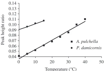
relationship from 0 to 40 °C (Fig. 1). As the membrane preparation from P. damicornis consistently reduced the spin probe and the unknown reducing agent was not responsive to addition of 5 mmol l−1 ferric cyanide, measurements were limited to temperatures from 0 to 15 °C (Fig. 1). These data also yield a linear relationship (r2=0.98 for each) indicating no membrane phase transition in plasma membranes from either
A. pulchella or P. damicornis (Fig. 1).
The effects on intact animals of pharmacological agents that perturb [Ca2+]
[image:4.612.330.537.536.678.2]We tested the possibility that [Ca2+]i could be altered by temperature stress by applying pharmacological agents to intact animals at 25 °C and observing the effect on release of host cells. These results were compared with results obtained from anemones held at their growth temperature (25 °C) and with results from cold-shocked anemones (12 °C). The results of treatment of the anemones with pharmacological agents are summarized in Table 1. To determine whether cold shock evoked entry of Ca2+through Ca2+channels, we treated intact anemones before and during cold shock with the Ca2+channel blockers nimodipine, verapamil, Ni2+and Co2+.None of these agents prevented release of host cells. Next, we treated intact anemones at 25 °C with the Ca2+ ionophores A23187 and ionomycin. Neither caused a significant release of host cells, nor did the calmodulin antagonists W-7 and TFP (Uyeda and Furuya, 1986).
Fig. 1. Peak height ratio (height of the lipid signal/height of the water signal) of the electron paramagnetic resonance spectrum generated from membranes from Aiptasia pulchella and Pocillopora
damicornis and labeled with 0.1µmol l−1 TEMPO over the
temperature range 0–40 °C. r2=0.98 for each.
0.04 0.05 0.06 0.07 0.08 0.09 0.10 0.11 0.12 0.13 0.14
0 10 20 30 40 50
Peak height ratio A. pulchella P. damicornis
To determine whether cold shock evoked release of Ca2+ from intracellular stores, we used the Ca2+-ATPase inhibitors thapsigargin and DTBHQ (Moore et al., 1987; Kass et al., 1989; Thastrup et al., 1989; Thastrup, 1990; Thastrup et al., 1990; Holliday et al., 1991; Lytton et al., 1991). Neither thapsigargin nor DTBHQ caused a significant release of host cells.
We then tested the effect of caffeine, which causes the
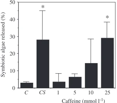
[image:5.612.46.570.86.337.2]release of Ca2+ from intracellular stores (Uyeda and Furuya, 1986; Klein et al., 1992; Lee, 1993). Fig. 2 shows that 10 mmol l−1caffeine evokes release of symbiotic algae at 25 °C, and at 25 mmol l−1the percentage release is equal to that of the cold-shock control (P<0.01). Moreover, the longer the anemones are exposed to caffeine, the more cells are released (Fig. 3). In addition, caffeine evokes the release of entire host cells
Table 1. The ability of various pharmacological agents to evoke host cell detachment in Aiptasia pulchella
Concentration range Added during
Agent (mol l−1) cold shock Effect
Ca2+channel blockers
Nimodipine 10−7to 10−6 Yes Release not prevented (P=0.42)
Verapamil 10−7to 10−5 Yes Release not prevented (P=0.31)
Ni2+ 10−5to 10−3 Yes Release not prevented (P=0.19)
Co2+ 5×10⁻⁴to 5×10⁻² Yes Release not prevented (P=0.24)
Ca2+ionophores
A23187 10−7to 10−5 No No release (P=0.45)
Ionomycin 10−7to 10−5 No No release (P=0.70)
Second messenger-pathway agents
W-7 10−6to 10−5 No No release (P=0.93)
TFP 10−7to 10−5 No No release (P=0.25)
Ca2+-ATPase inhibitors
Thapsigargin 10−8to 10−6 No No release (P=0.54)
DTBHQ 10−6to 10−4 No No release (P=0.92)
Ca2+store releasing agents
Caffeine 10−5to 2.5×10⁻² No Caused release (P=0.01)
Ryanodine 10−5to 3×10⁻⁴ No Release not prevented (P=0.33)
Procaine 10−5to 3×10⁻³ Yes Release not prevented (P=0.42)
Ca2+chelator
[image:5.612.328.556.463.646.2]BAPTA 10−5to 10−3 Yes Release not prevented (P=0.82)
Fig. 2. The effect of a 2.5 h treatment with different concentrations of caffeine on release of symbiotic algae from Aiptasia pulchella. The control (C) anemones were held at 25 °C, while the cold-shocked anemones (CS) were held at 12 °C for 2.5 h. Cell release is expressed as a percentage of the total number of symbiotic dinoflagellates in the anemone [(released/released+retained)×100] released into the medium. Values are means + S.E.M., N=15 in each condition. *Significantly different from the control value, P⭐0.01.
0 10 20 30 40 50
Caffeine (mmol l-1)
Sy
m
b
io
ti
c al
g
ae
relea
se
d
(
%
)
C CS 1 5 10 25
*
*
Fig. 3. The effect of varying the duration of exposure to 25 mmol l−1
caffeine on release of symbiotic algae from Aiptasia pulchella. The control anemones (C) were held at 25 °C, while the cold-shocked anemones (CS) were held at 12 °C for 2.5 h. Cell release is expressed as a percentage of the total number of symbiotic dinoflagellates in the anemone [(released/released+retained)×100] released into the medium. Values are means + S.E.M., N=5 in each condition. *Significantly different from the control value, P⭐0.01.
C CS 15 30 60 90 150
0 10 20 30 40 50
Duration of exposure (min)
Sy
m
b
ioti
c a
lg
ae re
lease
d
(
%
)
*
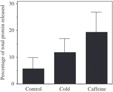
[image:5.612.76.268.477.645.2]containing the endosymbiotic algae (Fig. 4). When the cells released from anemones treated with 25 mmol l−1caffeine are stained with fluorescein diacetate, the esterases in the host cell cytoplasm cleave the dye to its active state (Fig. 4A). The host cell cytoplasm is revealed as yellow fluorescence in the interstices between the red fluorescent algae. When the released cells are stained with the DNA-specific dye Hoechst 33258, the host cell nucleus can be seen adjacent to the algal doublet (Fig. 4B), with the algae occupying most of the interior of the host cell. This unusual architecture is described in detail elsewhere (Gates et al., 1992). Finally, aposymbiotic anemones treated with caffeine tended to release more protein to the surrounding sea water than the 25 °C controls, although
this difference was not statistically significant (Fig. 5,
P<0.094). Increased protein release is interpreted as the result
of release and subsequent disintegration of endodermal cells. Because caffeine releases Ca2+ from ryanodine-sensitive stores (Uyeda and Furuya, 1986; Klein et al., 1992; Lee, 1993), we tested whether ryanodine could block the caffeine-induced release of cells. Ryanodine was not able to block the caffeine-induced release of cells (P=0.33). Cell release from anemones cold-shocked in the presence of procaine, which can block Ca2+ release from caffeine-sensitive stores (Kitamura et al., 1986; Klein et al., 1992), was also not affected (P=0.42) (Table 1).
Finally, to test generally whether Ca2+is involved, we added the Ca2+ chelator BAPTA during cold shock. BAPTA binds Ca2+ before it can exert a physiological effect (Dubinsky, 1993; Snow and Nuccitelli, 1993), and BAPTA did not prevent the release of host cells evoked by cold shock (P=0.82) (Table 1).
Observation of isolated host cells by Ca2+imaging
To gain further insight into the role of caffeine in evoking host cell release, we used the Ca2+-sensitive fluorochrome Fura-2AM to monitor [Ca2+]iin isolated host cells during the application of selected pharmacological agents. Addition of ionomycin, which does not cause bleaching in A. pulchella, increased [Ca2+]i from the resting level of 68±30 to 570.0±191.4 nmol l−1within 60 s of its application to isolated host cells (Fig. 6). In contrast, 12 min after the addition of caffeine, [Ca2+]iwas 114.7±61.5 nmol l−1(Fig. 6). These data provide direct evidence that caffeine, while able to induce bleaching in A. pulchella, does not cause an increase in [Ca2+]i in isolated host cells from A. pulchella.
[image:6.612.183.295.72.317.2]Attempts to measure [Ca2+]i as a function of temperature were thwarted because the fluorescence of Fura-2AM itself is temperature-sensitive (Fig. 7). Any change in [Ca2+]i we
Fig. 4. Photomicrographs of the product of release from Aiptasia
pulchella after treatment with
25 mmol l−1 caffeine. In A, the
[image:6.612.324.551.519.670.2]product was stained with 0.01 % fluorescein diacetate (×4000). Host cell cytoplasm is visible at the interstices of the algae; in B, the product was stained 0.01 % Hoechst 33258 (×4000). The host cell nucleus is visible adjacent to the algal doublet.
Fig. 5. The effect of a 2.5 h treatment with 25 mmol l−1caffeine on
the percentage of total protein released from aposymbiotic Aiptasia
pulchella. The control anemones were held at 25 °C, while the
cold-shocked anemones were held at 12 °C for 2.5 h. Values are means +
S.E.M., N=6 in each condition.
Control Cold Caffeine
0 10 20 30
P
er
ce
nt
ag
e
o
f
tot
al
p
rot
ei
n
r
el
ea
se
d
Fig. 6. The intracellular Ca2+ concentration in isolated host cells
from Aiptasia pulchella treated with either 10−6mol l−1ionomycin or
25 mmol l−1 caffeine. Ca2+ values were obtained from computer
analysis of stored images of cells labeled with Fura-2AM. Values are means + S.E.M., N=20 for ionomycin-treated cells; N=12 for caffeine-treated cells.
0 100 200 300 400 500 600 700 800
0 2 4 6 8 10 12 14
Time (min)
In
trac
ellular
[Ca
2
+]
(n
m
ol
l
-1)
[image:6.612.71.262.524.680.2]observed in isolated host cells was within the temperature-sensitive curve for Fura-2AM.
45Ca measurements on intact animals
Using the dye Fura-2AM, we were able to image Ca2+fluxes only over periods of 15 min or less because of quenching of the fluorescence. We used 45Ca to investigate Ca2+fluxes over longer periods. In anemones incubated in 45Ca during cold shock, the flux of 45Ca into the anemones did not differ from that of the control anemones. In the control animals, there was a 3.5 % decrease in [Ca2+], while in the cold-shocked anemones there was a 4 % decrease. In addition, the total amount of 45Ca in the cold-shocked anemones (animal tissue) was 5.7×10⁵±5.5×10⁵disints min−1, which is not significantly different from that in the control anemones (3.4×10⁵±2.1×10⁵disints min−1; P=0.28; unpaired t-test). In anemones preincubated in 45Ca, there was no difference between control and cold-shocked anemones either in release of Ca2+or in the amount of Ca2+in the anemones at the end of the cold shock (data not shown).
Measurement of cAMP concentration
Caffeine can affect protein phosphodiesterase activity as well as intracellular Ca2+ levels, so we next tested whether caffeine induced host cell detachment in the sea anemone A.
pulchella by inhibiting phosphodiesterases, leading to an
increase in the concentration of cAMP. The cAMP concentration in the animal homogenate was measured after a 2.5 h incubation in 25 mmol l−1caffeine and compared with the cAMP concentration in control anemones held at 25 °C, their growth temperature. Fig. 8 shows that caffeine induced an
increase in cAMP concentration compared with the control (P<0.05). To test whether temperature shock acted like caffeine and increased cAMP concentration, anemones were either heat-shocked (30 °C) nor cold-shocked (12 °C), and the cAMP concentration in the animal homogenate was assessed and compared with the cAMP concentration in control anemones. Fig. 8 shows that neither heat shock nor cold shock increased cAMP concentration compared with the control. Both the cold- and heat-shocked anemones had a lower intracellular concentration of cAMP than did the controls, although these differences were not statistically significant (P=0.38). This suggests that cAMP is not involved in the induction of host cell detachment by thermal stress.
The effects of pharmacological agents that affect protein phosphorylation in intact anemones
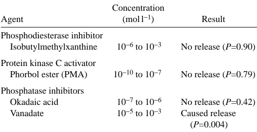
Caffeine, which increases cAMP concentrations by inhibiting phosphodiesterases, has a general effect on intracellular protein phosphorylation. We exposed A. pulchella to other pharmacological agents that affect protein phosphorylation (Table 2). IBMX, a phosphodiesterase inhibitor, had no effect on cell release from A. pulchella (P=0.90), nor did the protein kinase C activator PMA (P=0.79).
20.4 22 26 30 34 36.5 32 30 26 22
0 50 100 150 200 250 300
Temperature (°C)
Cellular
Buffer
In
tra
ce
ll
u
lar
[C
a
2
+] (
n
m
o
l
l
[image:7.612.51.300.72.265.2]-1)
Fig. 7. The intracellular Ca2+ concentration in isolated host cells
(N=30) from Aiptasia puchella and the Ca2+ concentration of a
buffer containing 150µmol l−1Ca2+obtained by Ca2+imaging using
the dye Fura-2AM during temperature stress. The cells were subjected to a temperature stress from 20.4 to 36 ° C and back to 22 °C, while the buffer was subjected to a temperature change from 21.6 to 36.5 °C and back to 22 °C.
0 10 20 30 40 50 60
Control Cold-shocked
Caffeine Heat-shocked
cAMP
co
n
te
n
t
(pm
o
l mg
-1 p
ro
te
in
[image:7.612.324.566.73.206.2])
*
Fig. 8. The amount of cAMP (pmol mg−1protein) in the sea anemone Aiptasia pulchella cold-shocked at 12 °C, held at 25 °C (Control),
incubated in 25 mmol l−1caffeine at 25 °C for 2.5 h or heat-shocked
at 30 °C for 14 h. Values are means + S.E.M., N=4. *Significantly different from the control value, P⭐0.05.
Table 2. The effects of pharmacological agents on cell host
release from the sea anemone Aiptasia pulchella
Concentration
Agent (mol l−1) Result
Phosphodiesterase inhibitor
Isobutylmethylxanthine 10−6to 10−3 No release (P=0.90)
Protein kinase C activator
Phorbol ester (PMA) 10−10to 10−7 No release (P=0.79)
Phosphatase inhibitors
Okadaic acid 10−7to 10−6 No release (P=0.42) Vanadate 10−5to 10−3 Caused release
[image:7.612.317.568.612.741.2]Of the two phosphatase inhibitors tested, okadaic acid did not affect release of cells (P=0.42), but 1 mmol l−1vanadate caused release of 15.5±2.7 % (P=0.004) of the symbiotic algae (Fig. 9). Higher concentrations of vanadate had no further effect, and vanadate was never as effective as temperature or caffeine in causing release of cells.
32P-phosphorylated proteins
To test directly whether temperature alters intracellular protein phosphorylation in A. pulchella, anemones were held at 25 °C, cold-shocked (12 °C), heat-shocked (30 °C) or incubated in 10 mmol l−1caffeine in the presence of 32P, and the animal protein was extracted and subjected to
two-dimensional gel electrophoresis (Fig. 10). To facilitate comparison among treatments, gels were superimposed using landmark proteins. Although the superimposition was not always perfect, many 32P-labeled proteins unique to each treatment were easily distinguished.
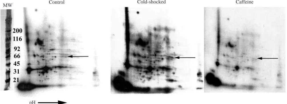
Comparison of the 32P-labeled proteins between control and cold-shocked, control and caffeine-treated and cold-shocked and caffeine-treated anemones revealed a number of proteins unique to each treatment (Fig. 10) (Sawyer, 1998). The pattern of protein phosphorylation in the control anemones differs from that in the cold-shocked anemones, and the patterns in both control and cold-shocked anemones differ from that in the caffeine-treated anemones. The protein phosphorylation pattern from the heat-shocked anemones was not determined because of consistently poor incorporation of 32P by these anemones. The molecular mass of the proteins that differed between the treatments ranged from 5 to 100 kDa (Fig. 10), and the number of unique proteins in the control and cold-shocked anemones ranged from 17 to 34. This is a conservative estimate; because of the slight offset in landmark proteins between gels, the number of unique proteins was deliberately underestimated.
Discussion
In the present study, we investigated the mechanism by which increased or decreased temperature causes loss of host cells and results in cnidarian bleaching. We initially tested the hypothesis that increased or decreased temperature induced a membrane thermotropic event in host cell membranes. This thermotropic event would cause an increase in [Ca2+]i, and this increase would induce cell adhesion dysfunction of the host cells (Gates et al., 1992). We saw no evidence of a phase transition, as measured by EPR, in isolated host cell membranes from the sea anemone A. pulchella and the coral
0 5 10 15 20 25 30 35 40
C CS 0.1 1
[Vanadate] (mmol l-1)
Sy
m
bi
ot
ic
al
g
ae
relea
sed (
%
)
*
[image:8.612.66.269.74.220.2]*
Fig. 9. The effect of a 2.5 h treatment with two concentrations of vanadate on the release of symbiotic algae from Aiptasia pulchella. The control anemones (C) were held at 25 °C, while the cold-shocked anemones (CS) were held at 12 °C for 2.5 h. Cell release is expressed as a percentage of the total number of symbiotic dinoflagellates in the anemone [(released/released+retained)×100] released into the medium. Values are means + S.E.M., N=4 in each
condition. *Significantly different from the control value, P⭐0.0004.
Fig. 10. Autoradiographs of 32P-phosphorylated proteins separated by two-dimensional gel electrophoresis from the control value anemones
held at 25 °C, anemones cold-shocked at 12 °C and anemones treated with 25 mmol l−1 caffeine for 2.5 h. The arrows point to a group of
proteins that are of equal molecular mass (MW) but are differentially phosphorylated. pH
[image:8.612.52.555.516.699.2]P. damicornis over the temperatures measured. Perturbing
intracellular Ca2+stores with pharmacological agents did not affect the bleaching response, and we did not observe an increase in [Ca2+]iusing either the calcium dye Fura-2AM or 45Ca during temperature shock. Caffeine, which can release Ca2+ from intracellular stores as well as affect protein phosphorylation levels (Uyeda and Furuya, 1986; Klein et al., 1992; Lee, 1993), can cause host cell detachment in A.
pulchella without affecting [Ca2+]i. In addition, vanadate, a protein phosphatase inhibitor, can induce bleaching in A.
pulchella. Measurement of protein phosphorylation using
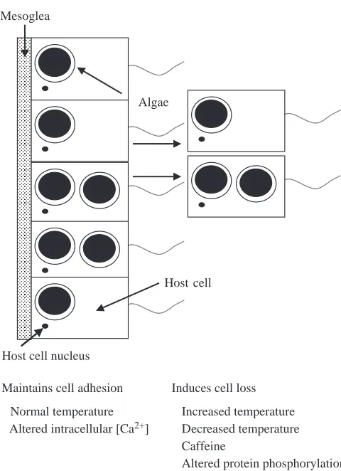
two-dimensional gel electrophoresis of 32P-labeled proteins demonstrates that cold-shocked, caffeine-treated and control anemones have different levels of phosphorylated host proteins. This suggests that temperature-induced cnidarian bleaching alters the protein phosphorylation of the host and, by this mechanism, induces the loss of host cells (Fig. 11).
EPR measurement of membrane fluidity
We tested the hypothesis that temperature-induced host cell detachment in symbiotic cnidarians begins with a membrane thermotropic event. Muscatine et al. (Muscatine et al., 1991) observed a break at 15 °C in the Arrhenius plot of rate of
release of symbiotic dinoflagellates versus temperature. We saw no evidence for a membrane phase transition between 0 and 40 °C (Fig. 1) in plasma membranes from A. pulchella. Isolated plasma membranes from P. damicornis labeled with TEMPO also show no evidence of a membrane phase transition (Fig. 1), although the temperature test range was restricted to 0–15 °C because of the unexplained reduction of the spin probe TEMPO. The addition of ferric cyanide did not halt this reduction, so we conclude that the reduction of the probe was not the result of mitochondrial activity, but of an unknown agent. The absence of a phase change in P. damicornis over this limited range of temperature is consistent with similar data from A. pulchella.
The absence of a phase transition in our data could be explained in several ways. First, phase transitions in plasma membranes are common only at very low temperatures (Blazyk and Steim, 1972; Quinn, 1988), so we may not have used a low enough temperature; however, 0 °C is below the lethal cold temperature for both A. pulchella and P. damicornis, so a phase transition at a temperature below that would be ecologically meaningless. In addition, plasma membranes often contain significant amounts of cholesterol, which stabilize membrane fluidity below the gel-to-liquid-crystalline transition temperature (Chapman et al., 1979; Yeagle, 1987). As cholesterol makes up approximately 17 % of the lipids in A.
pulchella (Doino, 1991), it may help to stabilize the
membranes during cold shock. Further, temperature-induced phase transitions in membranes have been reported mainly in specialized membranes, such as those from spermatozoa, chloroplasts and mitochondria (Raison et al., 1971; Blazyk and Steim, 1972; Quinn, 1989; Jurado et al., 1991; Ruiz-Sanz et al., 1992; Parks and Lynch, 1992; Drobnis et al., 1993). These specialized membranes contain a higher proportion of protein and also large amounts of specific phospholipids, making them more susceptible to a temperature-induced membrane phase transition (Quinn, 1988; Quinn, 1989). Thus, the absence of a phase transition in plasma membranes from A. pulchella and
P. damicornis is typical of other plasma membranes.
The effects on intact animals of pharmacological agents that perturb [Ca2+]
i
To determine whether changes in [Ca2+]i were involved in temperature-induced host cell detachment in A. pulchella, we used a variety of pharmacological agents to alter intracellular Ca2+pathways. If we could cause host cell detachment with an agent known to affect a Ca2+pathway, we might gain insight into how temperature causes loss of host cell adhesion. We used six different types of pharmacological agents: Ca2+ channel blockers, Ca2+ ionophores, second-messenger pathway agents, Ca2+-ATPase inhibitors, Ca2+ chelators and Ca2+ store releasing agents (Table 1). All have been used successfully to implicate Ca2+involvement in cells as diverse as melanoma metastatic cells to flagellates (Meissner, 1986; Uyeda and Furuya, 1986; Grega et al., 1987; Kass et al., 1989; Thastrup, 1990; D’Ancona et al., 1992; Klein et al., 1992; Dubinsky, 1993; Simasko and Yan, 1993; Snow and Nuccitelli,
Mesoglea
Host cell Algae
Host cell nucleus
Normal temperature Altered intracellular [Ca2+]
Increased temperature Decreased temperature Caffeine
Altered protein phosphorylation Induces cell loss
[image:9.612.54.294.67.398.2]Maintains cell adhesion
1993). In A. pulchella, the only pharmacological agent that induced host cell release was caffeine, an agent that can induce Ca2+release from stores in the endoplasmic reticulum (Uyeda and Furuya, 1986; Klein et al., 1992; Lee, 1993). The action of caffeine on A. pulchella is similar to that of temperature shock in several ways. Increasing concentrations of caffeine increased cell release (Fig. 2) just as more extreme (farther from the growth temperature) temperature shocks increase cell release (Muscatine et al., 1991). In addition, increased exposure time to caffeine increased cell release (Fig. 3) as does increased duration of temperature shock. Caffeine treatment induces release of host cells identical in appearance to those released after temperature shock (see Gates et al., 1992). Finally, aposymbiotic anemones are sensitive to caffeine (Fig. 4) just as they are to acute changes in temperature.
Besides causing Ca2+ release from the endoplasmic reticulum, caffeine can also inhibit the enzyme phosphodiesterase (Uyeda and Furuya, 1986; Klein et al., 1992; Lee, 1993). To determine caffeine’s action on A.
pulchella, we used two different pharmacological agents,
procaine and ryanodine, that can interact with caffeine-induced Ca2+release. Procaine can block Ca2+release from caffeine-sensitive stores (Kitamura et al., 1986; Klein et al., 1992), but did not affect cell release during cold shock. Ryanodine is capable of blocking Ca2+stores opened by caffeine (Meissner, 1986), but did not inhibit caffeine-induced cell release. These data, as well as the failure to see a change in release in response to any of the other Ca2+ pathway altering agents, call into question whether caffeine is inducing host cell detachment by inducing changes in intracellular Ca2+concentrations.
Ca2+imaging of isolated host cells
Using isolated host cells loaded with the intracellular Ca2+ dye Fura-2AM, we directly investigated the involvement of Ca2+ in temperature-induced bleaching. Cells treated with ionomycin (Fig. 6) showed a fivefold increase in [Ca2+] within 30 s, indicating that this is a viable method for measuring [Ca2+]i in isolated host cells, yet ionomycin did not induce bleaching. When cells were treated with 25 mmol l−1caffeine, no change in [Ca2+]iwas detected (Fig. 6) even after 12 min. This again suggests that caffeine does not act on A. pulchella by inducing Ca2+release from the endoplasmic reticulum.
We also used Fura-2AM to investigate how temperature affected [Ca2+]i; however, these results were inconclusive (Fig. 7) because the binding constant of Ca2+to the dye is affected by temperature (Groden et al., 1991; Owen, 1991).
Fang et al. (Fang et al., 1997), using Fura-2AM, observed an increase in Ca2+ concentration in isolated cells from the coral Acropora grandis during heat treatment; however, they used cells that did not contain symbiotic algae. Huang et al. (Huang et al., 1998) demonstrated that bleaching in A. grandis required intracellular Ca2+. Thus, while it is possible that some cell types in corals release Ca2+from intracellular stores with heat stress, in our hands, isolated host cells from the sea anemone A. pulchella showed no measurable change in [Ca2+]i during temperature stress.
45Ca measurements on intact animals
The duration of Ca2+ imaging with Fura-2AM is limited because of quenching of Ca2+fluorescence, so we used 45Ca to investigate long-term fluxes of Ca2+ in cold-shocked anemones. 45Ca has been used successfully to evaluate Ca2+ fluxes in both sea anemones and corals (Tambutte et al., 1995; Allemand and Benazet-Tambutte, 1996; Benazet-Tambutte et al., 1996). When anemones were incubated in 45Ca, to load Ca2+ stores with isotope, and then cold-shocked, release of 45Ca by the cold-shocked anemones did not differ from that of control anemones. In addition, when anemones were cold-shocked in the presence 45Ca to test for uptake of Ca2+, no difference between control and cold-shocked anemones was detected. These data suggest that temperature shock (either warm or cold) does not induce an alteration in intracellular Ca2+levels.
Measurement of cAMP
Temperature shock induces host cell detachment, i.e. bleaching, in the sea anemone A. pulchella by an unknown mechanism. Caffeine can also induce host cell detachment in this anemone, suggesting that caffeine and temperature may act on cell adhesion at a common locus. Inhibition of phosphodiesterase activity by caffeine can increase the concentration of cAMP, which can induce loss of cell adhesion in various mammalian cells (Hsie and Puck, 1971; Ortiz et al., 1973; Steinbach and Schubert, 1975; Miller et al., 1976; Li et al., 1977; Spruill et al., 1981; Kreisberg and Venkatachalam, 1986; Lamb et al., 1988; Glass and Kreisberg, 1993). Thus, temperature and caffeine could induce host cell detachment in
A. pulchella by causing an increase in cAMP concentration.
However, the data in Fig. 8 show that an increase in cAMP concentration in A. pulchella is evoked by caffeine, but not by heat or cold shock, suggesting that the modes of action of caffeine and temperature stress in causing cnidarian bleaching differ.
The effects on intact anemones of pharmacological agents that affect protein phosphorylation
An increased cAMP concentration induced by caffeine would stimulate protein kinase A activity and alter protein phosphorylation. Caffeine can enhance the tyrosine phosphorylation of proteins in cell culture (Aharoni et al., 1993). As phosphorylation of cell adhesion molecules controls adhesion (Volberg et al., 1991; Eriksson et al., 1992; Matsuyoshi et al., 1992; Romer et al., 1992; Volberg et al., 1992; Behrens et al., 1993; Hamaguchi et al., 1993) and phosphorylation of proteins is temperature-sensitive (Maher and Pasquale, 1989; Keyse and Emslie, 1992), temperature and caffeine may both act by altering the phosphorylation of cell adhesion proteins.
phosphodiesterase (Ishitani et al., 1995; Usachev and Verkhratsky, 1995), while caffeine may be a better phosphodiesterase inhibitor. The protein phosphatase inhibitor vanadate did induce release of cells (Fig. 9), while okadaic acid and PMA did not (Table 2). Vanadate is an inhibitor of tyrosine phosphatase, while both okadaic acid and PMA affect serine/threonine phosphorylation (Brautigan, 1992; Matsuyoshi et al., 1992; Walter and Mumby, 1993; Tsunoda, 1993). Thus, it is possible that vanadate and caffeine (Aharoni et al., 1993) can induce algal cell loss from A. pulchella by affecting tyrosine phosphorylation levels.
Patterns of 32P protein phosphorylation
The pattern of incorporation of 32P into proteins is different in cold-shocked and control anemones as determined by two-dimensional gel electrophoresis. This difference supports the hypothesis that temperature-induced host cell detachment results from altered protein phosphorylation. In addition, the labeling patterns of the 32P-labeled proteins in the two-dimensional gels from the caffeine-treated anemones are different from those from control anemones, supporting the hypothesis that caffeine can affect the phosphorylation of protein. However, the pattern of 32P-labeled proteins in the two-dimensional gels of the cold-shocked anemones is not the same as that obtained from the caffeine-treated anemones. This suggests that, while caffeine may induce host cell detachment by altering the phosphorylation of proteins, it may affect different proteins from those affected by temperature shock. On the basis of the pharmacological studies and the patterns of protein phosphorylation revealed by two-dimensional gel electrophoresis, it is clear that temperature-induced host cell detachment in A. pulchella is correlated with altered protein phosphorylation. It is not known which proteins are differentially phosphorylated during temperature shock in A.
pulchella.
We were unable to resolve phosphorylated proteins in heat-shocked anemones because incorporation of 32P during high-temperature stress was poor, nor did we determine whether the 32P was incorporated on tyrosine or serine/threonine residues. Caffeine, vanadate and temperature affect protein phosphorylation on tyrosine residues in other systems (Maher and Pasquale, 1989; Brautigan, 1992; Aharoni et al., 1993). Altered tyrosine phosphorylation of the cadherin and catenin cell adhesion molecules can perturb their adhesiveness (Matsuyoshi et al., 1992; Behrens et al., 1993; Hamaguchi et al., 1993). Thus, temperature shock in A. pulchella may alter the levels of tyrosine phosphorylated cell adhesion proteins.
Temperature could affect protein phosphorylation by affecting kinase and phosphatase activity. There is little information about the temperature-sensitivities of these enzymes; smooth muscle phosphatase and myosin-associated muscle phosphatase have Q10 values of 5.3 and 5.2, respectively (Mitsui et al., 1994). Such a high Q10 indicates that these enzymes would be very temperature-sensitive (Hochachka and Somero, 1982). Heat shock also induces the synthesis of a tyrosine phosphatase in cultured human skin
cells, suggesting that the control of phosphorylation levels is important during environmental stress (Keyse and Emslie, 1992).
Temperature stress is not the only environmental factor that induces bleaching in symbiotic cnidarians; ultraviolet radiation can also cause bleaching (Lesser et al., 1990; Gleason and Wellington, 1993; Brown et al., 1994; Le Tissier and Brown, 1996). Ultraviolet radiation can induce oxidative stress in sea anemones and corals (Shick et al., 1991; Dykens et al., 1992; Nii and Muscatine, 1997). Oxidative stress can alter protein phosphorylation and impair cell adhesion in cell culture (Heffetz et al., 1990; Gailit et al., 1993; Barchowsky et al., 1994; Sullivan et al., 1994; Zhang et al., 1994; Feng et al., 1995; Whisler et al., 1995), so it may do so in symbiotic cnidarians. Oxidative stress stimulates both tyrosine and serine/threonine phosphorylation by inhibiting the respective protein phosphatases (Heffetz et al., 1990; Sullivan et al., 1994; Whisler et al., 1995). In addition, oxidative stress stimulates the expression of a tyrosine phosphatase (Keyse and Emslie, 1992), which is also induced by temperature shock in cell culture. That protein phosphatases are differentially affected by oxidative stress and temperature stress in cell culture lends support to the hypothesis that these enzymes may be involved in both temperature and ultraviolet-radiation-induced cnidarian bleaching. Altered protein phosphorylation could be the common mechanism by which both temperature and ultraviolet radiation induce cnidarian bleaching.
The authors would like to thank Dr G. Baghdasarian and Dr R. D. Gates for their many helpful discussions. This research was supported by NSF grant 9115834.
References
Aharoni, D., Dantes, A. and Amsterdam, A. (1993). Cross-talk between adenylate cyclase activation and tyrosine phosphorylation leads to modulation of the actin cytoskeleton and to acute progesterone secretion in ovarian cells. Endocrinology 133, 1426–1436.
Allemand, D. and Benazet-Tambutte, S. (1996). Calcification in the Mediterranean red coral, Corallium rubrum (Linnaeus) (Cnidaria, Octocorallia). J. Exp. Zool. 276, 270–278.
Barchowsky, A., Williams, M. E., Benz, C. C. and Chepenik, K. P. (1994). Oxidant-sensitive protein phosphorylation in endothelial cells. Free Radical Biol. Med. 16, 771–777.
Bartlett, G. R. (1959). Phosphorus assay in column chromatography. J. Biol. Chem. 234, 466–468.
Behrens, J., Vakaet, L., Friis, R., Winterhager, E., VanRoy, F., Mareel, M. M. and Birchmeier, W. (1993). Loss of epithelial differentiation and gain of invasiveness correlates with tyrosine phosphorylation of the E-Cadherin/β-Catenin complex in cells transformed with a temperature-sensitive v-SRC gene. J. Cell Biol. 120, 757–766.
Benazet-Tambutte, S., Allemand, S. D. and Jaubert, J. (1996). Permeability of the oral epithelial layer in cnidarians. Mar. Biol. 126, 43–53.
Black, N. A., Voellmy, R. and Szmant, A. M. (1995). Heat shock protein induction in Montastraea faveolata and Aiptasia pallida exposed to elevated temperatures. Biol. Bull. 188, 234–240.
Blazyk, J. F. and Steim, J. M. (1972). Phase transitions in mammalian membranes. Biochim. Biophys. Acta 266, 737–741.
Bradford, M. (1976). A rapid and sensitive method for the quantitation of microgram quantities of protein utilizing the principles of protein-dye binding. Analyt. Biochem. 72, 248–254.
Brown, B. E., Dunne, R. P. and Chansang, H. (1996). Coral bleaching relative to elevated seawater temperature in the Andaman Sea (Indian Ocean) over the last 50 years. Coral Reefs 15, 151–152.
Brown, B. E., Dunne, R. P., Scoffin, T. P. and Le Tissier, M. D. A. (1994). Solar damage in intertidal corals. Mar. Ecol. Prog. Ser. 105, 219–230. Brown, B. E., Le Tissier, M. D. A. and Bythell, J. C. (1995). Mechanisms
of bleaching deduced from histological studies of reef corals sampled during a natural bleaching event. Mar. Biol. 122, 655–663.
Brown, B. E. and Suharsono (1990). Damage and recovery of coral reefs affected by El Nino related sea water warming in the Thousand Islands, Indonesia. Coral Reefs 8, 163–170.
Chapman, D., Gomez-Fernandez, J. C. and Goni, F. M. (1979). Intrinsic protein–lipid interactions. Physical and biochemical evidence. FEBS Lett. 98, 211–223.
Charles, A. C., Merrill, J. D., Dirksen, E. R. and Sanserson, M. J. (1991). Intercellular signaling in glial cells: communicated waves and asynchronous calcium oscillations in response to mechanical stimulation and glutamate. Neuron 6, 983–992.
Cheng, K. H. and Lepock, J. R. (1992). Inactivation of calcium uptake by EGTA is due to an irreversible thermotropic conformational change in the calcium binding domain of the Ca2+-ATPase. Biochemistry 31, 4074–4080. Coakley, W. T. (1987). Hyperthermia effects on the cytoskeleton and on cell
morphology. Soc. Exp. Biol. 41, 187–211.
Coles, S. L. and Jokiel, P. L. (1977). Effects of temperature on photosynthesis and respiration in hermatypic corals. Mar. Biol. 43, 209–216.
Cress, A. E., Majda, J. A., Glass, J. R., Stringer, D. E. and Gerner, E. W. (1990). Alteration of cellular adhesion by heat shock. Exp. Cell Res. 190, 40–46.
D’Ancona, S., Mazzo, M. and Pea, F. (1992). Increased spontaneous cell detachment of F10 cells induced in vitro by some calcium-channel blockers. J. Chemotherapy 4, 235–238.
Doino, J. A. (1991). Low temperature acclimation in the sub-tropical sea anemone, Aiptasia pulchella (Carlgren). Masters thesis, UCLA, USA. Drobnis, E. Z., Crowe, L. M., Berger, T., Anchordoguy, T. J., Overstreet,
J. W. and Crowe, J. H. (1993). Cold shock damage is due to lipid phase transitions in cell membranes: a demonstration using sperm as a model. J. Exp. Zool. 265, 432–437.
Dubinsky, J. M. (1993). Effects of calcium chelators on intracellular calcium and excitotoxicity. Neurosci. Lett. 150, 129–132.
Dykens, J. A., Shick, J. M., Benoit, C., Buettner, G. R. and Winston, G. W. (1992). Oxygen radical production in the sea anemone Anthopleura elagantissima and its endosymbiotic algae. J. Exp. Biol. 168, 219–241. Eriksson, J. K., Brautigan, D. L., Vallee, R., Olmsted, J., Fujiki, H. and
Goldman, R. D. (1992). Cytoskeletal integrity in interphase cells requires protein phosphatase activity. Proc. Natl. Acad. Sci. USA 89, 11093–11097. Fang, L., Huang, S. and Lin, K. (1997). High temperature induces the synthesis of heat-shock proteins and the elevation of intracellular calcium in the coral Acropora grandis. Coral Reefs 16, 127–131.
Feng, L., Xia, Y., Seiffert, D. and Wilson, C. B. (1995). Oxidative stress-inducible protein tyrosine phosphatase in glomerulonephritis. Kidney Int. 48, 1920–1928.
Fitt, W. K., Spero, H. J., Halas, J., White, M. W. and Porter, J. W. (1993). Recovery of the coral Montastrea annularis in the Florida Keys after the 1987 Caribbean ‘bleaching event’. Coral Reefs 12, 57–64.
Forscher, P. (1989). Calcium and polyphosphoinositide control of cytoskeletal dynamics. Trends Neural Sci. 11, 468–474.
Gailit, J., Coleflesh, D., Rabiner, I., Simone, J. and Goligorsky, M. S. (1993). Redistribution and dysfunction of integrins in cultured renal epithelial cells exposed to oxidative stress. Am. J. Physiol. 264, F149–F157. Gates, R. D. (1990). Seawater temperature and sublethal coral bleaching.
Coral Reefs 8, 193–197.
Gates, R. D., Baghdasarian, G. and Muscatine, L. (1992). Temperature stress causes host cell detachment in symbiotic cnidarians: implications for coral bleaching. Biol. Bull. 182, 324–332.
Gates, R. D. and Muscatine, L. (1992). Three methods for isolating viable anthozoan endoderm cell with their intracellular symbiotic dinoflagellates. Coral Reef. 11, 143–145.
Geiger, B., Ayalon, O., Ginsberg, D., Volberg, T., Rodriguez, J. L., Yarden, Y. and Ben-Ze’ev, A. (1992). Cytoplasmic control of cell adhesion. Cold Spring Harbor Symp. Quant. Biol. 57, 631–642.
Glass, W. F. and Kreisberg, J. I. (1993). Regulation of integrin-mediated adhesion at focal contacts by cAMP. J. Cell. Physiol. 157, 296–306. Gleason, D. F. and Wellington, G. M. (1993). Ultraviolet radiation and coral
bleaching. Nature 365, 836–838.
Gleason, M. G. (1993). Effects of disturbance on coral communities: bleaching in Moorea, French Polynesia. Coral Reefs 12, 193–201. Glynn, P. W. and D’Croz, L. (1990). Experimental evidence for high
temperature stress as the cause of El Nino – coincident coral mortality. Coral Reefs 8, 181–191.
Goreau, T. J. and Hayes, R. L. (1994). Coral bleaching and ocean hot spots. Ambio 23, 176–180.
Grega, D. S., Werz, M. A. and MacDonald, R. L. (1987). Forskolin and phorbol esters reduce the same potassium conductance of mouse neurons in culture. Science 235, 345–348.
Groden, D. L., Guan, A. and Stokes, B. T. (1991). Determination of Fura-2 dissociation constraints following adjustment of the apparent Ca-EGTA association constant for temperature and ionic strength. Cell Calcium 12, 279–287.
Grynkiewicx, G., Poenie, M. and Tsien, R. Y. (1985). A new generation of Ca2+ indicators with greatly improved fluorescence properties. J. Biol. Chem. 260, 3440–3450.
Hamaguchi, M., Matsuyoshi, N., Ohnishi, Y., Gotoh, B., Takeichi, M. and Nagai, Y. (1993). p60v-srccauses tyrosine phosphorylation and inactivation of the N-cadherin–catenin cell adhesion system. EMBO J. 12, 307–314. Hayes, R. L. and Bush, P. G. (1990). Microscopic observations or recovery
in the reef-building scleractinian coral, Montastrea annularis, after bleaching on a Cayman reef. Coral Reefs 8, 203–209.
Heffetz, D., Bushkin, I., Dror, R. and Zick, Y. (1990). The insulinomimetic agents H2O2and vanadate stimulate protein tyrosine phosphorylation in intact cells. J. Biol. Chem. 265, 2896–2902.
Hirano, S., Nose, A., Hatta, K., Kawakami, A. and Takeichi, M. (1987). Calcium-dependent cell–cell adhesion molecules (cadherins): subclass specificities and possible involvement of actin bundles. J. Cell Biol. 105, 2501–2510.
Hochachka, P. W. and Somero, G. N. (1984). Biochemical Adaptation. Princeton, NJ: Princeton University Press. 537pp.
Hoegh-Guldberg, O. and Smith, G. J. (1989). The effect of sudden changes in temperature, light and salinity the population density and export of zooxanthellae from the reef corals Stylophora pistillata Esper and Seriatopora hystrix Dana. J. Exp. Mar. Biol. Ecol. 129, 279–303. Holliday, J., Adams, R. J., Sejnowski, T. J. and Spitzer, N. C. (1991).
Calcium-induced release of calcium regulates differentiation of cultured spinal neurons. Neuron 7, 787–796.
Hsie, A. W. and Puck, T. T. (1971). Morphological transformation of Chinese hamster cells by dibutyl adenosine cyclic 3′,5′-monophosphate and testosterone. Proc. Natl. Acad. Sci. USA 68, 358–361.
Huang, S.-P., Lin, K.-K. and Fang, L.-S. (1998). The involvement of calcium in heat-induced coral bleaching. Zool. Studies 37, 89–94.
Hubbell, W. L., Froncisz, W. and Hyde, J. S. (1987). Continuous and stopped flow EPR spectrometer based on a loop gap resonator. Rev. Sci. Instrum. 58, 31–35.
Ishitani, K., Ikeda, K., Sunose, H., Wu, D., Honda, H. and Takasaka, T. (1995). Xanthine derivatives inhibit the increase in intracellular Ca2+ concentration induced by acetylcholine in nasal gland acinar cells of the guinea-pig. Eur. Resp. J. 8, 2114–2119.
Jokiel, P. L. and Coles, S. L. (1990). Response of Hawaiian and other Indo-Pacific reef corals to elevated temperature. Coral Reefs 8, 155–162. Jones, R. J., Hoegh-Guldberg, O., Larkum, A.W. D. and Schrieber, U.
(1998). Temperature-induced bleaching of corals begins with impairment of the CO2 fixation mechanism in zooxanthellae. Plant Cell Env. 21, 1219–1230.
Jurado, A. S., Pinheiro, T. J. and Madeira, V. M. (1991). Physical studies on membrane lipids of Bacillus stearothermophilus temperature and calcium effects. Arch. Biochem. Biophys. 289, 167–179.
Kass, G. E. N., Duddy, S. K., Moore, G. A. and Orrenius, S. (1989). 2,5-Di(tert-butyl)-1,4-benzohydroquinone rapidly elevates cytosolic Ca2+ concentration by mobilizing the inositol 1,4,5-trisphosphate-sensitive Ca2+ pool. J. Biol. Chem. 264, 15192–15198.
Keyse, S. M. and Emslie, E. A. (1992). Oxidative stress and heat shock induce a human gene encoding a protein-tyrosine phosphatase. Nature 359, 644–647.
Kitamura, K., Ueno, H., Kanmura, Y., Inque, R. and Kuriyama, H. (1986). The stabilization of vascular smooth muscle by procaine. Drugs Exp. Clin. Res. 9/10, 773–784.
Klein, M. G., Simon, B. J. and Schneider, M. F. (1992). Effects of procaine and caffeine on calcium release from the sarcoplasmic reticulum in frog skeletal muscle. J. Physiol., Lond. 453, 341–366.
![Poly[[tris(μ2 4,4′ bipyridyl κ2N:N′)bis(μ2 nitrato κ2O,O′)hexa μ2 oxo dioxodimolybdenumtricopper(II)] tetrahydrate]: a polymeric hybrid framework containing Cu2+, 4,4 bipyridine, [MoO4]2− and NO3− building units](data:image/gif;base64,R0lGODlhAQABAIAAAP///wAAACH5BAEAAAAALAAAAAABAAEAAAICRAEAOw==)