FLUID MECHANICAL VALVING OF AIR FLOW IN BIRD
LUNGS
BY DEAN O. KUETHE*
Department of Zoology, Duke University, Durham, NC 27706, USA
Accepted 18 November 1987
Summary
The unidirectional flow through the gas-exchanging bronchi of bird lungs is known to be effected by (1) the structure of the major bronchi and (2) a pressure difference between the cranial and caudal air sacs.
To study the effects of bronchial structure, simple physical models of bird lungs were constructed. They suggested that, to achieve unidirectional flow, air in the caudal portion of the primary bronchus must be directed towards the orifices of the mediodorsal bronchi.
To study the effect of air sac pressures, a controllable pressure difference was produced between the air sac orifices of fixed duck lungs. The cranial orifices had a higher pressure than the caudal ones during inhalation and vice versa during exhalation. There was a set of pressure differences for which the paleopulmo received the same flow rate during inhalation as during exhalation. High pressure differences caused more flow in the paleopulmo during exhalation than during inhalation; low pressure differences had the converse effect.
Introduction
Although birds inspire and expire, flow through most of their gas-exchanging bronchi is unidirectional. Obtaining unidirectional flow from bidirectional flow requires a mechanism equivalent to a set of valves. The nature and location of the valving mechanism of bird lungs, despite much investigation, remains enigmatic. Dotterweich (1936) proposed hydrodynamic valving, since he could not find the flap valves proposed by earlier authors (Brandes, 1924; Bethe, 1925; Vos, 1934). Hazelhoff (1943) advanced the hydrodynamic theory by producing unidirectional flow in dead birds and in a glass model of bird lungs. The details of the flow pattern in the major bronchi and air sacs (Fig. 1) were not elucidated until the work of Schmidt-Nielsen et at. (1969), Bretz & Schmidt-Nielsen (1971, 1972), Scheid & Piiper (1971), Brackenbury (1971) and Bouverot & Dejours (1971). A detailed history of the considerable body of additional literature is provided by Scheid (1979).
•Present address: Box 3808, Duke University Medical Center, Durham, NC 27710, USA.
INHALATION
/
Fig. 1. The standard flow pattern in bird lungs. During both inhalation (A) and exhalation (B), the air in the paleopulmo flows in the same direction (from the mediodorsal bronchi, MDB, to the medioventral bronchi, MVB). During inhalation there is no net flow through the proximal portion of the medioventral bronchi (small stars). During exhalation there is no net flow through the primary bronchus between the orifices of the mediodorsal and medioventral bronchi (large star). The air acts as if there are one-way valves at these locations even though there are none. Note: only a dozen of the hundreds of parabronchi are shown. Any air that enters one of the eight MDB must pass through the parabronchi on its way to one of the four MVB.
The valving mechanism appears to involve (1) some structural feature(s) of the major bronchi and (2) a difference in pressure between the cranial and caudal air sacs. The importance of structural features was demonstrated by Hazelhoffs (1943) glass model. This faithfully reproduced some structural features of bird bronchi, such as the guiding dam (Fig. 1) that he proposed as the cause of unidirectional flow, but omitted the cranial air sacs. The preserved lungs used by Scheid et al. (1972) also demonstrated the importance of structural features. They produced unidirectional flow with their air sac orifices all at the same pressure. In contrast to the work of these authors, Brackenbury (1971, I972a,b, 1979) maintained that the flow pattern is caused by pressure differences. Brackenbury (1971) found the pressure in the caudal air sacs to be lower during inhalation and higher during exhalation than in the cranial air sacs.
To study the effect of bronchial structure, a simple physical model that would reproduce the essential features of the standard flow pattern was developed. Aspects of the geometry in subsequent models were altered to see if changes in the flow pattern could suggest how structural features interacted with the passage of air to cause the unidirectional flow.
To study the effect of air sac pressure differences, a controllable pressure difference between the air sac orifices of fixed duck lungs was produced. Pressures in the caudal air sac orifices were lower during inhalation and higher during exhalation than in the cranial air sac orifices. To assess the resulting flow pattern, the flow rate through the paleopulmo was ascertained by measuring the pressure drop between a mediodorsal bronchus and a medioventral bronchus. The pressure drop between the two bronchi was calibrated against the flow rate through the whole paleopulmo.
Materials and methods Obtaining lungs
The lungs of four freshly killed adult Pekin ducks (Anas platyrhynchos, 2-7-2-9 kg in mass) were fixed in situ by blowing formaldehyde vapour through the trachea after the viscera had been removed and the air sacs slit. After fixation, the lungs, dorsal ribcage and associated vertebrae were isolated intact. The lung from only one side of a bird was used. All openings to the lung on the opposite side were blocked with silicone rubber (Dow Corning, cat. no. 8641). The diameters of the primary bronchi (measured 1 cm external to the lung) ranged from 5 to 7 mm.
Pressure and flow rate measurement
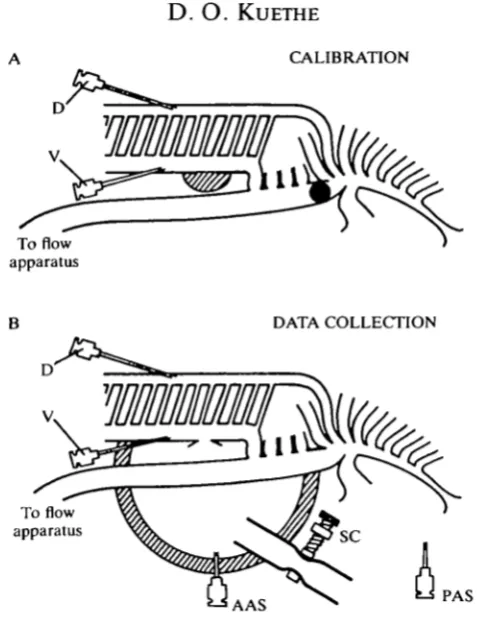
CALIBRATION
To flow apparatus
DATA COLLECTION
To flow apparatus
AAS PAS
Fig. 2. (A) During the calibration procedure, the cranial air sac openings were sealed shut (hatching) and the primary bronchus blocked between the orifices of the medioventral and mediodorsal bronchi with a balloon (shading). The measured flow of air that enters or leaves the primary bronchus must all pass through the paleopulmo. (B) During data collection, the air could flow through any bronchus. The flow through the paleopulmo was monitored by the pressure drop from D to V. The difference in air sac pressure was measured between PAS and AAS. A silicone rubber dome (hatching) isolated the cranial air sac orifices from the atmosphere while the caudal air sac orifices were open to the atmosphere. The pressure differences between PAS and AAS was a result of the resistance of a hose through the wall of the dome. The resistance of the hose was controlled with a screw clamp, SC.
the height difference between the openings of the cannulae. A constant air flow (between 1 and 30cm3s~1) was either pumped into the primary bronchus (inspiration) or withdrawn from it (expiration). The worst case error in measuring flow rates was less than 0-85cm3s~' (calculation appears in Kuethe, 1986).
Calibrating the paleopulmo for use as a flow meter
[image:4.451.107.346.50.359.2]1-5
1-25
10
2 0-75
0-5
0-25
0
-0 5 10 15 20 25 Flow rate (cm3s"')
[image:5.451.125.328.76.272.2]30
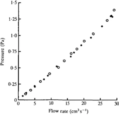
Fig. 3. A calibration graph. The vertical axis represents the absolute value of the pressure difference between D and V in Fig. 2A. The horizontal axis represents the absolute value of the flow rate into or out of the primary bronchus. Filled circles indicate data obtained when the flow was from medioventral to mediodorsal bronchi. Open circles indicate data obtained when the flow was from mediodorsal to medioventral bronchi. The 95 % confidence interval for the slope of a least-squares linear regression, assuming normally distributed data and a zero intercept, is 0-0450 ± 0-00055 Pa • s cm"3. N = 27.
The primary bronchus was blocked with a balloon between the orifices of the medioventral and mediodorsal bronchi (see Graf, Molony & Scheid, 1976), and the cranial air sac orifices were blocked with silicone rubber so that all the measured flow through the primary bronchus went through the paleopulmo (see Fig. 2A). The flow rate through the paleopulmo was plotted against the pressure drop from D to V to give the calibration graph (Fig. 3). For the purpose of this graph, so-called dynamic pressure effects which must be avoided when measuring resistances (as in Macklem, Bouverot & Scheid, 1979) were of no concern, as long as every time a given flow rate occurred in the paleopulmo the same pressure reading was obtained. The same pressure readings were obtained whether the caudal thoracic or abdominal sac orifice was blocked, or both were open.
Data collection from fixed lungs
pressure. Pressure differences were similar to those measured in live birds (Brackenbury, 1971; Kuethe, 1986).
Experimental data consisted of (1) the pressure drop from D to V (see Fig. 2B) obtained at different flow rates through the primary bronchus, which monitors flow through the paleopulmo, and (2) and pressure difference between AAS and PAS in Fig. 2B at different flow rates, which represents the difference in pressure between the cranial and caudal air sacs. During data collection, the primary bronchus was unobstructed (Fig. 2B).
The effects of three different air sac pressure differences on the flow in the paleopulmo were ascertained using three hose resistances. At each hose resistance flow data were collected for at least nine inspiratory and nine expiratory flow rates; air sac pressure data were collected for at least seven inspiratory and seven expiratory flow rates. The first resistance was such that the flow in the paleopulmo (indicated by the pressure drop from D to V in Fig. 2B) was the same during inhalation as during exhalation. The second resistance was higher than the first, giving more extreme air sac pressure differences. The third resistance was lower than the first, giving less extreme air sac pressure differences.
Since it was necessary to verify the position of the balloon (which was used only during calibration) by dissection, in practice the calibration was performed after the data collection.
Models
Clear plastic models were constructed using polymethylmethacrylate. Cut pieces were sandwiched between two solid sheets so that passages had rectangular cross-sections. The primary bronchus of the models was 9 mm x 10 mm. The models had one orifice for the cranial air sacs, one for the caudal air sacs and one for the primary bronchus. The parabronchi of the paleopulmo were represented by one passage connecting a single mediodorsal bronchus to a single medioventral bronchus.
A short hose was attached to the cranial sac orifice to create a pressure difference just as for the fixed lungs. The models were submerged in a basin of water and water was passed through their primary bronchi (steady flows of 0-1-15cm3s~J). The flow pattern was observed with Evans blue dye. Flow rates were estimated to within a factor of two.
For flow in the models to represent flow in the lungs, the Reynolds number (Lup//i) must be the same for both situations (see Batchelor, 1967). For both fixed lungs and models, the length (L) was taken as the square root of the cross-sectional area of the primary bronchus; the speed, u, was the flow rate through the primary bronchus divided by the cross-sectional area of the primary bronchus; p was the density; and pi was the viscosity of the fluid.
/ 1 0 0 % 50%
r
B i / • / / , i i / f 100% o • • •r
ci / t ,0 ° I o 1 /100% * s** O °
I i i
«o
5 10 15 2 0 2 5 3 0 0 5 10 15 2 0 2 5 3 0 Flow rate in primary bronchus (cm3s~')
Fig. 4. Experimental data. The top graphs, A-C, show the flow rate through the paleopulmo and its direction (i.e. the pressure drop from D to V divided by 0-0450Pa-scm"3, the slope of the calibration curve) versus the absolute value of the flow rate through the primary bronchus. A positive ordinate indicates that flow in the paleopulmo is in the correct direction. The straight lines indicate 100 % and 50 % of the flow rate in the primary bronchus going through the paleopulmo in the conect direction. The bottom graphs, D-F, display the absolute value of the pressure difference between PAS and AAS (Fig. 2B) versus the absolute value of the flow rate through the primary bronchus. The graphs are arranged in vertical pairs so that, for each pair, the conditions that produced the flow rate data in the top graph also produced the air sac pressure differences in the bottom graph. Filled circles represent data collected when air was pumped into the primary bronchus (inhalation); the cranial air sac pressures were higher than the caudal ones. Open circles represent data collected when air was withdrawn from the primary bronchus (exhalation); the caudal air sac pressures are higher than the cranial ones.
Results
Fixed lungs
bronchus. The air flow was unidirectional in the paleopulmo for all the flow rates and air sac pressure differences, except for flow rates below 8cm3s~1 in Fig. 4C.
To be consistent with the standard flow pattern, the flow rate through the paleopulmo during inhalation plus the flow rate through the paleopulmo during exhalation should equal the flow rate on the 100 % line (their average should fall on the 50 % line). In Fig. 4A-C, when the flow rate in the primary bronchus was below 15cm3s~x, not enough air was passing through the paleopulmo to be consistent with the standard flow pattern. As the flow rate increased towards 30cm3s~1, the average of the paleopulmo flow rates during inhalation and exhalation came closer to the 50 % line.
The difference in the air sac pressures affected the relative amount of flow in the paleopulmo during inhalation and exhalation. For the pressure differences in Fig. 4D, the flow rates were approximately equal. For pressures only 5 % higher (Fig. 4E rather than Fig. 4D), there was noticeably more air going through the paleopulmo during exhalation than during inhalation (Fig. 4B). When the pressure differences were lower (Fig. 4F rather than Fig. 4D), the flow through the paleopulmo was higher during inhalation than during exhalation (Fig. 4C).
Models
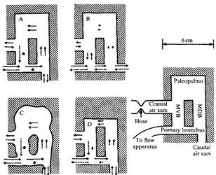
Row patterns obtained in four different models at a flow rate of 10cm3s"1 of water (Re = 930) are shown in Fig. 5. The model in Fig. 5A had the standard flow pattern. Modifications of this model either maintained the standard flow pattern (as in Fig. 5C) or failed to produce the standard flow pattern (as in Fig. 5B,D).
Changing the resistance of the hose to the cranial air sac orifice, and thus changing the pressure difference between the air sac orifices, had a similar effect as in the fixed lungs. When the hose was constricted relative to the position resulting in the flow pattern shown in Fig. 5A,C, more water passed through the paleopulmo during exhalation than during inhalation. When it was loosened, more water passed through the paleopulmo during inhalation than during exhalation.
As in the fixed lungs, the model shown in Fig. 5A became less effective at valving at slower flow rates. However, with the model, it was possible to see the alterations in flow pattern. At 7cm3s~1, water 'leaked' through the proximal medioventral bronchus during inhalation and through the primary bronchus during exhalation. This leakage became more severe as the flow rate was slowed, until at 1-5 cm3s"1, there was no flow through the paleopulmo during inhalation. At 0-5 cm3 s~l, water was observed passing through the paleopulmo in the wrong direction during inhalation.
Discussion
Fig. 5. Flow patterns in models. Arrows indicate the flow of water in plastic models at a rate of 10cm3s~1 when the resistance of the hose on the cranial air sac orifice was such that the paleopulmo received the same flow rate during inhalation as during exhalation. Solid arrows represent inhalation, dashed arrows represent exhalation. The length of the arrows indicates the relative amount of water flowing through a given passage. Stars indicate no net flow (at this flow rate, stationary vortices). Model A has the standard flow pattern. Model B has the caudal air sac orifice in line with the primary bronchus. The small amount of flow in the paleopulmo is bidirectional. Model C is a wavy-edged version of A and has the standard flow pattern. Model D has a paleopulmo of higher resistance. While the flow is unidirectional, the flow in the paleopulmo is reduced as some of the water takes alternative paths.
rate during inhalation, and the same as the primary bronchus flow rate minus the paleopulmo flow rate during exhalation. Even though the complete flow pattern is not known, the data (Fig. 4) support Brackenbury's (1971, 1972a,b, 1979) claim that the flow pattern is dependent on the pressure difference between the cranial and caudal air sacs. The relative flow in the paleopulmo during inhalation compared with exhalation appears to be controlled by this pressure difference.
thoracic and clavicular sacs of an anaesthetized goose, 2-4—4-7 kg in mass; Kuethe (1986), 0-200 Pa between caudal thoracic and clavicular sacs, and 0-100 Pa between the abdominal and clavicular or between the cranial thoracic and caudal thoracic sacs of conscious ducks, 2-7-2-9 kg in mass], one cannot be sure that the pressure differences reported for the fixed lung would occur in a live duck, even if at a given instant the duck had the same flow rates and flow pattern as the fixed lung.
The valving demonstrated by the crude models bears a remarkable functional similarity to the unidirectional flow in birds lungs, although the models did not valve as effectively as the fixed lungs. Despite the similarities of the flow patterns, there are obvious differences in geometry and compliance between the models and bird lungs, and obvious differences between the steady flow employed in this study and the tidal flow in birds. Given these differences, it must remain unclear whether the models, or even the fixed lungs, operate with the same mechanisms as live birds.
However, the models suggest that the fluid in the caudal portion of the primary bronchus needs to be guided by some structural feature towards the orifices of the mediodorsal bronchi, as suggested by Hazelhoff (1943). However, Hazelhoffs dam per se may not be responsible for the valving. When it was excised from one of the fixed duck lungs, the lung was still capable of valving. The only difference in its behaviour was that for the same relative flow rates in the paleopulmo, a different set of air sac pressure differences was required (data appear in Kuethe, 1986). Apparently, when Hazelhoffs dam is removed from a duck lung, the air is still guided towards the mediodorsal bronchi.
The models and fixed lungs suggest how fluid momentum and air sac pressures might interact to produce the flow pattern in the absence of Hazelhoffs dam. During inhalation, air must flow around a bend to enter the caudal air sacs, whether Hazelhoffs dam is present or not. In general, the pressure on the outside of a bend is higher than the pressure on the inside. The pressure difference represents the force which changes the fluid's momentum. The openings of the mediodorsal bronchi are on the outside of the bend. The pressure difference around this bend, together with other manifestations of the air's inertia, might explain the valving. During inhalation the pressure difference created by the bend opposes the pressure difference between the air sacs. The bend pressure difference is high enough to create a net force driving fluid from the mediodorsal bronchial orifices towards the medioventral bronchi. The inertia of the air in the primary bronchus tends to prevent it from changing momentum and entering the medioventral bronchi directly. During exhalation the pressure difference between the air sacs favours the flow from mediodorsal bronchi to medioventral bronchi. The inertia of the air from the caudal sacs tends to prevent it from changing momentum and flowing directly into the primary bronchus.
I thank Knut Schmidt-Nielsen for philosophical advice, Steven Vogel for helping me transform my dissertation into a paper and Knut Schmidt-Nielsen, Sharon Gowan and Ira Warrenfelt for help with earlier versions. This work was supported by NIH grant no. HL02228.
References
BATCHELOR, G. K. (1967). An Introduction to Fluid Dynamics. Cambridge: Cambridge University Press. 615pp.
BETHE, A. (1925). AUgemeines und Vergleichendes. In Handbuch der Normalen und
Pathologischen Physiologie, vol. 2, Atmung (ed. A. Bethe, G. V. Bergmann, G. Embden &
A. Ellinger), pp. 1-36. Berlin: J. Springer. 552pp.
BOUVEROT, P. & DEJOURS, P. (1971). Pathway of respired gas in the air sacs-lung apparatus of fowl and ducks. Respir. Physiol. 13, 330-342.
BRACKENBURY, J. H. (1971). Airflow dynamics in the avian lung as determined by direct and indirect methods. Respir. Physiol. 13, 319-329.
BRACKENBURY, J. H. (1972a). Lung-air-sac anatomy and respiratory pressures in the bird. /. exp. Biol. 57, 543-550.
BRACKENBURY, J. H. (19726). Physical determinants of air flow patterns within the avian lung.
Respir. Physiol. 15, 384-397.
BRACKENBURY, J. H. (1979). Corrections to the Hazelhoff model of airflow in the avian lung.
Respir. Physiol. 36, 143-154.
BRANDES, G. (1924). Beobachtungen und Reflexionen uber die Atmung der Vogel. Pflugers
Arch. ges. Physiol. 203, 492-511.
BRETZ, W. L. & SCHMIDT-NIELSEN, K. (1971). Bird respiration: flow patterns in the duck lung.
J. exp. Biol. 54, 103-118.
BRETZ, W. L. & SCHMIDT-NIELSEN, K. (1972). The movement of gas in the respiratory system of the duck. J. exp. Biol. 56, 57-65.
DOTTERWEICH, H. (1936). Die Atmung der Vogel. Z. vergl. Physiol. 23, 744-770.
DUNCKER, H.-R. (1971). The lung air sac system of birds. Ergebn. Anat. EntwGesch. 45, no. 6, 1-171.
GRAF, W., MOLONY, V. & SCHEID, P. (1976). A technique for study of lung function in birds by blocking the primary bronchus. Respir. Physiol. 26, 325-331.
HAZELHOFF, E. H. (1943). Bouw en Functie van de Vogellong. Versl. gewone Vergad. Afd.
Naturk. K. ned. Akad. Wet. 52, 391-400. English translation: (1951). Structure and function
of the lung of birds. Poult. Sci. 30, 3-10.
KUETHE, D. O. (1986). Fluid mechanical valving of air flow in bird lungs. Ph.D. dissertation. Duke University. Durham, NC. 72 pp. University Microfilms International, order no. DA8629322.
LASIEWSKI, R. C. & CALDER, W. A. (1971). A preliminary allometric analysis of respiratory variables in resting birds. Respir. Physiol. 11, 152-166.
MACKLEM, P. T., BOUVEROT, P. & SCHEID, P. (1979). Measurement of the distensibility of the parabronchi in duck lungs. Respir. Physiol. 38, 23-35.
POWELL, F. L., GEISER, J., GRATZ, R. K. & SCHEID, P. (1981). Airflow in the avian respiratory tract: variations of O2 and CO2 concentrations in the bronchi of the duck. Respir. Physiol. 44,
195-213.
SCHEID, P. (1979). Mechanisms of gas exchange in bird lungs. Rev. Physiol. Biochem. Pharmac. 86, 138-186.
SCHEID, P. & PIIPER, J. (1971). Direct measurement of the pathway of respired gas in duck lungs.
Respir. Physiol. 11, 308-314.
SCHMIDT-NIELSEN, K., KANWISHER, J., LASIEWSKI, R. C , COHN, J. E. & BRETZ, W. E. (1969). Temperature regulation and respiration in the ostrich. Condor 71, 341-352.
VOGEL, S. (1981). Life in Moving Fluids: The Physical Biology of Flow. Princeton: Princeton University Press. 352 pp.