VENTILATORY ACTION OF THE HYPAXIAL MUSCLES OF THE LIZARD IGUANA IGUANA: A FUNCTION OF SLOW
MUSCLE
BY DAVID R. CARRIER*
Department of Biology, The University of Michigan, Ann Arbor, MI 48109, USA
Accepted 30 January 1989
Summary
Patterns of muscle activity during lung ventilation, patterns of innervation and some contractile properties were measured in the hypaxial muscles of green iguanas. Electromyography shows that only four hypaxial muscles are involved in breathing. Expiration is produced by two deep hypaxial muscles, the transversalis and the retrahentes costarum. Inspiration is produced by the external and internal intercostal muscles. Although the two intercostal muscles are the main agonists of inspiration, neither is involved in expiration. This conflicts with the widely held notion that the different fibre orientations of the two intercostal muscles determine their ventilatory action.
Several observations indicate that ventilation is produced by slow (i.e. non-twitch) fibres of these four muscles. First, electromyographic (EMG) activity recorded from these muscles during ventilation has an unusually low range of frequencies (<100Hz). Such low-frequency signals have been suggested to be characteristic of muscle fibres that do not propagate action potentials (i.e. slow fibres). Second, during inspiration, EMG activity is restricted to the medial sides of the two intercostal muscles. Muscle fibres from this region have multiple motor endplates and exhibit tonic contraction when immersed in saline solutions of high potassium content. Like the intercostals, the transversalis and retrahentes costarum muscles also contain fibres with multiple motor endplates. Thus, although breathing is a phasic activity, it is produced by tonic (i.e. slow) muscle fibres. The intercostal muscles are also involved in postural and locomotor movements of the trunk. However, such movements employ twitch as well as slow fibres of the intercostal muscles.
Introduction
Recent observations suggest that lizards are unable to run and breathe at the same time (Carrier, 1987a). In the four species that have been studied {Iguana
* Present address: Department of Zoology, 4079 LSB, University of California, Berkeley, CA
94720, USA.
1981; Butler, 1982; Hornicke etal. 1983; Bramble & Carrier, 1983; Baudinette et al. 1987; Jenkins etal. 1988; Bramble, 1989). In birds and mammals, the locomotor movements may actually facilitate breathing. Thus, diminished breathing in running lizards is an unexpected finding.
A physical conflict between the locomotor and ventilatory systems may be responsible for the reduced breathing in running lizards (Carrier, 1987a,b). The actions of the ventilatory muscles might differ, in an opposing fashion, from the actions of the locomotor muscles. If this were true, locomotion and ventilation would place conflicting demands on the thorax, limiting the capacity for venti-lation whenever the animals walked or ran.
The possibility of a physical constraint on simultaneous running and breathing cannot be addressed directly because very little is known of how lizards ventilate their lungs. Lizards are known to be aspiration breathers (Milsom, 1984). That is, they draw air into their lungs by actively deforming the walls of the thoracic cavity to create a subatmospheric pressure. However, the axial muscles responsible for these ventilatory movements have not been identified in lizards. In contrast, the muscles responsible for ventilation have been studied extensively in mammals (De Troyer & Loring, 1986) and have received limited attention in turtles (Gans & Hughes, 1967; Gaunt & Gans, 1969), crocodilians (Naifeh et al. 1970; Gans & Clark, 1976) and birds (Kadono et al. 1963; Fedde et al. 1964a,b). Hence, Lepidosauria (i.e. squamates and Sphenodon; Gauthier et al. 1988) is the one remaining group of amniotic tetrapods for which the basic mechanism of lung ventilation has not been determined.
This study used electromyography to identify those muscles responsible for ventilation in the lizard, Iguana iguana. The aim was to increase our understand-ing of how lizards breathe and to provide a basis for further investigations of locomotor-ventilatory coupling in lizards. During the course of the investigation, questions arose concerning the contractile physiology of the muscles responsible for ventilation. Consequently, in addition to characterizing the activity patterns of the ventilatory muscles, data are presented that are relevant to the function of slow muscle.
Materials and methods Specimens
(Iguana iguana, 600-1735 g) that were obtained from local animal suppliers and by courtesy of Dr Dagmar Werner of the Smithsonian Tropical Research Institute. They were housed in large cages with a photothermal gradient on a 12h:12h light: dark photoperiod, and were fed a diet of Romaine lettuce and Iguana Chow (Zeigler Bros, Inc.).
For experiments requiring surgery, the lizards were anaesthetized by an initial intramuscular injection of 140 mg of Ketamine per kilogram body mass, followed by one-third the initial dosage, as needed. Throughout the various experiments body temperature of the lizards was monitored with a cloacal thermometer and maintained at 30-35 °C.
Pressure and air flow recordings
Thoracic pressure was measured in anaesthetized animals, with a Statham PM5 pressure transducer. To accomplish this, an incision was made in the skin of the throat and the muscles overlying the trachea were reflected. The trachea was then cannulated with a small-diameter tube (<one-third the diameter of the trachea) the end of which was advanced into the lung.
Inspiratory air flow was monitored in both anaesthetized and fully active animals, with a thermistor flow meter (Thomas, 1981). This provided simultaneous and independent measurements of the direction of air flow and tidal volume. The probe of the flow meter was attached over one of the external nares and did not appear to annoy the animals in any way (Carrier, 1987a).
Changes of thoracic pressure resulting from contraction of individual muscles were measured in four anaesthetized animals. In these animals, the skin on one side of the trunk was reflected. Starting with the most superficial layers, successive muscles were stimulated directly (1-ms square wave pulses between 0-2 and 1-0 V, just above threshold level) with pin electrodes held on the surface of the muscle. Low voltages were used in an effort to restrict contraction to those muscle fibres closest to the stimulation site. Although the observed changes in thoracic pressure were quite small, the direction of pressure change could be recorded. A response profile for each muscle was obtained by moving the electrode from site to site.
Muscle contractile properties and histochemistry
Slow muscle can be differentiated from twitch muscle by its response to immersion in depolarizing solutions and by its pattern of innervation, although there are exceptions (Lannergen, 1979; Johnston, 1985). In depolarizing solutions, twitch muscle either does not contract or contracts and then relaxes quickly. In contrast, sustained depolarization of slow muscle results in a prolonged contrac-tion, lasting many minutes (Morgan & Proske, 1984). Differences in innervation are equally distinct. Twitch muscle fibres generally have a single, centrally located endplate, whereas slow fibres have multiple, small endplates spaced at 0- to 2-mm intervals along the cell (Morgan & Proske, 1984).
original voltage and then recorded on a Gould Brush 481 recorder and stored on magnetic tape in a Honeywell 5600 medium band-path tape recorder.
Contractile properties were measured after 5-10 min of thermal equilibration. The muscles were first stimulated through two aluminium plate electrodes using a Grass S44 stimulator. Twitch contractions were elicited with single pulses of 1-ms duration and 40-80 V. The capacity of the intercostals to contract tonically was then measured by depolarizing the cells with high extracellular potassium. The Ringer's bath was drained and quickly replaced by Ringer's solution with 155 mmol I"1 KC1 and no NaCl. Tensions are reported as g cm"2 of cross-sectional area. Cross-sectional area was estimated by dividing muscle mass by muscle length.
The distribution of motor endplates in the hypaxial muscles was determined by staining whole muscles with the cholinesterase stain of Karnovsky & Roots (1964). Freshly dissected whole muscles were pinned at resting length in an incubation chamber. The muscles were immersed in the acetylthiocholine iodide medium for 2 - 4 h and then placed in 1 % ammonium sulphate for 1-2min.
Electromyography
The axial muscles responsible for ventilation were determined electromyogra-phically. The hypaxial muscles are thin sheets and in some cases very difficult to reach surgically. Consequently, a variety of barbed bipolar and patch electrodes (Loeb & Gans, 1986) were employed. In one set of experiments the animals were anaesthetized, the skin and external oblique were reflected on one side of the body and up to 34 bipolar electrodes (75 nm diameter stainless-steel wire, Teflon coated) were inserted in the intercostal and underlying muscle layers. Bared electrode tips 0-5 mm long were placed approximately 1-2mm apart within the muscle. These electrodes were monitored four at a time, along with thoracic pressure and air flow in the anaesthetized animal.
Electrode wires passed percutaneously to exit points along the midline of the back. At the exit point the wires were glued to the skin and soldered to gold connector pins. The animals were allowed to recover from the anaesthesia, and muscle activity and air flow were monitored during both quiet breathing and heavy breathing induced by vigorous locomotor activity.
The EMG signals were passed to Tektronix FM 122 preamplifiers, amplified 1000 times and filtered below 8 Hz and above 10 kHz. Signals were then passed to Honeywell 117 d.c. amplifiers, and simultaneously stored on a Honeywell 5600 tape recorder and printed out on a Gould chart recorder. To provide a comparison of the amplitude of the EMG activity and tidal ventilation, some recordings were digitized by an IBM AT microcomputer through a Keithley 570 analog-to-digital converter. Data were collected at 2500 Hz, and analysed with a program that recorded the number and amplitude of individual spikes (Beach et al. 1982). Multiplication of the number and average amplitude of the spikes occurring in a given interval provided a measure of the EMG activity.
Fast Fourier transformations (FFT) were performed on selected electromyo-grams to determine the range of dominant frequencies. Data were digitized at 2500 Hz, maintaining a frequency sensitivity of 1250 Hz based on Nyquist sampling criteria. The Cooley & Tukey (1965) algorithm was used in a program that calculates an FFT using a sample size of any power of two.
Results Anatomy
The axial musculoskeletal system of Iguana iguana is described elsewhere (Carrier, 1988). Only those muscles and bones that play a role in lung ventilation are noted here.
Thoracic ribs
The anterior 10 ribs enclose the thoracic cavity (Fig. 1). The first three thoracic ribs lack costal cartilages and do not articulate with the sternum. The next seven ribs (sternal ribs) are composed of a bony element that articulates with the vertebrae and a costal cartilage that articulates with the sternum. The vertebral segments extend laterally and ventrally to the mid-body wall and have a circular cross-section. In contrast, the costal cartilages are composed of fibrocartilage that is readily deformable.
External intercostal muscle
Fig. 1. Diagrams of the axial musculoskeletal system of Iguana iguana. (A) The epaxial and external oblique muscles have been removed to illustrate the underlying hypaxials: external intercostal (ei), quadratus lumborum (ql), external intercostal abdominus {eia), rectus abdominus (ra), internal oblique (io) and internal intercostal («). (B) The intercostal and internal oblique muscles have been removed to illustrate the deep muscle layers: retrahentes costarum (re) and transversalis (/).
Internal intercostal muscle
The internal intercostal muscle lies medial to the external intercostal and occupies a more ventral position in the thorax (Fig. 1). Fibres run from rib to rib, directed anteriorly and ventrally. The muscle extends dorsoventrally from the mid-portion of the vertebral ribs to the ventral aspect of the costal cartilages. Dorsal to the bend in the costal cartilages the fibres lie at an angle of about 60° to the horizontal. Fibres below the bend have a more shallow orientation of about 20° to the horizontal. Posterior to the last true rib a slip of this muscle extends dorsally to attach to the lumbodorsal fascia.
Transversalis muscle
The transversalis is the innermost muscle of the lateral body wall (Fig. 1). It is quite thin, but continuous from girdle to girdle. Dorsally, it attaches at the same sites as the internal oblique: lumbodorsal fascia and the mid-portion of the vertebral ribs. The fibres run ventrally and slightly caudally to insert on the dorsal aspect of the rectus abdominus and in the thoracic region on the ventral-most portions of the costal cartilages.
Retrahentes costarum muscle
to the intercostal musculature (Fig. 1). Fibres attach to the ventral aspect of the centra of the vertebrae. They course anteriorly and ventrally to insert on the mid-portion of the vertebral element of each sternal rib.
Thoracic pressure
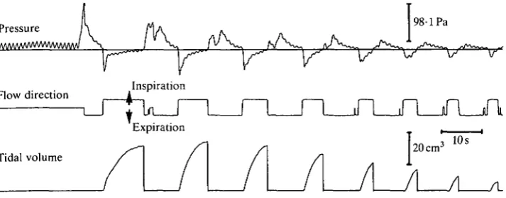
Green iguanas are aspiration breathers. They pump air out of their lungs by increasing thoracic pressure above atmospheric (Fig. 2). They then fill their lungs by the generation of a subatmospheric thoracic pressure. Pressure changes during regular breathing were of quite low magnitude, generally less than 98 Pa (1 cmH2O). However, thoracic pressures could be much higher (621 ± 37-3 Pa in a 1060 g individual) when the lungs were filled during routine periods of apnoea.
The ribs could be seen to swing posteriorly during expiration and anteriorly during inspiration. Manual simulation of these rib displacements produced changes in thoracic pressure in anaesthetized animals or fresh cadavers. Anterior rotation of the ribs decreased thoracic pressure, whereas posterior rotation increased pressure. This held for each of the seven sternal ribs.
In anaesthetized iguanas, direct unilateral stimulation of small portions of individual axial muscles produced changes in thoracic pressure that were muscle-and/or site-specific. The direction and amplitude of these changes of pressure were largely independent of the phase of ventilation. Most thoracic muscles increased thoracic pressure when they were stimulated individually. Stimulation of those muscles that lie lateral to the ribs (i.e. external oblique and iliocostalis) as well as those medial to the ribs (i.e. internal oblique, transversalis, retrahentes costarum) increased thoracic pressure. The external oblique, iliocostalis and retrahentes costarum appeared to effect this increase in thoracic pressure by
Pressure
Ay
98-1 PaFlow direction
\r v v v
[image:7.451.48.405.411.550.2]Inspiration
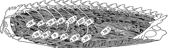
Fig. 3. Changes in thoracic pressure that results from stimulation of small segments of the intercostal musculature in an individual of Iguana iguana. The numbers represent the average change in thoracic pressure (Pa) that resulted when each site was electrically stimulated with pin electrodes. The upper sites stimulated the external intercostal, and the lower sites stimulated the internal intercostal.
rotating the vertebral portion of the ribs caudally. However, stimulation of the thoracic portions of the internal oblique and transversalis complex could be seen to pull the costal cartilages medially, actually deforming these flexible skeletal elements. In these five muscles stimulation produced increased thoracic pressure, no matter which portion of the muscle was activated.
In contrast, stimulation of the intercostal muscles produced either increased or decreased thoracic pressure, depending on the site of stimulation. For any given intercostal segment, separate stimulation of the external or internal intercostal muscles usually had the same effect on thoracic pressure, and this response was largely independent of the phase of ventilation. Stimulation of anterior segments decreased thoracic pressure, whereas stimulation of posterior segments increased thoracic pressure (Fig. 3). However, there was a significant amount of variation among individuals. In some, decreased thoracic pressure resulted from activation of only the anterior three intercostal segments, whereas in others activation of the anterior five segments decreased thoracic pressure.
Those appendicular muscles which originate on the ribs also influenced thoracic pressure. Stimulation of the pectoralis and latissimus dorsi increased thoracic pressure, whereas stimulation of the serratus ventralis decreased it.
Distribution of multiply innervated fibres
configuration, generally associated with slow fibres. Interspersed among these multiply innervated fibres were a few singly innervated fibres. In summary, both the external and the internal intercostal muscles were composed of a lateral portion of singly innervated fibres and a medial portion of predominantly multiply innervated fibres.
Three other hypaxial muscles, the external oblique superficialis, transversalis and retrahentes costarum, also contained multiply innervated fibres. As in the intercostal muscles, these three muscles were stratified, with the multiply innervated fibres located only on the medial side. The internal oblique and the rectus abdominus contained only singly innervated fibres. The epaxial muscles and the quadratus lumborum also appeared to be composed of singly innervated fibres, but only superficial surfaces of these muscles were examined.
[image:9.451.44.363.257.567.2]I ,'
Fig. 5. Comparison of the distribution of motor endplates on the lateral and medial surfaces of the intercostal muscles of Iguana iguana. (A) Lateral view of ribs and intercostal muscles, showing the centrally located sites of innervation. Each muscle fibre has a single motor endplate. The anterior end of the body wall is on the right. (B) Medial view of ribs and intercostal muscles showing the multiply innervated fibres. The muscles shown are the external intercostal (ei), iliocostalis (;'), internal intercostal (») and retrahentes costarum (re).
Response of the intercostal muscles to depolarization
Table 1. Means and standard errors of the isometric contractile properties of the intercostal muscles between sternal ribs 2 and 3 of Iguana iguana
Twitch contraction time (s)
Tonic contraction time (s)
Twitch force (gem"2) Tonic force
(gem-2)
Internal intercostal (ventral)
0-051 ± 0-015 (3) 170 ±53-5 (3) 111 ±48 (3) 67 ±24 (3) Internal intercostal (lateral)
0-038 ± 0007 (5) 112 ±31-3
(5) 101 ± 39
(5) 198 ± 57
(5)
External intercostal
0-032 ± 0-045 (2) 58 ±0-5
(2) 228 ± 99
(3) 148 ± 58
(3)
Twitch contractions were elicited by electrical stimulation. Tonic contractions were elicited by immersion in a bath of Ringer's solution that depolarized the tissue.
Contraction time represents the time from stimulation to peak force. Sample size is indicated in parentheses in each case.
taken from the lateral body wall. This is consistent with the distribution of multiply innervated fibres in the internal intercostal (Fig. 5).
Muscle activity during ventilation
Preliminary attempts to record muscle activity during lung ventilation were largely unsuccessful. Occasionally, low-amplitude activity was recorded during expiration from one or more of the deep hypaxial muscles, but none of the hypaxials displayed any activity during inspiration. This posed a problem because the ribs move anteriorly during inspiration. Analysis of fibre orientations of the various hypaxial muscles indicated that the only muscles mechanically capable of moving the ribs anteriorly were the intercostals. However, repeated attempts to record electrical activity from the intercostals suggested that they did not play a role in any phase of ventilation.
inspiration, electrical activity in the anterior intercostals was bilaterally symmetri-cal (Fig. 6C), and positively correlated with breath amplitude (Fig. 7). Thus, deeper inspiration required greater muscular activity from the intercostal muscles. In contrast, neither intercostal muscle was active during expiration. Expiration was associated with activity in the retrahentes costarum (Fig. 6A) and in the internal oblique-transversalis complex (Fig. 6B). Activity of the inspiratory and expiratory muscles showed little or no temporal overlap.
Most of the axial muscles were not involved in lung ventilation. The epaxial muscles and the greater portion of the hypaxial musculature displayed no electromyographic activity, even during the most vigorous ventilation. In particu-lar, the rectus abdominus, external oblique, quadratus lumborum and the major portion of both intercostal muscles were silent during ventilatory activity. Although the multiply innervated fibres of the medial surfaces of the anterior intercostal segments were active during inspiration, analogous fibres in the more posterior rib segments (5-9) were not (Fig. 8).
The initiation and cessation of muscular activity was closely correlated with the phases of ventilation (Fig. 9). Activity in the two intercostal muscles began roughly 100-230 ms before the start of inspiratory air flow, ending some 120-250 ms before inspiration stopped. The two expiratory muscles became active 40-150 ms before expiratory air flow began, activity ceasing 50-110 ms before the end of expiration.
The relatively long delays between muscle activity and the initiation and cessation of air flow appeared to result from a slow development of muscular force. The long delays were not a result of the response time of the recording equipment. The response time of the flow direction circuitry was of the order of a few milliseconds (compare the change in flow direction to the change of sign of thoracic pressure in Fig. 2). Furthermore, during rapid breathing (e.g. Fig. 6), the glottis was held open and did not affect air flow. This suggests a relatively long interval between activation of the ventilatory muscles and the generation of forces sufficient to affect thoracic pressure.
whereas the maximum amplitude of the postural signal was roughly 1-2 mV. Fourier transformations revealed differences in the frequencies of the two signals. The dominant frequencies during ventilation were below 100 Hz, with greatest activity around 30 Hz. These low frequencies were also produced during postural
A a
U
-*#-B ii
C eir
eil
«-i"jrr I-IT^YT
I s
uuw
JU
10 cm3
C 6
10 20 30 EMG activity
[image:14.451.155.329.73.296.2]40 50
Fig. 7. Plot of the total electromyographic activity (i.e. number of spikes x average amplitude x 100) from a patch electrode positioned on the medial surface of the internal intercostal versus tidal volume for Iguana iguana.
[image:14.451.97.385.368.554.2]ei\ t re
Flow
"1
in
/
°"'
1 1 L
Is
Fig. 9. Ventilatory activity patterns of the hypaxial muscles that effect lung ventilation in Iguana iguana. Bars represent the mean and standard deviation of the time of onset and cut-off of muscular activity relative to the beginning of inspiration and expiration. For each muscle, data were collected from 30 breaths of a single specimen. Activity in the internal and external intercostals (ii, ei, respectively) is associated with inspiration, whereas activity in the transversalis (/) and retrahentes costarum (re) is associated with expiration.
or locomotor movements. However, the dominant frequencies during postural adjustments were much higher, ranging from 100 to 700 Hz.
Discussion
The basic pattern of ventilatory air flow in lizards has been described for a number of species (Gans & Clark, 1978; Cragg, 1978; Milsom, 1984). Green iguanas conform to these published accounts. In resting lizards, ventilation begins with expiration, which is followed immediately by inspiration. Usually a single ventilatory cycle, or a few cycles, will be followed by a prolonged period of breath-holding. This periodic pattern of breathing has been shown to minimize the work of breathing in the Tokay gecko (Milsom & Vitalis, 1984; Milsom, 1984). When metabolic demand is increased, as it is after a bout of exercise, periods of breath-holding are less common and may be absent (Milsom, 1984; Carrier, 1987a).
Inspiration
100 ms
Inspiration
^ W w ^
Posture
400 600 Frequency (Hz)
1000
Fig. 10. Comparison of amplitude and dominant frequencies of electromyographic signals recorded during ventilatory and postural movements in Iguana iguana. The top two traces are digitized EMG signals from the internal intercostal muscle between sternal ribs 1 and 2, recorded during inspiration and a brief adjustment of body posture. The two signals were recorded from a single electrode within a few seconds of each other. The two graphs plot the Fourier transformations of the above signals, and show the different range of EMG frequencies that characterize these movements.
The electromyographic signals produced during inspiration are lower in amplitude and frequency than the signals produced during postural or locomotor movements of the trunk. The low-amplitude, low-frequency signals resemble those produced by the slow myotomal muscle of elasmobranch fishes (Bone, 1966) and the slow opercularis muscle of frogs (Hetherington & Lombard, 1983). Such signals have been suggested to be characteristic of the junctional potentials produced by slow (non-twitching) fibres (Roberts, 1969; Fetcho, 1987).
and the snake Thamnophis sp. (Ridge, 1971). In these species, intracellular recordings show that the amplitudes of the junctional potentials of slow fibres are roughly 6-10 times lower than the amplitudes of action potentials (10-20 mV compared to 120mV). Furthermore, the time of half-decay of potentials is 10-20 times longer in slow fibres than in fibres that propagate action potentials (40-70 ms compared to 3-10ms). These differences seem to provide an explanation for the two types of electrical signals produced by the intercostal muscles. The electro-myographic signals produced during inspiration have amplitudes and frequencies that are consistent with intracellular recordings of junctional potentials of slow fibres. In constrast, signals produced during postural movements appear to be recordings of action potentials.
Several additional observations suggest that inspiration is produced by slow fibres rather than twitch fibres. First, electrical activity can only be recorded from the multiply innervated side (i.e. medial) of either intercostal muscle. Second, these multiply innervated fibres produce a tonic contraction when immersed in a depolarizing solution. Finally, the latency period between activation of the muscles and detectable changes in thoracic pressure is longer than would be expected for twitch fibres. Thus, although inspiration is a phasic activity, it is produced by tonic (i.e. slow) muscle fibres.
Expiration
During expiration, electromyographic activity was recorded from only the retrahentes costarum and the internal oblique-transversalis complex. This activity was consistently of low frequency and amplitude. The low-frequency (<50Hz) signals indicate that slow, and not twitch, muscle fibres are responsible for expiration. The internal oblique, however, is composed entirely of twitch fibres. Therefore, the EMG signals recorded from the internal oblique-transversalis complex must have been produced by the transversalis.
Two additional observations indicate that the ventilatory action of the retra-hentes costarum and transversalis is to decrease thoracic volume. First, their fibre orientation is such that contraction will act to decrease thoracic volume. The retrahentes costarum decreases thoracic volume by pulling the vertebral ribs posteriorly. In contrast, because the ventral body wall is more compliant than the dorsal body wall, contraction of the transversalis pulls the deformable costal cartilages medially. Second, direct electrical stimulation of either of these muscles produces an increase in thoracic pressure. Thus, in green iguanas, the transversalis and retrahentes costarum are the main agonists of expiration.
move the ribs anteriorly will be greater than those tending to rotate the ribs posteriorly. In contrast, the caudodorsal orientation of the internal intercostal results in larger moments tending to rotate the ribs posteriorly than those tending to rotate them anteriorly. Thus, Hamberger suggested that the external intercos-tals should increase thoracic volume and be inspiratory in function, whereas the internal intercostals should deflate the thorax. Experimental support for this hypothesis has been provided by work on mammals (Bronk & Ferguson, 1935; Taylor, 1960).
However, there is increasing evidence that the two intercostal muscles do not display the antagonistic pattern predicted by Hamberger. In mammals (Gesell, 1936) and birds (Kadono et al. 1963; Fedde et al. 1964a), anterior portions of external and internal intercostals are active during inspiration, whereas posterior portions of the two muscles are active during expiration. In green iguanas, this study found that anterior portions of both intercostal muscles are active during inspiration, but neither is involved in expiration. During ventilation in all these animals, the external and internal intercostal muscles act as synergists to pump air in and out of the lungs. Thus, in contrast to Hamberger's model, the different fibre orientations of the two intercostal muscles seem of little significance to ventilatory function.
Recent experiments in dogs suggest that the ventilatory action of the intercostal muscles is determined by the relative resistance of the ribs to anterior versus posterior displacement (De Troyer et al. 1985). In dogs, separate stimulation of the external and internal intercostal muscles causes similar displacement of the ribs. When either of these two muscles is stimulated at low thoracic volume there is a net anterior displacement of the ribs. In contrast, at high thoracic volume, stimulation causes a net posterior displacement of the ribs. De Troyer et al. (1985) concluded that the relative anterioposterior stiffness of the body wall is more important to intercostal action than their respective fibre orientations.
The present study shows that, in green iguanas, stimulation of either external or internal intercostal muscles from anterior segments decreases thoracic pressure, whereas stimulation of either muscle from posterior segments increases thoracic pressure. These observations are consistent with the findings in dogs, and are inconsistent with the older literature.
Action of axial slow fibres
broad categories: twitch and slow (Hess, 1970; Morgan & Proske, 1984). A certain level of confusion exists in the literature because twitch fibres are also divided into fast- and slow-contracting categories. In addition, there are also intermediate fibres which display characteristics of both slow and twitch fibres (Lannergren, 1979; Morgan & Proske, 1984; Johnston, 1985). However, slow fibres are a distinct class that differ from twitch fibres in internal structure, pattern of innervation and contractile physiology. The physiology of slow fibres differs from that of twitch fibres in three principle ways (Morgan & Proske, 1984). First, slow fibres are unable to propagate an action potential. Thus, they cannot be stimulated by a single pulse and do not twitch. Second, slow fibres have a much slower isotonic shortening speed. Third, when immersed in depolarizing solutions, slow fibres contract tonically, maintaining tension for prolonged periods (i.e. minutes). Twitch fibres, in contrast, contract only briefly or do not contract at all. These differences in contractile physiology suggest that slow and twitch muscle might serve separate functions.
Unfortunately, the function of slow muscle has received only limited attention. Studies of contractile and energetic properties led early workers to suggest that slow muscle is ideally suited to the maintenance of body posture (Fulton, 1926). Slow muscle shortens very slowly, can maintain tension for prolonged periods with only minimal energy expenditure (Kuffler & Williams, 1953) and is reported to be grossly inefficient at performing isotonic work (Goldspink et al. 1970). These characteristics suggest that slow muscle is ill-suited to the production of active movements. However, in several cases, slow muscle has been shown to contract phasically to produce isotonic work. Among these are slow swimming in fish (Bone, 1978) and aerial hearing or possibly buccal pumping in frogs (Hetherington & Lombard, 1983). The present study shows that green iguanas use slow fibres of the hypaxial muscles to pump air in and out of their lungs.
The contractile physiology of slow muscle may explain why it is used to ventilate the lungs of lizards. First, in tetrapods, breathing is a relatively slow process. Only during thermoregulatory panting (Richards, 1970) or during locomotion in birds and mammals (Butler, 1982; Bramble & Carrier, 1983) do ventilatory frequencies exceed 2Hz in animals heavier than 100g (Lasiewski & Calder, 1971; Stahl, 1967). Ectotherms breathe particularly slowly. The highest ventilatory frequencies of lizards occur immediately following vigorous activity and usually do not exceed 0-5 Hz (Carrier, 1987a). These low frequencies indicate that ventilation could be accomplished by slow fibres. Second, contrary to the observations of Goldspink et al. (1970), there is evidence that slow-contracting muscle can be very efficient at doing work (Woledge, 1968; Morgan & Proske, 1984). Finally, ventilatory muscles must be nonfatigable. Although there appear to be no studies of the relative fatigability of slow muscle, the low metabolic rate of slow muscle (Morgan & Proske, 1984) suggests that it should be highly resistant to fatigue. Thus, slow muscle appears to be well suited to the requirements of aspiration breathing in Jizards.
intercostal muscles show that during ventilation the intercostal muscles produce only low-frequency signals. As suggested above, such low-frequency signals appear to be characteristic of the junctional potentials produced by slow fibres. In contrast, postural changes (i.e. locomotor movements) are associated with much higher frequency EMG signals in these same muscles. Such high-frequency signals (i.e. 100-700Hz) are typical of the electrical activity of muscle fibres that propagate action potentials (i.e. twitch fibres). These observations suggest that the intercostal muscles are important to both ventilation and locomotion, but that different categories of fibres are used to produce these movements.
Slow muscle fibres are widely distributed in both the axial and appendicular muscles of most ectothermic tetrapods (Hess, 1970; Guthe, 1981; Morgan & Proske, 1984). The extent to which slow muscle is used by these animals to produce active movements deserves further investigation. If slowly contracting muscle is particularly efficient at doing work, as Woledge (1968) suggests, then slow muscle might be employed much more commonly than we now realize in behaviour that requires slow movements. The fact that slow muscle is generally not associated with active movements may be more a reflection of the frequency at which our electrical generators create alternating current than biological reality. Biologists attempting to record electrical signals from active muscles typically set their high-pass filters at 80 or 100Hz. This effectively removes the ubiquitous 50 or 60 Hz noise originating from the electrical systems in modern buildings. Unfortu-nately, filtering below 100 Hz also eliminates the low-frequency signals produced by contracting slow muscle. In many electromyographic studies, the activity of slow muscle may simply have been hidden.
References
BAUDINETTE. R. V., GANNON, B. J., RUNCIMAN, W. R., WELLS, S. & LOVE, J. B. (1987). Do cardiorespiratory frequencies show entrainment with hopping in the tammar wallaby? /. exp.
Biol. 129, 251-263.
BEACH, J. C , GORNIAK, G. C. & GANS, C. (1982). A method for quantifying electromyograms (technical note). J. Biomech. 15, 611-617.
BENNETT, A. F. (1982). The energetics of reptilian activity. In Biology of the Reptilia, vol. 13 (ed. C. Gans & F. H. Pough), pp. 155-199. London: Academic Press.
BONE, Q. (1966). On the function of the two types of myotomal muscle fibre in elasmobranch fish. J. mar. biol. Ass. U.K. 46, 321-349.
BONE, Q. (1978). Locomotor muscle. In Fish Physiology, vol. 7 (ed. W. S. Hoar & D. J.
Randall), pp. 361-424. New York: Academic Press.
BRAMBLE, D. M. (1989). Axial-appendicular dynamics and the integration of breathing and gait in mammals. Am. Zool. 29, 171-186.
BRAMBLE, D. M. & CARRIER, D. R. (1983). Running and breathing in mammals. Science 219, 251-256.
BRONK, D. W. & FERGUSON, L. K. (1935). The nervous control of intercostal respiration. Am. J.
Physiol. 110, 700-707.
BUTLER, P. J. (1982). Respiration during flight and diving in birds. In Exogenous and
Endogenous Influences in Metabolic and Neural Control (ed. A. D. F. Addink & N. Sprank),
pp. 103-114. Oxford: Pergamon Press.
CARRIER, D. R. (1987a). Lung ventilation during walking and running in four species of lizards.
Exp. Biol. 47, 33-42.
CARRIER, D. R. (19876). The evolution of locomotor stamina in tetrapods: circumventing a
mechanical constraint. Paleobiology 13, 326-341.
CARRIER, D. R. (1988). Locomotor-ventilatory coupling in lizards and early tetrapods. PhD
thesis, Department of Biology, University of Michigan, Ann Arbor. University Microfilms, Ann Arbor.
COOLEY, J. W. & TUKEY, J. W. (1965). An algorithm for the machine computation of complex Fourier series. Math. Comput. 19,297-301.
CRAGG, P. A. (1978). Ventilatory patterns and variables in rest and activity in the lizard, Lacerta.
Comp. Biochem. Physiol. 60A, 399-410.
DE TROVER, A., KELLY, S., MACKLEM, P. T. & ZIN, W. A. (1985). Mechanics of intercostal space and actions of external and internal intercostal muscles. J. clin. Invest. 75, 850-857.
DE TROYER, A. & LORING, S. H. (1986). Action of the respiratory muscles. In Handbook of
Physiology, The Respiratory System, section 3, vol. Ill (ed. P. T. Macklem & J. Mead),
pp. 443-561. Bethesda: American Physiological Society.
FEDDE, M. R., BURGER, R. E. & KITCHELL, R. L. (1964a). Electromyographic studies of the effects of bodily position and anesthesia on the activity of the respiratory muscles of the domestic cock. Poultry Sci. 43, 839-846.
FEDDE, M. R., BURGER, R. E. & KITCHELL, R. L. (19646). Anatomic and electromyographic studies of the costopulmonary muscles of the cock. Poultry Sci. 43, 1177-1184.
FETCHO, J. R. (1987). A review of the organization and evolution of motoneurons innervating the axial musculature of vertebrates. Brain Res. Rev. 12, 243-280.
FULTON, J. F. (1926). Muscular Contraction and the Reflex Control of Movement. 644pp.
Baltimore: The Williams & Wilkins Company.
GANS, C. & CLARK, B. (1976). Studies on ventilation of Caiman crocodilus (Crocodilia: Reptilia). Respir. Physiol. 26, 285-301.
GANS, C. & CLARK, B. (1978). Air flow in reptilian ventilation. Comp. Biochem. Physiol. 60A, 453-457.
GANS, C. & HUGHES, G. M. (1967). The mechanism of lung ventilation of the tortoise Testudo
graeca Linne\ J. exp. Biol. 47, 1-20.
GAUNT, A. S. & GANS, C. (1969). Mechanics of respiration in the snapping turtle, Chelydra
serpentina (Linn<5). J. Morph. 128, 195-228.
HETHERINGTON, T. E. & LOMBARD, R. E. (1983). Electromyography of the opercularis muscle of
Rana catesbeiana: an amphibian tonic muscle. J. Morph. 175, 17-26.
HORNICKE, H., MEIXNER, R. M. & POLLMANN, U. (1983). Respiration in exercising horses. In
Equine Exercise Physiology (ed. D. H. Snow, S. G. B. Person & R. J. Rose). Cambridge,
England: Burlington Press.
JENKINS, F. A., DIAL, K. P. & GOSLOW, G. E. (1988). A cineradiographic analysis of bird flight: the wishbone in starlings is a spring. Science 241, 1495-1498.
JOHNSTON, I. A. (1985). Sustained force development: specializations and variations among the vertebrates. J. exp. Biol. 115, 239-251.
KADONO, H., OKADA, T. & ONO, K. (1963). Electromyographic studies on the respiratory muscles of the chicken. Poultry Sci. 42, 121-128.
KARNOVSKY, M. J. & ROOTS, L. (1964). A "direct coloring" thiocholine method for cholinesterases. J. Histochem. Cytochem. 12, 219-221.
KUFFLER, S. W. & WILLIAMS, E. M. V. (1953). Properties of the "slow" skeletal muscle fibres of the frog. J. Physiol., Lond. 121, 313-340.
LANNERGREN, J. (1979). An intermediate type of muscle fibre in Xenopus laevis. Nature, Lond. 279, 254-256.
LASIEWSKI, R. C. & CALDER, W. A. (1971). A preliminary allometric analysis of respiratory variables in resting birds. Respir. Physiol. 11, 152-166.
LOEB, G. E. & GANS, C. (1986). Electromyography for Experimentalists. Chicago: The University of Chicago Press. 373pp.
MILSOM, W. K. (1984). The interrelationship between pulmonary mechanics and the
spontaneous breathing pattern in the Tokay lizard, Gekko gecko. J. exp. Biol. 113, 203-214.
MILSOM, W. K. & VITALIS, T. Z. (1984). Pulmonary mechanics and the work of breathing in the lizard, Gekko gecko. J. exp. Biol. 113, 187-202.
MORGAN, D. L. & PROSKE, U. (1984). Vertebrate slow muscle: Its structure, pattern of innervation, and mechanical properties. Physiol. Rev. 64, 103-169.
NAIFEH, K. H., HUGGINS, S. E. & HOFF, H. E. (1970). The nature of the ventilatory period in crocodilian respiration. Respir. Physiol. 10, 338-348.
OTIS, A. B. (1986). History of respiratory mechanics. In Handbook of Physiology, section 3,
The Respiratory System, vol. Ill (ed. P. T. Macklem & J. Mead), pp. 1-12. Bethesda:
American Physiological Society.
PROSKE, U. & VAUGHAN, P. (1968). Histological and electrophysiological investigation of lizard skeletal muscle. J. Physiol., Lond. 199, 495-509.
RICHARDS, S. A. (1970). The biology and comparative physiology of thermal panting. Biol. Rev. 45, 223-264.
RIDGE, R. M. A. P. (1971). Different types of extrafusal muscle fibres in snake costocutaneous muscles. /. Physiol., Lond. 217, 393-418.
ROBERTS, B. L. (1969). Spontaneous rhythms in the motorneurons of spinal dogfish (Scyliorhius
canicula). J. mar. biol. Ass. U.K. 49, 33-49.
STAHL, W. R. (1967). Scaling of respiratory variables in mammals. J. appl. Physiol. 22,453-460.
TAYLOR, A. (1960). The contribution of the intercostal muscles to the effort of respiration in man. J. Physiol., Lond. 151, 390-402.
THOMAS, S. P. (1981). Ventilation and oxygen extraction in the bat Pteropus gouldii during rest and steady flight. J. exp. Biol. 94, 231-250.