George Krol1 Gordon Sze Mark Malkin Russell Walker
This article appears in the July/August 1988 issue of AJNR and the September 1988 issue of AJR.
Received August 1 0, 1987; accepted after revi-sion November 27,1987.
Presented at the annual meeting of the American Society of Neuroradiology, New York, May 1987.
1 All authors: Department of Medical Imaging and Neurology, Memorial Sloan-Kettering Cancer Cen -ter, 1275 York Ave., New York, NY 10021. Address reprint requests to G. Krol.
AJNR 9:709-714, July/August 1988 0195-6108/88/0904-0709
© American Society of Neuroradiology
MR
of Cranial and Spinal
Meningeal
Carcinomatosis:
Comparison
with
CT
and Myelography
709
Thirty-nine patients with histologically proved primary neoplasms, focal neurologic deficits, and positive CSF cytology were evaluated by enhanced cranial CT and MR, or complete myelography and MR of the spine. Intracranial abnormalities were noted on CT in 56% of cases and included abnormal enhancement of subarachnoid space and ventricular walls, ventricular dilatation, obliteration of cortical sulci, and enhancing nodules within the subarachnoid cisterns and lumen of the lateral ventricles. Although the degree of ventricular enlargement and intraventricular tumor deposits were equally well seen on CT and MR, involvement of ventricular walls, tentorium, subarachnoid cisterns, or subarachnoid space interpreted as abnormal enhancement on CT was not readily appreciated on routine
n-
and T2-weighted spin-echo sequences. Forty-four percent of CT and 65% of MR studies were interpreted as normal.There was high correlation of myelographic findings with clinical diagnosis, and no false-negative myelograms. Nodular filling defects within the subarachnoid space, thickening and crowding of roots of the cauda equina, irregularity of individual roots, and scalloping of the subarachnoid membranes were demonstrated. MR was rather insensitive in detecting these changes, revealing a definite abnormality of the subarach-noid space in 27% of patients with positive myelograms. False-negative interpretation of MR of the spine was made in 44% of cases.
Neoplastic involvement of the leptomeningeal membranes may occur as a complication of neoplasms arising within the CNS or as a metastatic process originating from a distant primary tumor. Clinical presentation of intracranial involve -ment is usually that of a severe headache and change of mental status associated with focal deficits related to cranial nerve palsies. Involvement of the spinal subarachnoid space results in spinal root signs and symptoms including back and extremity pain, urinary and bowel incontinence, and motor deficits. A cytologic examination of CSF establishes the diagnosis. Conventional radiographic assess-ment of the extent of the disease includes cranial CT without and with contrast and complete myelography with water-soluble contrast medium.
MR has demonstrated superiority over other imaging methods in evaluating numerous cranial and spinal abnormalities. In this report we examine the contribu-tion of MR to the diagnostic evaluacontribu-tion of patients with meningeal carcinomatosis.
Materials and Methods
710 KROL ET AL. AJNR:9, July/August 1988
TABLE 1: Primary Neoplasms Metastasizing to the Leptomeninges
No. of Cases
Head Spine
Medulloblastoma 6 2
Ependymoma 1 3
Lymphoma, leukemia 4 1
Breast carcinoma 2 0
Lung carcinoma 4 5
Melanoma 2 1
Chordoma 0 2
Other 4 2
Total 23 16
intervals of 600/20 and 2000/40/80 were obtained in 20 patients and of 800/25 and 2000/40/80 in three patients. Slice thickness was 1 cm with a 5-mm interslice gap in seven patients and 0.5 cm with a 2-mm gap in 16 patients.
CSF intensity within the lateral ventricles (frontal horns) was meas-ured on MR using the region of interest (ROI) option in 10 patients from this series and compared with measurements of the same regions in healthy volunteers and patients without evidence of neo-plasm.
Sixteen patients with clinical evidence of involvement of the spinal axis had complete myelography.lohexol240 was used in 10 patients, Amipaque (diluted to a concentration of 250 mg %) in four, and Pantopaque in two. Both lumbar and lateral C1-C2 approaches were used in patients with canal obstruction. MR of the spine was limited to the region of clinical interest. There were three cervical, one thoracic, and 12 lumbar examinations. Fourteen studies were per -formed on a GE 1 .5-T Signa scanner in sagittal and axial planes. A rectangular surface coil measuring 11 x 5 in. was used in eight examinations. Slice thickness was 0.5 cm with a 2-mm interslice gap. T1- (600/20) and T2-weighted (2000/40/80) spin-echo sequences were obtained routinely. For comparison, MR of the lumbar region was obtained in sagittal and axial planes in five healthy volunteers, using the same technique. Additional heavily T2-weighted images of the lumbar spine (2500/40/80, 3000/40/80) were also obtained in two patients who were willing to undergo a prolonged examination. Two patients were examined on the same scanner with a body coil using the routine timing sequences and 0.5-cm slice thickness. Ex-amination of the lumbar spine in sagittal and axial planes was obtained in four patients using intermediate TR values of 1000/20/70, 0.5-cm slice thickness, and a 2-mm interslice gap. Good-quality MR studies of the lumbar spine performed on 0.35-T Diasonic and 0.5-T Techni-care scanners were available for another two patients. The sequences used were 1000/40/80 and 1600/32/64/96, 500/32, respectively.
CT, myelography, and MR studies were interpreted independently and evaluated for evidence of meningeal involvement. The results were categorized into three groups: consistent with meningeal car-cinomatosis, equivocal, and negative (Table 2).
Results
Nine of 23 CT scans of the head demonstrated findings consistent with meningeal carcinomatosis. Abnormal contrast enhancement of basal cisterns, tentorium, peripheral cisterns, and cortical sulci was identified in six, eight, six, and five cases, respectively. The cortical sulci were obliterated in nine cases and intraventricular tumor masses were seen in three patients. Nodular lesions were identified within the subarach -noid space in another two cases (Table 3).
Only five MR studies in this group were considered to be consistent with meningeal metastases. Three demonstrated nodular lesions within the ventricles and two patients had tumor masses in the subarachnoid space (subfrontal, cere-bellopontine angles, superior cerebellar cistern; Fig. 1). There was no alteration of MR signal intensity of abnormally en-hancing basal and peripheral cisterns and cortical sulci as seen on CT (Fig. 2). Intraventricular tumor nodules were equally well seen on CT and MR. Intracranial lesions were seen to the better advantage on T1-weighted images in three patients and in two they were equally well demonstrated on T1- and T2-weighted sequences.
Four CT examinations were considered equivocal on the basis of the configuration of the ventricular system or the borderline enhancement pattern. Ten CT scans of the head were normal. The features of early communicating hydro-cephalus without appreciable signal alterations from sub-arachnoid cisterns or ventricles were noted in three MR studies, which were categorized as equivocal. Fifteen MR examinations of the head were normal.
A comparison was made of the intensity values of the intraventricular CSF in 1 0 patients with meningeal carcino-matosis against those of healthy volunteers and patients without intracranial disease. The differential values of meas-urements of CSF intensity within the frontal horns and back-ground of patients with no history of cancer and normal volunteers (group 1) and patients with meningeal carcinoma-tosis (group 2) were compared. The variance and mean of the abnormal group were higher, as determined by the F test for equity of variances and the Student two-sample t test.
TABLE 2: Comparison of MR, CT, and Myelographic Findings
Findings consistent with meningeal carcinomatosis Head Spine Findings equivocal Head Spine Findings normal Head Spine MR 5 4 3 5 15 7
CT Myelography
9
15
4
10
o
TABLE 3: Regional CT and MR Abnormalities
RegionjPathology
Abnormal contrast enhancement or MR signal
Basal cisterns Tentorium Peripheral cisterns Cortical sulci Obliteration of sulci Intraventricular masses Subarachnoid space masses Enlarged ventricles
No. of Cases
CT MR
[image:2.612.58.298.105.231.2] [image:2.612.315.560.454.567.2] [image:2.612.317.560.614.741.2]AJNR:9, July/August 1988 MR OF MENINGEAL CARCINOMATOSIS 711
Fig. 1.-Recurrent medulloblastoma with dif-fuse meningeal metastases.
A and B, Contrast-enhanced CT scans. En-hancing tumor fills lateral ventricles, basal cys-terns, and sylvian fissures. Abnormal enhance-ment of tentorium is also present.
C and D, T1-weighted MR images, 800/25. Ventricular lumen is occupied by lobulated tumor mass. Basal cisterns appear normal.
E, T2-weighted MR image, 2000/80, through posterior fossa. There is normal signal from ten-torium and peri mesencephalic cisterns.
The difference was borderline (p = .005 and .062, respec-tively).
Fifteen myelograms demonstrated findings consistent with meningeal carcinomatosis, and one case was equivocal. Mul-tiple intradural filling defects of various sizes were demon-strated in nine patients. Scalloping of subarachnoid space was seen in two, and thickening of the nerve roots of the cauda equina was seen in four patients. The equivocal mye-logram revealed slight prominence of the roots of the cauda equina. These high-yield myelographic findings are probably the result of careful selection of symptomatic patients only,
with well-documented cytology of CSF. Only four MR studies
revealed multiple nodular lesions within the subarachnoid
space. These were slightly hyperintense on relative
T1-weighted images, suggestive of subarachnoid tumor (Fig. 3).
Five MR studies of the lumbar region were considered equiv-ocal. In this group, a nonuniformity of the signal from the
subarachnoid space was noted on the T1-weighted image in
two patients (Fig. 4). There was poor definition of the conus
medullaris as compared with studies in normal volunteers, all
of which showed sharp outline of the conus within the canal.
In another two cases there was a suggestion of prominence
of the roots of the cauda equina. In one case the subarachnoid
space was asymmetrically positioned within the canal but no signal abnormalities were seen. No obvious abnormalities on T2-weighted images were observed in this group. Further investigation was considered warranted in these patients.
Six MR examinations of the lumbar spine and one cervical study revealed no abnormalities of the subarachnoid space.
Myelography demonstrated crowding and thickening of the roots of the cauda equina with narrowing of the distal
sub-arachnoid space in two of these patients and nodular
intraar-achnoid lesions in five. Two patients from the equivocal group
and one patient from the negative group had high-degree or
complete block of the spinal canal on myelography (Fig. 5).
Subarachnoid lesions in positive and equivocal cases were
depicted to better advantage on images obtained with relative
[image:3.614.59.557.72.511.2]712 KROL ET AL. AJNR:9, July/August 1988
A
on T2-weighted images. Heavily T2-weighted (TR 2500 and 3000) images were obtained in two patients. With all other
imaging parameters remaining the same, they were
consid-ered superior to routine TR timing (2000) in one patient.
Discussion
Leptomeningeal carcinomatosis represents diffuse involve-ment of the subarachnoid space by metastases. The met-astatic process may originate from tumors arising primarily within the CNS or in a distant organ. Primary CNS neoplasms that most frequently disseminate into the CSF space are
medulloblastomas, ependymomas, and glioblastomas [1].
Solid tumors metastasize to the leptomeninges infrequently, whereas breast carcinoma, lung carcinoma, and lymphoma are the most common offenders [2, 3]. The mode of spread of tumor cells into CSF is not fully explained, although ana-tomicopathologic studies suggest extension through the cra-nial and spinal foramina via perivascular and perineural
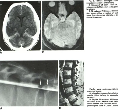
Iym-Fig. 2.-Diffuse meningeal carcinomatosis
(rhabdomyosarcoma of the orbit).
A, Enhanced CT scan. There is prominent
enhancement of tentorium and superior cerebel-lar cistern.
B, T2-weighted MR image, 2000/80. No defi-nite abnormalities in region of tentorium are seen. There is normal intensity of brain paren-chyma throughout.
Fig. 3.-Lung carcinoma, metastatic to
sub-arachnoid space.
A, lohexol myelogram, lateral view. There are
nodular filling defects in subarachnoid space (arrows).
B, Sagittal T1-weighted MR image, 1000/20, of lumbar spine. Several small slightly hyperin-tense nodules are identified within central as-pect of spinal canal at L2 and L31evels (arrows).
phatics [4]. Clinically, the patient presents with a variety of
symptoms, usually multifocal. Headache, change of mental
status, leg and lower back pain, cranial and spinal nerve deficits, and gait disturbances are most frequently encoun-tered [5, 6]. The most specific diagnostic test is a lumbar puncture and CSF cytology. Abnormalities of CSF are re-ported to occur in 20-100% of the patients with meningeal carcinomatosis [6-8]. Typical intracranial changes may be observed on contrast-enhanced CT. Communicating hydro-cephalus and abnormal enhancement of the tentorium, sylvian fissures and basal cisterns, cortical subarachnoid space, and ventricular walls may be noted [9]. Myelographic abnormali-ties of meningeal carcinomatosis include intraarachnoid nod-ular filling defects, longitudinal striations, prominent and crowded nerve roots of the cauda equina, and scalloping of
the subarachnoid space [10]. Both myelography and
contrast-enhanced CT are rather insensitive in detecting meningeal
tumor [6, 9].
The advantages of MR over CT in the examination of
[image:4.615.57.525.74.507.2]AJNR:9. July/August 1988
A
A
MR OF MENINGEAL CARCINOMATOSIS 713
Fig. 4.-Metastatic lung carcinoma.
A, Myelogram showing prominence of roots of cauda equina.
Band C, MR images, 1000/20 (B) and 2000/80 (C). Upper lumbar subarachnoid space appears
patchy and inhomogeneous (arrows in B).
B
B
c
Fig. 5.-Metastatic rhabdomyosarcoma. A, Complete intraarachnoid block is noted at level of L3. Roots of cauda equina are thickened and there is enlargement of conus medullaris.
[image:5.615.53.556.57.751.2]714 KROL ET AL. AJNR:9, July/August 1988
comparative studies [11-13], and the higher sensitivity of MR
in detecting disease in the parenchyma of the nerve tissue
has been noted. However, a recent article by Davis et al. [14]
calls attention to the rather poor performance of MR in
detecting meningeal neoplasm caused by disseminated
pri-mary lesions of the CNS. Our experience with meningeal
metastases to the cranial and spinal meningeal membranes
from primary tumors within the CNS as well as from distant
primary neoplasm also demonstrates the strikingly low
sen-sitivity of MR. The findings considered to be consistent with
intracranial meningeal spread of tumor were noted on CT in
39% (nine) of 23 patients with positive CSF cytology and
focal neurologic deficits. The diagnosis of meningeal
involve-ment on MR could be elicited only in five patients in this group
on the basis of the presence of nodular masses in the
ven-tricular lumen or subarachnoid cisterns. No definite alteration
of MR signal characteristics could be detected in the regions
of abnormal membrane or ependymal enhancement as
dem-onstrated on contrast CT. Unenhanced MR of the spine was
also insensitive, revealing definite abnormalities in 27% (four
out of 15) of positive myelographic studies. However, there
was rather a large number of equivocal lumbar spine MRs
(five of 16, or 31%), indicating nonspecific abnormality of the
subarachnoid space and thus requiring further investigation.
One could generally describe the abnormality as poor
defini-tion of subarachnoid space components with inability to
dif-ferentiate between the distal spinal cord, CSF, and roots of
cauda equina, accounting for the "ground glass" appearance
of the subarachnoid space.
The ratio of false-negative MR examinations was high in
both intracranial and spinal groups (15 of 23, or 65%, and
seven of 16, or 44% of cases, respectively). No definite
abnormalities were seen in these patients even on
retrospec-tive evaluation.
Measurements of T1 and T2 relaxation times are not
relia-ble [15, 16] and measurements of calculated relaxation times
of CSF spaces contribute little to the identification of
neoplas-tic changes within the subarachnoid space [14]. Intensity
measurements of intraventricular CSF in patients with
men-ingeal carcinomatosis are inconclusive. T1-weighted images
are considered superior in depicting changes of meningeal
metastases [14, 17]. In our experience T1-weighted images
provided superior information as compared with T2 weighting,
which frequently obscured the subarachnoid nodules.
In conclusion, nonenhanced MR imaging of the head and
spine is a rather insensitive method of evaluating
leptomen-ingeal neoplasms. In our study it detected significantly fewer
lesions than were demonstrated on contrast-enhanced CT
and myelography. Time restrictions did not permit an
assess-ment of the entire spinal column, and lesions located outside
the examined area were not detected on MR. The abnormal
enhancement of the tentorium, ventricular walls, cisternal
spaces, and cortical subarachnoid sulci seen on CT scans
apparently is not seen as a signal alteration on routine MR
images. Intraventricular and subarachnoid tumor deposits,
indicating advanced disease, represented the only positive
MR evidence of meningeal seeding in the CSF space.
Dem-onstration of tumor nodules within spinal subarachnoid space
in patients with cancer appears to be highly suggestive of
meningeal spread of tumor. However, this is expected to be
found in a small percentage of patients (27% of those with
positive myelography). Normal MR findings do not exclude
the possibility of meningeal metastasis; even high-degree obstruction or complete block of the spinal canal may result in no apparent alteration of MR signal within the spinal canal. Of interest was the presentation of meningeal involvement in
the lumbar region with patchy signal intensity overlying the
cauda equina. Poor definition of roots and asymmetric
posi-tion of the conus and cauda equina within the canal were
probably due to adhesions.
Contrast-enhanced CT and complete myelography with water-soluble contrast material are the methods of choice in evaluating the CNS for presence and extent of neoplastic involvement of the leptomeninges. Nonenhanced MR of the head and spine is significantly less sensitive in depicting these changes, although recent experience with gadolinium (gado-linium-DTPA meglumine) is promising [18].
ACKNOWLEDGMENTS
We gratefully acknowledge the assistance of Jerome Posner in reviewing the manuscript and of Howard Thaler in preparing the statistical data. We also thank Patricia Dudley for typing the paper.
REFERENCES
1. Ascherl GF, Hilal SK, Brisman R. Computed tomography of disseminated meningeal and ependymal malignant neoplasms. Neurology 1981;31:
567-574
2. Sorenson SC, Egan RT, Scott M. Meningeal carcinomatosis in patients with primary breast or lung cancer. Mayo Clin Proc 1984;59:91-94
3. Dubois JP, Martinez JA, Myerowitz LR, et al. Subependymal and lepto-meningeal spread of systemic malignant lymphoma demonstrated by cra-nial computed tomography. J Comput Assist Tomogr 1978;2:218-221
4. Gonzales Vitale J, Garcia Bunuel R. Meningeal carcinomatosis. Cancer
1976;37: 2906-2911
5. Little J, Dale AJ, Okasaki H. Meningeal carcinomatosis: clinical manifesta-tions. Arch Neuro/1974;30:138-143
6. Olson M, Chernik LN, Posner JB. Infiltration of the leptomeninges by systemic cancer. Arch Neuro/1974;30: 122-137
7. Parsons M. The spinal form of carcinomatous meningitis. Q J Med
1972;41 :509-518
8. Grain GO, Karr JP. Diffuse leptomeningeal carcinomatosis: clinical and pathological characteristics. Neurology 1955;5:706-722
9. Lee YY, Glass JP, Geoffray A, Wallace S. Cranial computed tomographic abnormalities in leptomeningeal metastasis. AJNR 1984;5:559-563
10. Kim KS, Ho SU, Weinberg P, Lee C. Spinal leptomeningeal infiltration by systemic cancer: myelographic features. AJR 1982;139: 361-365 11. Brant Zawadzki M, Badami P, Mills C, et al. Primary intracranial tumor
imaging: a comparison of magnetic resonance and CT. Radiology 1984;150:435-440
12. Bradley W, Wallu Z, Yarley R. Comparison of CT and MRI in 400 patients with suspected disease of the brain and cervical spinal cord. Radiology
1984;152:695-702
13. Brant Zawadzki M, Davis PL, Crooks LE, et al. NMR demonstration of cerebral abnormalities: Comparison with CT. AJNR 1983;4: 117-124, AJR
1983;140:847-854
14. Davis PC, Freedman NC, Fry SM. Leptomeningeal metastases: MRI Im-aging. Radiology 1987;163:449-454
15. Kjos BO, Ehman RL, Brant-Zawadzki M. Producibility of Tl and T2 relaxation times calculated from routine MR imaging sequences: phantom study. AJNR 1985;6:277-283
16. Kjos BO, Ehman RL, Brant-Zawadzki M, et al. Reproducibility of relaxation times and spin density calculated from routine MR imaging sequences:
clinical study of CNS. AJNR 1985;6:271-276
17. Barloon TJ, Yuh WT, Yang CJ, et al. Spinal subarachnoid tumor seeding from intracranial metastases: MR findings. J Comput Assist Tomogr
1987;11 :242-244
18. Sze G, Abramson A, Krol G. Gadolinium-DTPA in evaluation of intradural extramedullary neoplasm. Presented at the 25th annual meeting of the


![Polymorphic form II of 2 methyl 4 (4 methyl 1 piperazinyl) 10H thieno[2,3 b][1,5]benzodiazepine](data:image/gif;base64,R0lGODlhAQABAIAAAP///wAAACH5BAEAAAAALAAAAAABAAEAAAICRAEAOw==)