Evolutionary, Embryologic, Anatomic, and MR Analysis
E. Leon Kier and Charles L. Truwit
PURPOSE: To define the normal and abnormal genu of the corpus callosum by examining its evolution and embryology and by analyzing its normal and abnormal appearance on MR images. METHODS: A reference line was drawn from the mamillary body through the anterior commissure and corpus callosum—the MAC line. This line was used to evaluate the genu in adult mammal brains, in human fetal brains, on MR images of 1800 patients with normal corpora callosi, and on MR images of 113 patients with callosal anomalies. RESULTS: In primates, increased frontal lobe size is associated with an anteriorly shifted genu. In human fetal development, the anterior body of the corpus callosum develops before the definitive genu. The normal human genu always projects in front of the MAC line. In none of the 113 patients with callosal anomalies was there only a normal genu. CONCLUSIONS: The human corpus callosum develops bidirectionally, not from front to back. The MAC line is a useful frame of reference to study the evolution and embryology of the genu and to distinguish the normal from the abnormal genu of the human corpus callosum.
Index terms: Animal studies; Brain, growth and development; Corpus callosum, abnormalities and anomalies; Corpus callosum, anatomy
AJNR Am J Neuroradiol17:1631–1641, October 1996
The normal sequence of formation of the components of the corpus callosum has a bear-ing on anomalous development and impacts on the examination of anomalies by magnetic res-onance (MR) imaging. Knowledge of the normal developmental sequence permits distinguishing developmental from destructive lesions of the corpus callosum. Correct analysis of anomalies of the corpus callosum depends on precise identification of its components. Before the era of computed tomography (CT), the compo-nents of the corpus callosum were not identified
readily. With cross-sectional imaging, the cal-losal components became visible. With this came interest on the part of neuroradiologists in the embryology of the corpus callosum and, in particular, in the sequence in which its compo-nents developed. Many descriptions of the de-velopment of the corpus callosum have been published in the neuroradiologic literature since the advent of CT and MR imaging. In general, the corpus callosum has been described as de-veloping in an anterior to posterior direction, starting with the genu, followed by the body, splenium, and finally the rostrum (1–9).
The MR imaging finding of a callosal body without a recognizable genu is not explained by the anterior-to-posterior theory of callosal de-velopment. If the genu were the first structure to develop, the body of the corpus callosum could not be present without a genu. As the genu is pivotal in the above-described developmental schema, it was decided to undertake a mul-tiphasic investigation of the genu of the corpus callosum by using anatomic landmarks. It was hypothesized that two ancient and permanent evolutionary and embryologic structures could serve as landmarks to identify and study the Received January 5, 1996; accepted after revision April 30.
Presented in part at the annual meeting of the Radiological Society of North America, Chicago, Ill, November 1989; at the XIV Symposium Neu-roradiologicum, London, England, June 1990; at the annual meeting of the American Society of Neuroradiology, Chicago, Ill, April 1995; and at the annual meeting of the Radiological Society of North America, Chicago, Ill, November 1995.
From the Department of Diagnostic Radiology, Yale University School of Medicine, New Haven, Conn (E.L.K.) and the Department of Radiology, University of Minnesota School of Medicine, Minneapolis (C.L.T.).
Address reprint requests to E. Leon Kier, MD, Section of Neuroradiol-ogy, Department of Diagnostic RadiolNeuroradiol-ogy, Yale University School of Med-icine, PO Box 208042, New Haven, CT 06520.
AJNR 17:1631–1641, Oct 1996 0195-6108/96/1709 –1631 qAmerican Society of Neuroradiology
normal and abnormal genu. A line connecting the mamillary body, anterior commissure, and corpus callosum was used as a frame of refer-ence. With the use of this line, the first phase of the investigation examined the evolutionary changes of the genu. After the presentation of the evolutionary material in 1989 (E. L. Kier, “MR Patterns of Corpus Callosum Abnormali-ties: Phylogenetic Clarification of Current Ra-diologic Concepts,” presented at the annual meeting of the Radiological Society of North America, Chicago, Ill, November 1989), the next two phases of the study were done. The second phase examined the human embryol-ogy of the genu. The third phase analyzed MR images of the normal and abnormal corpus cal-losum.
Materials and Methods
The corpus callosum of adult mammals and normal human fetuses and the normal and abnormal corpora cal-losi in children and adults were studied by means of dis-section and MR imaging. A line connecting the mamillary body and anterior commissure was extended to the corpus callosum. This line was termed themamillary body–ante-rior commissure– corpus callosum line, or the MAC line
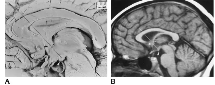
(Fig 1). The MAC line delineates the region of the genu– body interface in the normal human corpus callosum. The genu is in front of the line and the body of the corpus callosum behind it. The MAC line was established by plac-ing a line on the photographs of the dissected brain spec-imens. For the MR cases, the line was drawn on the films or generated on the MR scanner console. Sagittal T1-weighted MR images of 1800 patients with a normal cor-pus callosum were analyzed during a 6-year period using the MAC line to establish the position of the genu of the corpus callosum. Retrospective analysis of sagittal T1-weighted MR images of 113 patients with abnormalities of the corpus callosum was also performed by using the MAC line to determine the presence or absence of a normal genu of the corpus callosum.
The anterior commissure is a compact, vertically oval bundle of myelinated fibers (10). It crosses the midsagittal plane, in the anterior wall of the third ventricle, at the upper end of the lamina terminalis, above the optic chiasm. In the region of the third ventricle, the fornix is just posterior and superior to the anterior commissure. The fornix, near the foramen of Monro, can be identified as a small dot just superior to the anterior commissure (Fig 1). The MAC line was traced through the anterior commissure whenever the fornix could be identified as a separate structure. The mamillary bodies are two rounded structures located side by side on the floor of the third ventricle behind the optic chiasm and tuber cinereum and in front of the posterior perforated substance (11). They are situated at the front end of the interpeduncular fossa.
[image:2.612.175.544.90.237.2]The brains of adult mammals including three rats, three rabbits, three cats, two dogs, two monkeys, and six hu-mans were examined. In some of the adult mammals, the cerebral ventricles were injected before fixation in 10% formaldehyde with a barium gelatin mixture (12). Addi-tionally, brains from eight human fetal specimens ranging from 10 to 30 weeks’ gestational age were examined. Fetal age was determined by measuring crown-rump length and occipitofrontal diameter (13, 14). As the tables used for assessing fetal age show a 10- to 20-mm difference be-tween the smallest and largest specimen in each age group, the ages of the specimens may vary by 1 to 2 weeks from their true gestational age. To verify fetal age further, the occipitofrontal diameter of the fetal brains was com-pared with the occipitofrontal diameter of photographs of fetal brains in the literature (15, 16). The specimens ap-peared normal and the dissected brains did not show vious abnormalities. The midsagittal sections were ob-tained with the brain in situ within the calvaria. The sagittal suture was opened and the superior sagittal sinus, falx, arachnoid, and pia removed by dissection. Sagittal sec-tioning of the younger fetal heads, done to visualize the corpus callosum, could not be performed with the pia and arachnoid intact in the interhemispheric fissure. The ex-tensive midline venous vasculature had to dissected away from the interhemispheric fissure, as it was pulled while being sectioned, destroying the underlying corpus callo-sum. The partially myelinated fetal brain with its high water Fig 1. Photograph of the
content does not fix well, even after being preserved in 10% formaldehyde for 20 years. Of the eight specimens (16 cerebral hemispheres), useful dissections were obtained from two hemispheres in fetuses less than 13 weeks old, from three hemispheres in fetuses between 14 and 18 weeks old, and from seven hemispheres in fetuses be-tween 18 and 30 weeks old. Finally, six adult human brains, obtained from the anatomy and pathology depart-ments and fixed in 10% formaldehyde, were examined.
To identify the developing corpus callosum on the me-dial surface of the cerebral hemisphere, previous studies of human embryology and anatomy were used (11, 14 –25). Images of 20 fetuses that were obtained on 1.5-T scanners were compared with the dissections. The specimens that were imaged but not dissected ranged in age from 13 to 30 weeks, as determined by crown-rump length and by oc-cipitofrontal diameter. Images in the sagittal plane were acquired with a three-dimensional spoiled gradient-echo sequence that accentuates T1 weighting. Sagittal T1-weighted MR images of 1800 patients with a normal cor-pus callosum were analyzed using the MAC line to estab-lish the position of the normal genu of the corpus callosum. These patients, comprising all ages and both sexes, were studied over a 6-year period. Retrospective analysis of
sagittal T1-weighted MR images of 113 patients with ab-normalities of the corpus callosum was performed using the MAC line to evaluate the genu and anterior body of the corpus callosum. Initially, 141 patients with abnormalities of the corpus callosum were included in the study; but 28 patients with total absence of the corpus callosum were excluded from the study group.
Results
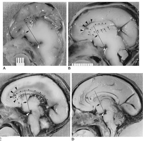
significant anterior curvature around the genu before continuing toward the subcallosal area. In the 12-week-old human fetal specimen, the rudimentary corpus callosum is present below the callosal sulcus and behind the MAC line (Fig 4A). In the 16-week-old specimen, a rudimen-tary genu is now present in front of the MAC line; the cingulate gyrus has a minimal anterior curvature before continuing toward the future subcallosal area (Fig 4B). In the 20-week-old specimen, a prominent genu is present; the cin-gulate gyrus shows a marked anterior curvature around the genu before continuing into the sub-callosal region (Fig 4C). In the 30-week-old specimen, the entire corpus callosum is thicker and shows increased flexion; the cingulate gy-rus shows a prominent anterior curvature around the larger genu before reaching the sub-callosal area (Fig 4D). An MR image of a 13-week-fetal specimen shows the presence of the corpus callosum behind the MAC line (Fig 5A). The corpus callosum is not seen in front of the MAC line. An MR image of a 14-week-old spec-imen shows the presence of a rudspec-imentary genu in front of the MAC line (Fig 5B). An MR image of a 22-week-old fetal specimen shows a larger genu in front of the MAC line (Fig 5C).
Sagittal T1-weighted MR images of 1800 pa-tients with a normal corpus callosum were an-alyzed using the MAC line to evaluate the posi-tion of the genu of the corpus callosum. In all 1800 cases the genu always projected anterior
to the MAC line (Fig 1). In a small number of infants less than 1 month of age, it was some-times difficult to identify the anterior end of the corpus callosum and to determine if the genu was in front of the MAC line. MR images of 113 patients with anomalies of the corpus callosum were reviewed retrospectively. The components of the corpus callosum were analyzed as to their presence, absence, size, and relationship to the MAC line. Review of the abnormal cases re-vealed no cases in which only the genu was present in its expected normal position in front of the MAC line and in which the body and splenium were absent. In 18 cases, however, the entirety of the abnormal corpus callosum was behind or at the MAC line.
Discussion
[image:4.612.48.541.91.255.2]Fig 4. Photographs of the medial surface of brains of human fetal specimens show the growth of the corpus callosum.
A, In a 12-week-old specimen, a rudimentary corpus callosum (betweensmall arrowsandsmall arrowheads) is present below the callosal sulcus (small arrowheads) and behind the MAC line.
B, In a 16-week-old specimen, a rudimentary genu (betweenshort arrowsandsmall arrowheads) is present below the callosal sulcus (small arrowheads) and in front of the MAC line. The callosal body (betweenlong arrowsandsmall arrowheads) is behind the MAC line. The cingulate gyrus (CG), below the cingulate sulcus (large black arrowheads), has a minimal anterior curvature before continuing toward the future subcallosal area.
C, In a 20-week-old specimen, a prominent genu (betweensmall arrowsandsmall arrowheads) is present below the callosal sulcus (small arrowheads). The cingulate gyrus (CG) and cingulate sulcus (large black arrowheads) now show a prominent anterior curvature, around the larger genu, before continuing toward the subcallosal area (S). The body and splenium (betweenlong arrowsandsmall arrowheads) are elongated.
D, In a 30-week-old specimen, the entire corpus callosum is thicker and shows increased flexion. The cingulate gyrus (CG) shows a prominent anterior curvature, around the larger genu (G), before reaching the subcallosal area (S).
upwards and backwards and the genu develops, followed by the body and finally the splenium” (26). This was subsequently amended to sug-gest that the rostrum is the final component to form (1–9). The anterior-posterior view of cal-losal embryology was temporarily revised to suggest that the posterior genu forms first, fol-lowed by growth in a posterior direction to fill out the body and splenium and in an anterior direction to complete the remaining genu and rostrum (27). However, the anterior-to-poste-rior direction of callosal growth remains the pre-vailing notion in the neuroradiologic literature.
As the purpose of this investigation was to evaluate the genu of the corpus callosum, an anatomic landmark to distinguish the genu from the body was needed. The mamillary body–an-terior commissure– corpus callosum line, the MAC line, was selected for this investigation because the mamillary body and the anterior commissure can always be identified on an ad-equate sagittal MR image of the brain. The line is simple to apply, and the MAC pneumonic is an easy reminder of the structures through which the line passes. The MAC line is an ap-propriate landmark from evolutionary, embryo-logic, anatomic, and MR points of view. The mamillary body and anterior commissure are ancient and permanent evolutionary and em-bryologic structures (12, 28, 29). The corpus callosum develops in the hippocampal primor-dium, near and superior to the anterior commis-sure. Thus, the continuation of a line connecting
these two ancient and permanent structures is an appropriate frame of reference for evaluating developmental changes in the genu of the cor-pus callosum. This is especially true when com-paring the corpora callosi of various mammals. The marked changes in brain size and shape require a stable frame of reference.
This investigation revealed the usefulness of assessing the normal corpus callosum by means of the MAC line. In 1800 clinical MR images that showed a normal corpus callosum that were reviewed over a 6-year period, the genu always projected anteriorly to the MAC line. The clinical images reviewed were not pre-selected and probably included patients with varying head size and shape and differing brain size. These patients most likely had different anteroposterior size of the corpus callosum, which could explain why the MAC line passed in slightly different locations in the region of the boundary of the genu and anterior body. As the genu always projected anteriorly to the MAC line, this line is a valid landmark for the position of the normal genu.
[image:6.612.44.542.90.258.2]The finding in the normal corpus callosum makes sense in view of the evolutionary phase of this study, which showed that as the frontal lobes expand, the anterior edge of the corpus callosum moves progressively forward (Figs 2 and 3). The forward movement of the genu is confirmed by the increased anterior curvature of the cingulate gyrus (Fig 3). Without the MAC line, it would be difficult to compare the position Fig 5. Sagittal T1-weighted MR images of human fetal specimens show the growth of the corpus callosum.
A, A 13-week-old fetal specimen shows the presence of a rudimentary body of the corpus callosum (small arrows) behind the MAC line. The genu is not present in front of the MAC line.
B, A 14-week-old fetal specimens shows the presence of a rudimentary genu (double arrow) in front of the MAC line. C, A 22-week-old fetal specimen shows a much larger genu (G) in front of the MAC line.
of the genu in mammals with differing brain shapes and sizes. The more anterior position of the genu in the monkey and its larger size is linked to the marked expansion of the frontal lobe in primates (Fig 3). A review of the evolu-tionary studies of Smith (30) and Abbie (31) and the use of the MAC line on the specimens of the present study permit one to speculate that the anterior end of the corpus callosum—the genu of the rat, rabbit, and cat—is incorporated within the body of the corpus callosum in the primate.
The embryologic growth of the corpus callo-sum is also the subject of controversy in the comparative anatomic literature. Tilney (32) states that in pig, rat, guinea pig, cat, and hu-man embryos, the earliest fibers of the corpus callosum appear in front of the intraventricular foramen and the anterior commissure. Studies in cat embryos (33) have shown that at the 38th embryonic day only part of the body had formed. At the 53rd and 58th embryonic days, the genu, body, and splenium have formed. Ex-periments in mouse embryos (34, 35) found that the region of the genu forms before the splenium, in contradistinction to the study in cat embryos. Wahlsten (36) noted that in the mouse embryo, after the initial callosal fibers cross the midline, the corpus callosum under-goes rapid growth, adding fibers anterior to the existing corpus callosum and forming a distinct genu. He points out the interesting fact that the first callosal fibers in the rat embryo appear just dorsal to the hippocampal commissure in the same region in which the corpus callosum is present in adult bats, which have the smallest corpus callosum among all mammals. Ozaki and Wahlsten (35) make the important state-ment that the “genu and splenium are terms
most appropriate for the gross morphology of the adult.” This validates the approach taken throughout this investigation to relate the loca-tion of fetal structures to the localoca-tion of the components of the corpus callosum in the adult. The earliest fibers of the human corpus cal-losum appear during the 11th to 12th week of fetal development (22). In embryos aged 6 to 8 weeks, the thin rostral wall of the telencephalon, anterior to the chiasmatic plate, shows a rapid increase in the thickness of its dorsal part near the paraphysis. The anterior part near the chi-asmatic ridge, the lamina terminalis, remains thin. The thicker dorsal part represents the lam-ina reuniens of His, a region of active cellular proliferation. The ventral part of the lamina re-uniens, near the lamina terminalis, develops into the area precommissurals. Later it will be-come the septal area with the anterior commis-sure within it. The hippocampal primordium and the future archaeocortex arise within the dorsal part of the lamina reuniens. As the hemi-spheric vesicles expand bilaterally, the dorsal part of the lamina reuniens begins to fold into the median groove. The banks of the median groove (sulcus medianus telencephali medii) approximate and come into juxtaposition, form-ing the massa commissuralis. In embryos of 11 to 12 weeks, the earliest fibers of the future corpus callosum begin to penetrate the massa commissuralis in the region of the hippocampal primordium.
[image:7.612.45.410.89.257.2]callosal sulcus can be seen in front of the MAC line on MR images. The development of the callosal sulcus was described previously (37). The forward movement of the human fetal genu is confirmed by the increased anterior curvature of the cingulate gyrus (Fig 4). The observation that the region of the future human genu devel-ops after the region of the future anterior body of the corpus callosum is confirmed by superim-posing the MAC lines on the figures in the stud-ies by Retzius (19) and by Feess-Higgins and Larroche (16). The corpora callosi in some of Retzius’s figures of the third month are very similar in appearance to the fetal MR images in Figs 5A and B and the clinical MR images in Fig 6 of the present study. Thus, in human embry-ology, the region of the anterior body develops before the region of the genu. In fact, that the human anatomic references show the anlage of the corpus callosum to be near and superior to the lamina terminalis precludes a unidirectional growth. Several publications are explicit about the bidirectional growth of the corpus callosum (14, 21, 38). Rakic and Yakovlev (22) in dia-gram 13 of their frequently cited article show the genu in a progressively more anterior posi-tion from the 18th to 40th week of fetal devel-opment. According to Loeser and Alvord (39), the earliest callosal fibers can be seen at the 74th gestational day, whereas the splenium and genu are recognizable at the 84th day. In their growth curve charts (39, 40), these authors list the primordial corpus callosum as developing before the genu and splenium. In addition, as recently as 1982, Sidman and Rakic (25), when discussing the formation of the cavum septi pel-lucidi, state that “the zone of fusion of the atten-dant callosal decussation extend[s] first for-ward, then down, and finally back again to form
the genu and rostrum of the corpus callosum.” Clearly, the neuroanatomic literature has re-tained the concept of bidirectional callosal for-mation.
Another source of possible confusion is the nomenclature of the genu. In the human adult
and other primates, the term genu is used to
describe the anterosuperior segment of the cor-pus callosum. The fibers crossing the genu in the rhesus monkey and the human connect the prefrontal motor and association cortical re-gions (41– 43). However, the term genuis also used in connection with other mammals in whom the prefrontal regions of the primate are not present (Figs 2 and 3). The genu of these vertebrates would be considered the anterior body region of the primate corpus callosum. In addition, in the older embryologic literature
(17–19), the term genu has been used to
de-scribe the anterior– knee form– end of the hu-man corpus callosum during in the early stages of fetal development. Retzius (19), in describing the developing corpus callosum in a 3-month-old embryo, stated, “the knee shaped (Kniefo ¨r-mig) anlage of the corpus callosum is visible above the lamina terminalis.” However, this area is within the region of the future definitive body. Thus, the termgenuhas been used in the literature to denote three different anatomic re-gions: the nonprimate genu, the knee-shaped anterior end of the early human fetal corpus callosum, and the callosal genu of the devel-oped primate.
[image:8.612.181.542.88.256.2]With the information gained by this investiga-tion, a more precise classification of anomalies of the corpus callosum can be made. In the 113 MR studies of patients with abnormalities of the corpus callosum that were reviewed retrospec-tively, the MAC line was used as a landmark. Fig 7. Sagittal T1-weighted
Two particular findings underscore our dis-agreement with the concept of unidirectional, front-to-back growth of the corpus callosum. First, no abnormal cases were found that con-sisted of only a genu in its expected position in front of the MAC line with all the other segments of the corpus callosum absent. Second, in 18 cases, the entirety of the abnormal corpus cal-losum was behind or at the MAC line.
A review of the cases of hypogenesis of the corpus callosum showed that when a rudimen-tary corpus callosum is present, it lies at or behind the MAC line (Fig 6). In these cases, the rudimentary corpus callosum is generally in the position where it originally developed in the re-gion of the fetal massa commissuralis, superior to the anterior commissure. In some cases it may lie anterior to the anterior commissure, but in all cases it lies at or behind the MAC line. The fetal genu may be incorporated within these rudimentary hypogenetic corpora callosi. In cases in which the genu was present, it was always associated with the presence of the an-terior body (Fig 7). This study found no cases in which a normal genu was present without the anterior body.
Cases with no corpus callosum in front of the MAC line have been described as showing the presence of a genu. This study demonstrated that in cases similar to those described in the literature, the normal genu is absent (Fig 8). Only the anterior body of the corpus callosum is present with a genulike anterior end, which may represent the genu remaining in its early fetal position. Our analysis of 113 cases of callosal hypogenesis confirms that the region of the body of the corpus callosum is the first segment to develop, and that a normal genu is unlikely to
be present without a segment of the anterior body. This study also confirms the bidirectional growth of the normal corpus callosum. Rubin-stein and coauthors (44) were also concerned by cases such as the ones shown in Figure 8. They stated that previous reports did not con-sider the position of the corpus callosum rela-tive to the anterior commissure and fornices and that every anterior portion of the corpus callosum that was formed was called in the lit-erature either a genu or a genu and body.
[image:9.612.45.410.89.258.2]explained by the phenomenon of arrested growth of the corpus callosum.
Conclusions
The contention that the corpus callosum de-velops normally in an anterior-to-posterior di-rection and that the genu is the first to form, followed by the body, splenium, and rostrum is not supported by an evolutionary, embryologic, anatomic, or MR imaging analysis. Rather, our data corroborate that the corpus callosum de-velops first in the region of the future anterior body and grows bidirectionally.
Acknowledgments
We thank Helmuth Gahbauer and Heinz Schlemmer for their help with the German literature, and Douglas H. Yock, Jr, for sharing his case material.
References
1. Byrd SE, Harwood-Nash DC, Fitz CR. Absence of the corpus callosum: computed tomographic evaluation in infants and chil-dren.J Can Assoc Radiol1978;29:108 –112
2. Davidson HD, Abraham R, Steiner RE. Agenesis of the corpus callosum: magnetic resonance imaging. Radiology
1985;155:371–373
3. Barkovich A, Norman D. Anomalies of the corpus callosum: cor-relation with further anomalies of the brain.AJNR Am J Neurora-diol1988;9:493–501
4. Barkovich AJ, Kjos BO. Normal postnatal development of the corpus callosum as demonstrated by MR imaging.AJNR Am J Neuroradiol1988;9:487– 491
5. Barkovich AJ. Apparent atypical callosal dysgenesis: analysis of MR findings in six cases and their relationship to holoprosen-cephaly.AJNR Am J Neuroradiol1990;11:333–339
6. Truwit CL, Barkovich AJ. Pathogenesis of intracranial lipoma: an MR study in 42 patients.AJNR Am J Neuroradiol1990;11:665– 674
7. Georgy BA, Hesselink JR. MR imaging of the corpus callosum.
AJR Am J Roentgenol1993;160:949 –955
8. Hansen PE, Ballesteros MC, Soila K, Garcia L, Howard JM. MR imaging of the developing brain, 1: prenatal development. Radio-graphics1993;13:21–36
9. Oba H, Barkovich AJ. Holoprosencephaly: an analysis of callosal formation and its relation to development of the interhemispheric fissure.AJNR Am J Neuroradiol1995;16:453– 460
10. Naidich TP, Daniels DL, Pech P, Haughton VM, Williams A, Poju-nas K. Anterior commissure: anatomic-MR correlation and use as a landmark in three orthogonal planes.Radiology1986;158:421– 429
11. Williams PL, Warwick R, Dyson M, Bannister LH, eds. Gray’s Anatomy. 37th ed. London, England: Churchill Livingstone; 1989:190 –194, 1063–1064
12. Kier EL. The cerebral ventricles: a phylogenetic and ontogenetic study. In: Newton TH, Potts DG, eds.Radiology of the Skull and Brain.St Louis, Mo: Mosby; 1977:2787–2914
13. Scammon RE, Calkins LA.The Development and Growth of the External Dimensions of the Human Body in the Fetal Period.
Minneapolis, Minn: University of Minnesota Press; 1929:96 –99 14. Patten BM.Human Embryology.3rd ed. New York, NY:
McGraw-Hill; 1968:139 –145, 289 –294
15. Barbe A.Recherches sur L’Embrylogie du Systeme Nerveux Cen-tral de L’Homme.Paris, France: Masson; 1938:59 –160 16. Feess-Higgins A, Larroche JC.Development of the Human Fœtal
Brain: An Anatomical Atlas.Paris, France: Masson; 1987:88 –166 17. Blumenau L. Zur Entwicklungsgeschichte und Feineren Anatomie
des Hirnbalkens.Arch Mikroskop Anat1891;37:1–15
18. Marchand F. Ueber die Entwickelung des Balkens im Menschli-chen Gehirn.Arch Mikroskop Anat1891;37:298 –334
19. Retzius G. Das Menschenhirn.Stockholm, Sweden: Norstedt & Son; 1896:5–7, plate 4
20. Streeter GL. The development of the nervous system. In: Keibel F, Mall FP, eds.Manual of Human Embryology.Philadelphia, Pa: Lippincott; 1912:82–95
21. Hamilton WJ, Boyd JD, Mossman HW.Human Embryology.3rd ed. Baltimore, Md: Williams & Wilkins; 1962:354 –357
22. Rakic P, Yakovlev PI. Development of the corpus callosum and cavum septi in man.J Comp Neurol1968;132:45–72
23. Probst FP. Congenital defects of the corpus callosum.Acta Radiol Suppl1973;331:1–152
24. Probst FP. The prosencephalies.Morphology, Neuroradiological Appearances and Differential Diagnosis.New York, NY: Springer-Verlag; 1979:3–13
25. Sidman RL, Rakic P. Development of the human nervous system. In: Haymaker W, Adams RD, eds.Histology and Histopathology of the Nervous System.Springfield, Ill: Charles C. Thomas; 1982: 41– 49
26. Bull J. The corpus callosum.Clin Radiol1967;18:2–18 27. Barkovich AJ, Gilles L, Evrard P. Formation, maturation, and
disorders of white matter.AJNR Am J Neuroradiol1992;13:447– 461
28. Sarnat HB, Netsky MG.Evolution of the Nervous System.2nd ed. New York, NY: Oxford University Press; 1981:308 –309, 370 –372 29. Kier EL. Comparative anatomy of the third ventricular region. In:
Apuzzo MLJ, ed.Surgery of the Third Ventricle.Baltimore, Md: Williams & Wilkins; 1987:37–91
30. Smith GE. The morphology of the true “limbic lobe,” corpus callosum, septum pellucidum and fornix.J Anat Physiol1895;30: 157–205
31. Abbie AA. The origin of the corpus callosum and the fate of the structures related to it.J Comp Neurol1939;70:9 – 44
32. Tilney F. The hippocampus and its relationship to the corpus callosum.J Nerv Ment Dis1939;89:433–513
33. Berbel P, Innocenti GM. The development of the corpus callosum in cats: a light- and electron-microscopic study.J Comp Neurol
1988;276:132–156
34. Silver J, Lorenz SE, Wahlsten D, Coughlin J. Axonal guidance during development of the great cerebral commissures: descrip-tive and experimental studies, in vivo, on the role of preformed glial pathways.J Comp Neurol1982;210:10 –29
35. Ozaki HS, Wahlsten D. Prenatal formation of the normal mouse corpus callosum: a quantitative study with carbocyanine dyes.J Comp Neurol1992;323:81–90
36. Wahlsten D. Prenatal schedule of appearance of mouse brain commissures.Dev Brain Res1981;1:461– 473
37. Kier EL, Fulbright RK, Bronen RA. Limbic lobe embryology and anatomy: dissection and MR of the medial surface of the fetal human cerebral hemisphere.AJNR Am J Neuroradiol1995;16: 1847–1853
38. Carpenter MB, Sutin J.Human Neuroanatomy.8th ed. Baltimore, Md: Williams & Wilkins; 1983:83
39. Loeser JD, Alvord EC Jr. Agenesis of the corpus callosum.Brain
40. Lemire RJ, Loeser JD, Leech RW, Alvord EC Jr. Normal and Abnormal Development of the Human Nervous System. Hagers-town, Md: Harper & Row; 1975:260 –265
41. de Lacoste MC, Kirkpatrick JB, Ross ED. Topography of the human corpus callosum. J Neuropathol Exp Neurol 1985;44: 578 –591
42. Pandya DN, Rosene DL. Some observation on trajectories and topography of commissural fibres. In: Reeves AG, ed.Epilepsy
and the Corpus Callosum.New York, NY: Plenum Press; 1985: 21–39
43. LaMantia A-S, Rakic P. Axon overproduction and elimination in the corpus callosum of the developing rhesus monkey.J Neurosci
1990;10:2156 –2175
44. Rubinstein D, Youngman V, Hise JH, Damiano TR. Partial devel-opment of the corpus callosum.AJNR Am J Neuroradiol1994; 15:869 – 875