David L. Daniels'
John F
.
Schenck2
Thomas Foster2
Howard Hart, Jr.2
Steven J. Millen
3Glenn A. Meyer4
Peter Pech'
Victor M. Haughton'
Received November 28, 1984; accepted after
revision February 28, 1985.
Presented at the annual meeting of the American
Society of Neuroradiology, New Orleans, February 1985.
This work was supported by a grant from
Gen-eral Electric Medical Systems.
1 Department of Radiology, Medical College of Wisconsin, Froedtert Memorial Lutheran Hospital, 9200 W. Wisconsin Ave., Milwaukee, WI 53226. Address reprint requests to D. L. Daniels.
2 General Electric Research and Development
Center, Schenectady, NY 12301.
3 Department of Otolaryngology, Medical Co
l-lege of Wisconsin, Milwaukee, WI 53226.
4 Department of Neurosurgery, Medical College of Wisconsin, Milwaukee, WI 53226.
AJNR 6:699-703, September/October 1985 0195-6108/85/0605-0699
© American Roentgen Ray Society
Magnetic Resonance
Imaging of the Jugular
Foramen
699
The jugular foramen in normal volunteers was studied with 1.5 T magnetic resonance (MR) systems in 3-mm-thick head- and surface-coil images. Anatomic sections through cadaver heads were correlated with the MR images to identify the jugular bulb and the course of cranial nerves IX-XI. Sagittal images were more useful than coronal or axial to show the course of these nerves through the skull base. MR demonstrates the anatomic relations of the jugular foramen (except its osseous margins) such that its primary use in evaluating this region can be anticipated.
Although computed tomography (CT) is useful for demonstrating the jugular
foramen, it fails to demonstrate cranial nerves IX-XI consistently. Since effective
magnetic resonance (MR) demonstration of cranial nerves
in
the internal auditory
canals and
cavernous sinuses has been reported [1, 2]
,
its demonstration of
cranial
nerves IX-XI can be predicted. MR imaging in the jugular foramen is facilitated by
the lack of signal from flowing blood and
bone.
The purpose of our article is to
describe the MR appearance of the normal contents of the jugular foramen.
Materials and Methods
Four fresh, frozen cadaver heads were embedded with carboxymethyl cellulose gel in styrofoam boxes. The jugular foramina were sectioned using a heavy-duty sledge cryomicro-tome (LKB 2250, Gaithersburg, MO) either parallel to the sagittal plane or to planes forming an angle of 00 or +300 to the canthomeatal line (CML) [3]. Anatomic images were obtained by photographing the surfaces of the specimens as they were sectioned.
Five normal volunteers were studied in research General Electric MR scanners with superconducting 1.5 T magnets [4] and head or surface coils. All images were obtained with a spin-warp spin-echo technique [5]. Head-coil imaging included an initial sagittal image for localizing and thin axial (parallel to the CML) and parasagittal sections. A flexible-surface receiver coil 13.5 cm in diameter and tuned to 63.9 MHz was placed over the jugular foramina of the normal volunteers for surface-coil imaging. A 54-cm-diam cylindrical coil, used for body imaging, was used for the radiofrequency transmitter. Surface-coil images were obtained using planes parallel or +300 to the CML and using parasagittal planes. Technical factors for
head-coil images included 300-400 msec TR; 14 msec TE; 128
x
256 or 256x
256 matrices;1.9 x 1.0 mm and 1.0 x 1.0 mm, respectively, pixel sizes; 25 cm field of view; and 3 and 5 mm slices. Technical factors for surface-coil images included 200-400 msec TR; 19 msec TE; 128 x 256 or 256 x 256 matrices; 1.0 x 0.5 mm and 0.5 x 0.5 mm, respectively, pixel sizes; 12.8 cm field of view; and 3-mm-thick slices.
The MR images of the volunteers and photographs of the anatomic sections (figs. 1-5) were correlated with published anatomic and CT literature [6-12] to identify cranial nerves
700
DANIELS ET AL.
AJNR:6, Sept/Oct 1985A
c
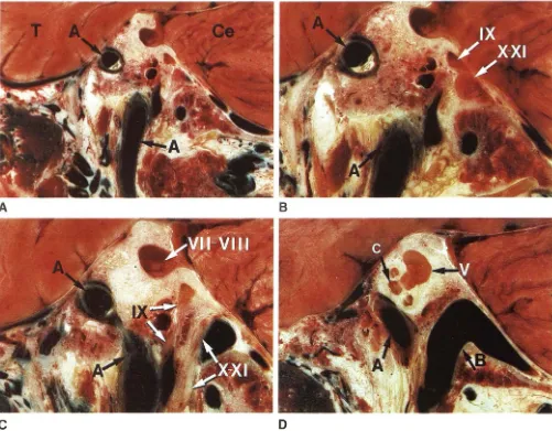
Fig. 1 . -Parasagitlal cryomicrotomic sections of jugular foramen from medial to lateral. A, Cranial nerve IX is in pars nervosa, and cranial nerves X and XI together, in pars vascularis. Segments of internal carotid artery (A) are below and within carotid canal. B, Magnification of A. C, Cranial nerves IX and X-XI,
Results and Discussion
The jugular foramen consists
of the pars nervosa
(glosso-pharyngeal [IX]
nerve
and inferior
petrosal
sinus)
and
,
poste
-rior to it
,
the
pars vascularis
(vagus [X] and
spinal accessory
[XI]
nerves
and
jugular bulb). A plane of
section
of
+30
0to
the
CML
and
dynamic
scanning
techniques
are
used with CT
to
identify
cranial
nerves
in the
jugular
foramen [6].
In
cryomicrotomic sections, cranial nerves IX-XI
have
a
constant position in the
jugular
foramen (figs. 1 and 4).
In
a
plane of
+30
0to the CML (nearly parallel to the clivus), these
nerves have an almost straight
lateral
course from the
medulla
to the endocranial opening of the jugular foramen (fig
.
4).
In
the foramen
,
IX
is
anterior and X and XI
posterior
.
Sagittal
sections show
cranial nerve IX in a dural sheath in the jugular
foramen and
then from the skull base anteroinferiorly to the
tongue and pharynx (fig
.
1)
.
Cranial nerves X and XI
,
in a
B
o
separated by thin spicule of bone, have oblique and nearly parallel course through skull base. D, Jugular bulb (8) and internal carotid artery. T = temporal lobe; Ce = cerebellum; VII-VIII = cranial nerves in the internal auditory canal; V = vestibule; C = cochlea.
common
dural sheath behind IX
,
lie anteromedial to the jugular
bulb
and then course anteroinferiorly below the skull base.
They may appear
slightly
wider at the endocranial opening of
the
foramen
.
Through
the
skull base they have a slightly
oblique
course
nearly parallel to cranial nerve IX
.
Cranial nerve
X and one branch of XI terminate in the carotid sheath; one
branch of XI terminates in
the
neck muscles.
On MR images
structures
that have a signal intensity
intermediate between
fat
and cerebrospinal fluid (figs. 2
,
3
,
and
5) have been identified that correspond to cranial nerves
IX-XI in
the
cryomicrotomic sections
.
The jugular bulb can be
demonstrated on parasagittal images
.
Surface-coil
images demonstrate
the
cranial nerve anatomy
better
than
do head-coil
images. The
higher signal-to-noise
ratio offered
by
surface
coils permits smaller pixel
sizes.
The
[image:2.615.59.560.86.477.2]AJNR:6, Sept/Oct 1985 MR OF THE JUGULAR FORAMEN 701
c
Fig. 2.- Parasagittal surface-coil MR scans, 400 msec TR, 19 msec TE, 256 x 256 matrix, 205 sec scan time, 3 mm thick, from medial to lateral corresponding to anatomic sections in fig. 1. A and B, Segments of internal carotid artery (A) have negligible signal. Ns = cranial nerves. C, Cranial nerves
Fig. 3.-Parasagittal surface-coil MR images, 400 msec TR, 19 msec TE, 256 x 256 matrix, 205 sec scan time, 3 mm thick. Cranial nerves IX and X-XI in skull base (A) and jugular bulb (8) just lateral to cranial nerves IX-XI (B). C = cochlea; V
=
vestibule; JV=
jugular vein.A
D
IX and X-XI, osseous foramen (negligible signal), and fat (high-intensity signal). Cranial nerves VII and VIII are identified in internal auditory canal. D, Internal carotid artery and jugular bulb (8) and vein have negligible signal. C = cochlea; V = vestibule.
702
DANIELS ET AL.
AJNR:6, Sept/Oct 1985Fig. 4.-Cryomicrotomic sections of jugular foramen from posterosuperior (A) to anteroinferior (C) in plane of +300 to CML (nearly parallel to clivus). A, Cranial nerves IX-XI (Ns) have straight course from medulla (M) to pars vascularis (Le., X and XI) and pars nervosa (Le., IX). B, Magnification of A. C, X and XI lie medial to jugular bulb (8) and slightly posteromedial to IX. P = pons.
8
shown
in the
axial
plane
,
the endocranial segments of
the
nerves
in a plane of +30
0to the
CML. Parasagittal
surface-coil
images
consistently show the intraforaminal and
extra-cranial course of these
nerves
(figs
.
2 and 3)
.
High-resolution
surface-coil
MR images
provide
consist-ently better demonstration of cranial
nerves IX-XI
in
the
jugular foramen and in extracranial tissues without a contrast
agent than do CT
images
[6].
Because it demonstrates the
individual
nerves
,
MR
should effectively e
x
clude tumor in
the
jugular foramen when
IX
,
X
,
or XI nerve signs are
present.
CT reliably demonstrates
intraforaminal
tumors only when the
osseous margins are eroded
.
A series of foraminal tumors
should be studied with
MR
to determine if
T1
and
T2 meas
-urements or different pulse sequences will
increase
the spec
-ificity of neuroradiologic diagnosis
in jugular
foramen
lesions.
ACKNOWLEDGMENTS
We thank Cecil Hayes, William A. Edelstein, Ann Shimakawa, and
Laurie Dunk for help in this study.
REFERENCES
1. Daniels DL, Herfkins R, Koehler PR, et al. Magnetic resonance
imaging of the internal auditory canal. Radiology 1984; 151 : 1
05-108
2. Daniels DL, Pech P, Mark L, Pojunas K, Williams AL, Haughton
VM. Magnetic resonance imaging of the cavernous sinus. AJNR
1985;6: 187-192, AJR 1985;144: 1 009-1 014
3. Rauschning W, Bergstrom K, Pech P. Correlative craniospinal
anatomy studies by computed tomography and cyromicrotomy.
AJNR:6, Sept/Oct 1985
MR OF THE JUGULAR FORAMEN
703
A
Fig. 5.-Surface-coil MR images, 200 msec TR, 19 msec TE, 128 x 128 matrix, 52 sec scan time, 3 mm thick, of jugular foramen in plane of +300 to
CML corresponding to anatomic sections in fig. 4. A, Endocranial course of
4. Bottomley PA, Hart HR, Edelstein WA, et al. Anatomy and metabolism of the normal human brain studied by magnetic resonance at 1.5 Tesla. Radiology 1984;150:441-446
5. Edelstein WA, Bottomley PA, Hart HR, Smith LS. Signal, noise and contrast in nuclear magnetic resonance (NMR) imaging. J
Comput Assist Tomogr 1983;7:391-401
6. Daniels DL, Williams AL, Haughton VM. Jugular foramen:
ana-tomic and computed tomographic study. AJNR 1983;4: 1227-1232, AJR 1984;142: 153-158
7. Lo WWM, Solti-Bohman LG, Lambert PRo High-resolution CT in the evaluation of glomus tumors of the temporal bone. Radiology
1984;150:737-742
B
cranial nerves IX-XI (Ns). IX lies in pars nervosa and X-XI in pars vascularis.
Jugular bulb is not seen well. B, Slightly anteroinferior to A. Again, X-XI can be seen. P = pons; M = medulla.
8. Lo WWM, Solti-Bohman LG. High-resolution CT of the jugular
foramen: anatomy and vascular variants and anomalies. Radiol
-ogy 1984;150:743-747
9. Strickler JM. New and simple techniques for demonstration of
the jugular foramen. AJR 1966;97:601-606
10. Gray H. Anatomy of the human body. Philadelphia: Lea & Febiger,
1966
11. Rhoton AL, Buza RC. Microsurgical anatomy of the jugular foramen. In: Rand RW, ed. Microneurosurgery, 2d ed. St. Louis:
Mosby, 1978:252-264
12. Di Chiro G, Fisher RL, Nelson KB. The jugular foramen. J
[image:5.612.57.556.97.296.2]