Myoglobin, a protein with a rich and varied history, has recently become the object of renewed interest regarding its potential roles beyond those previously characterized. Distinguished as the first protein for which a three-dimensional structure was determined, myoglobin has been studied extensively in relation to its roles in O2storage, PO∑(oxygen
partial pressure) buffering and facilitated O2diffusion. More
recently, however, transgenic and knockout technologies have promoted renewed interest regarding the regulation of myoglobin, both in terms of its expression and its necessity for organismal survival and function. This Commentary provides a brief review of this important hemoprotein in skeletal muscle, including studies relating to its ‘classical’ roles and more recent work that provides intriguing findings regarding its role in normal muscle function. Recent reviews have reassessed myoglobin’s role in oxygen delivery and utilization (Wittenberg and Wittenberg, 2003) and summarized the structural, molecular and physiological roles for myoglobin in the heart (Garry et al., 2003).
Structural elegance
Myoglobin is a cytoplasmic hemoprotein consisting of a single polypeptide chain of 154 amino acids. Expressed solely
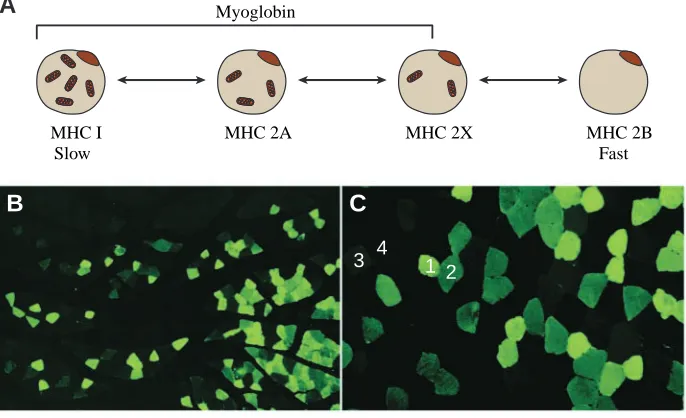
in cardiac myocytes and oxidative skeletal muscle fibers (types I>2A@2X) (Fig.·1), myoglobin was so named because of its functional and structural similarity to hemoglobin (Kendrew et al., 1954; Wittenberg and Wittenberg, 1989). Evolutionarily, myoglobin and hemoglobin arose from a common ancestral gene over 500 million years ago. Like hemoglobin, myoglobin reversibly binds O2and thus may facilitate O2transport from
red blood cells to mitochondria during periods of increased metabolic activity or serve as an O2 reservoir during
hypoxic or anoxic conditions. Unlike hemoglobin, however, monomeric myoglobin with a single O2-binding site has a
hyperbolic O2-saturation curve characteristic of normal
Michaelis–Menten enzyme kinetics rather than the sigmoid-shaped curve seen with tetrameric hemoglobin (Fig.·2).
The structure of myoglobin was first delineated by John Kendrew and colleagues over 40·years ago (Kendrew et al., 1958, 1960; Kendrew, 1963). Subsequent work has shown that the myoglobin backbone is a polypeptide chain that consists of eight α-helices assigned the letters A–H (Fig.·3A). Myoglobin binds oxygen by its heme residue, a porphyrin ring:iron ion complex. The polypeptide chain is folded and cradles the heme prosthetic group, positioning it between two histidine residues, His64 and His93. The iron ion interacts with six ligands, four of which are provided by the nitrogen atoms of the four pyroles
doi:10.1242/jeb.01172
Myoglobin is a cytoplasmic hemoprotein, expressed solely in cardiac myocytes and oxidative skeletal muscle fibers, that reversibly binds O2 by its heme residue, a
porphyrin ring:iron ion complex. Since the initial discovery of its structure over 40·years ago, wide-ranging work by many investigators has added importantly to our understanding of its function and regulation. Functionally, myoglobin is well accepted as an O2-storage protein
in muscle, capable of releasing O2 during periods of
hypoxia or anoxia. Myoglobin is also thought to buffer intracellular O2 concentration when muscle activity
increases and to facilitate intracellular O2 diffusion by
providing a parallel path that augments simple diffusion
of dissolved O2. The use of gene targeting and other
molecular biological techniques has revealed important new insights into the developmental and environmental regulation of myoglobin and provided additional functions for this hemoprotein such as scavenging nitric oxide and reactive O2 species. These recent findings, coupled with
additional emerging technologies and the discovery of other tissue globins, provide a framework for addressing new questions about myoglobin and readdressing old ones.
Key words: myoglobin, hemoprotein, skeletal muscle, cardiac myocyte, function, regulation, gene targeting.
Summary
Commentary
Myoglobin: an essential hemoprotein in striated muscle
George A. Ordway
1,* and Daniel J. Garry
2,3Departments of 1Physiology, 2Internal Medicine and 3Molecular Biology, University of Texas Southwestern Medical
Center, Dallas, TX 75390, USA
*Author for correspondence (e-mail: george.ordway@utsouthwestern.edu)
Accepted 30 June 2004
and share a common plane (Fig.·3B). The imidazole side chain of His93 provides the fifth ligand, stabilizing the heme group and slightly displacing the iron ion away from the plane of the heme. The sixth ligand position, unoccupied in deoxymyoglobin, serves as the binding site for O2, as well as
for other potential ligands such as CO or NO. When O2binds,
the iron ion is partially pulled back toward the porphyrin plane. Although this displacement is of little consequence in the function of monomeric myoglobin, it provides the basis for the conformational changes that underlie the allosteric properties of tetrameric hemoglobin. In addition, studies using X-ray diffraction and xenon-binding techniques have identified four highly conserved internal cavities within the myoglobin molecule that may serve to concentrate and orient molecules for binding to the heme residue (Frauenfelder et al., 2001).
Functional roles Oxygen storage
Myoglobin is perhaps best known as an O2-storage protein
in muscle. This role is especially evident in marine mammals and birds that undergo extended periods of apnea when diving. In the absence of inspired O2, stored O2 (oxymyoglobin;
Kooyman and Ponganis, 1998) becomes available to supply locomotor muscles involved in diving-related activities. The role of myoglobin as a store of O2 is supported by the
observation that diving mammals and birds can have muscle myoglobin contents that are increased 10- to 30-fold compared with those seen in animals that do not experience prolonged apnea (Table·1; Noren and Williams, 1999). Thus, when oxygen delivery ceases during breath-hold diving, O2 bound
to myoglobin is released to sustain aerobic metabolism in active muscles. Skeletal muscle myoglobin concentration is positively and significantly correlated with dive duration in some species (Kooyman and Ponganis, 1998; Noren and Williams, 1999). Myoglobin concentration in skeletal muscle is also increased in humans and other species living at high altitude (Gimenez et al., 1977; Terrados, 1992). In addition, myoglobin expression is increased in response to chronic contractile activity in animal models (Neufer et al., 1998; Underwood and Williams, 1987).
PO∑buffering
Related to its role as a tissue reservoir of O2, myoglobin has
been proposed to also serve as a buffer of intracellular PO∑in
a number of species including the human, rodent and bovine models. Similar to the role of creatine phosphokinase, which functions to buffer ATP concentrations when muscle activity increases, myoglobin functions to buffer O2 concentrations
under similar conditions (Hochachka, 1999; Meyer et al., 1984). As a result, the intracellular concentration of O2remains
relatively constant and homogeneous despite dramatic
activity-Table 1. Myoglobin content in skeletal muscle
Content
Model (mg·g–1wet mass) Tissue Reference
Northern elephant seal 64 Longissimus dorsi m. Noren et al. (2001) Bottlenose dolphin 26 Longissimus dorsi m. Noren and Williams (1999) New Zealand white rabbit 8 Skeletal muscle Noren and Williams (1999) Mouse (129) 1.9 Skeletal muscle Perkoff and Tyler (1958)
Human 2.1 Psoas m. Perkoff and Tyler (1958)
1 2 3 4
B
C
MHC 2A MHC 2X MHC I
Slow
MHC 2B Fast Myoglobin
[image:2.595.202.545.101.310.2] [image:2.595.26.555.678.766.2]induced increases in O2flux from capillary to mitochondria.
Myoglobin saturation has been shown to decrease rapidly at the onset of muscle activity and reach its nadir (30–60%) at approximately half-maximal levels of work (Richardson et al., 1995). As work increased to maximal effort, however, myoglobin saturation remained relatively constant, indicating that O2 concentration was likewise relatively constant
(Richardson et al., 1995). By contrast, Molé et al. (1999) showed that, although myoglobin saturation was approximately 48% at peak muscle O2 consumption, the
degree of desaturation increased linearly as a function of muscle work output. Irrespective of this difference, these studies indicate that myoglobin may provide a source of readily available O2 at the onset of exercise and increase the PO∑
gradient from capillary to muscle cell even at low levels of activity, suggesting that myoglobin has a role that is intermediate between two other functions, O2 storage and
facilitated O2diffusion.
Facilitated O2diffusion
A third role purported for myoglobin is facilitated or myoglobin-mediated O2 diffusion. As indicated, myoglobin
desaturates rapidly at the onset of muscle activity, increasing the PO∑ gradient from capillary blood to cytoplasm.
Furthermore, it has been proposed that desaturated myoglobin close to the cell membrane then binds O2and diffuses to the
mitochondria, providing a parallel path that supplements simple diffusion of dissolved O2 (Wittenberg, 1959, 1970).
Compelling theoretical and experimental evidence has been presented for (Conley and Jones, 1996; Merx et al., 2001; Murray, 1971; Salathe and Chen, 1993) and against (Jürgens et al., 1994; Papadopoulos et al., 2001) this purported role for myoglobin, so its contribution to overall O2flux in exercising
muscle remains equivocal.
Genetic regulation and expression patterns
The recent review by Garry et al. (2003) provides a succinct account of the genomic organization and transcriptional regulation of myoglobin. In addition, a number of studies have focused on developmental aspects of the expression of this hemoprotein (Fig.·4). In newborn mice, myoglobin is expressed at low levels in hindlimb muscles; however, as development proceeds and locomotor activity increases, myoglobin expression increases dramatically in oxidative, fatigue-resistant fibers (Garry et al., 1996). Neonatal dolphins, penguins and seals have myoglobin levels in locomotor muscles that are a fraction of those seen in adults (Noren et al., 2001). As the young mature and spend increasing amounts of time swimming and diving, myoglobin content in their muscles increases accordingly, approaching adult levels coincident with weaning and independent activity. Thus, although genetics plays an important role in establishing inherent levels of muscle myoglobin content, developmental programs and/or environmental cues and stresses such as physical activity, temperature and O2availability play at least equally important
roles in determining functional levels of this protein.
It’s nice to have but is it necessary?
Transgenic technologies provide unique opportunities to study the effects of gain-of-function or loss-of-function of proteins of interest on animal development and performance. Eliminating a protein by gene targeting or knockout techniques can produce valuable information regarding the predicted role
100
80
60
40
20
0
100 80
60 40
20
0 120
PO2 (torr)
Percent O
2
saturation
Myoglobin
Hemoglobin
Fig.·2. Myoglobin avidly binds oxygen. Myoglobin and hemoglobin function as oxygen transporters. Myoglobin displays a hyperbolic-shaped oxygen-binding curve whereas hemoglobin displays a sigmoidal-shaped oxygen-binding curve.
Fe C
N N
O N
CH
N H HC
C
His93 His64
A
B
of the protein but can also reveal unexpected findings and unanticipated functions. Although previous work had shown that cardiac and skeletal muscle function were significantly impaired by pharmacological or chemical agents that reduced the levels of oxymyoglobin (Wittenberg and Wittenberg, 1975; Doeller and Wittenberg, 1991), additional non-specific effects of the agents (e.g. CO) were a concern. To address this concern, mice that lack myoglobin were engineered using gene targeting strategies that deleted exon 2 of the myoglobin gene (Garry et al., 1998; Gödecke et al., 1999), which encodes almost half of the 154 amino acids that make up the protein (amino acids 31–105), including the essential heme-binding domain (Fig.·5A). Immunohistochemical and western analysis confirmed that the myoglobin protein could not be detected in cardiac or skeletal muscles of the knockout mice. While the mutation resulted in a number of lethal cardiovascular defects in embryos between days E9.5 and E10.5, those that survived this critical period showed no additional problems but revealed critical cellular and molecular adaptive responses (see below; Meeson et al., 2001).
Following birth, the myoglobin knockout mice showed no apparent phenotype other than depigmentation of the heart and soleus muscles (Fig.·5B; Garry et al., 1998; Gödecke et al., 1999). They grew normally, were able to perform exhaustive treadmill exercise and responded normally to a hypoxic challenge (Garry et al., 1998). In addition, skeletal muscles and hearts isolated from the knockout mice were equal to those from their wild-type counterparts in studies of contractile function in the presence or absence of O2(Garry et al., 1998;
Gödecke et al., 1999). Although there were no apparent disruptions of sarcomere structure or mitochondrial content, a number of cardiac adaptations were seen that favored improved
O2 delivery in the absence of myoglobin (Gödecke et al.,
1999). These included increases in capillarity, coronary flow and hematocrit. Subsequent studies have confirmed these cardiac adaptations and shown that they occur in skeletal muscle as well (Grange et al., 2001; Meeson et al., 2001). In addition, myoglobin-deficient mice demonstrate increased expression of a number of hypoxia-inducible genes (hypoxia-inducible factors 1 and 2, vascular endothelial growth factor, nitric oxide synthase, etc.) that may provide the molecular basis for the cellular adaptations observed in the muscles of these knockout animals (Grange et al., 2001; Meeson et al., 2001). Studies are in progress to determine whether previously undescribed tissue hemoproteins may further compensate and preserve contractility in the absence of myoglobin.
Emerging functional roles
A number of recent studies have demonstrated that myoglobin may have important functions beyond those associated with O2binding. One of these is its ability to bind
NO, a molecule whose effects can be either beneficial or detrimental to cellular function. In addition to its role as a potent vasodilator, NO has been shown to inhibit cytochrome c oxidase and thus impair mitochondrial respiration (Brunori, 2001a; Poderoso et al., 1998; Shiva et al., 2001). Brunori proposed recently that, based on its ability to bind NO, myoglobin may serve as an important scavenger of NO in heart and oxidative skeletal muscle (Brunori, 2001a,b). A subsequent report by Flögel et al. (2001b) provided convincing experimental evidence supporting this proposal. Additional support comes from studies showing NO-related alterations in skeletal and cardiac muscle function in myoglobin-deficient
Fig.·4. Myoglobin is temporally expressed during muscle differentiation. (A) Immunohistochemical localization of myoglobin in differentiated C2C12 myotubes. Note that myoglobin (green) is uniformly expressed in the cytoplasm of differentiated myotubes and absent in the nuclear compartment (propidium iodide stains red and demarcates the nuclear
compartment). (B) Western blot analysis of myoglobin expression following differentiation of C2C12 myotubes. Myoglobin (Mb) and myosin heavy chain (MHC) proteins increase with exposure of C2C12 myogenic cell line to differentiation media (DM).
Mb
MHC Days in DM
1 2 3 4
A
B
neo
1 2 3
Exon
neo
1
Exon 3
Native Mb gene
Target vector
Disrupted Mb gene
A
B
mice (Grange et al., 2001; Mammen et al., 2003). Myoglobin is also known to have peroxidase activity (George and Irvine, 1955; Cadenas, 1989; Khan et al., 1998), and a similar additional role for this protein as a scavenger of reactive O2
species has recently been demonstrated (Flögel et al., 2001a,b). Future studies will be necessary to further define the functional role(s) for myoglobin in oxidative skeletal muscle. For example, important questions regarding myoglobin biology that remain unanswered include: what are the genetic factors that regulate myoglobin expression in response to an acute or chronic hypoxic stimulus; is the transcriptional regulation of myoglobin a hypoxia inducible factor (HIF-1α )-dependent mechanism; does the induction of myoglobin expression in response to hypoxia require muscle activity (i.e. swimming, running, etc.)? Moreover, the recent identification of additional tissue hemoglobins – neuroglobin and cytoglobin – suggests a physiological model in skeletal muscle that is increasingly complex and fluid regarding the role of tissue hemoglobins and muscle biology.
Nice to have and necessary
In the nearly 50·years since John Kendrew described the three-dimensional structure of myoglobin, this important tissue hemoglobin has revealed a number of diverse and important functions supporting the notion that it’s not only nice but necessary. These include providing a reservoir of readily accessible O2, buffering intracellular O2 concentration,
facilitating intracellular O2 transport, inactivating NO and
scavenging reactive O2 species. Gene targeting and other
molecular biological techniques have added importantly to our understanding of the overall role of myoglobin in O2delivery
and utilization by answering new questions and enabling old ones to be revisited. In addition, the embryonic lethality and compensatory adaptations associated with myoglobin-deficient mice give further evidence of the necessity of myoglobin in normal muscle development and function. These and other emerging technologies will provide scientists with powerful tools to address additional fundamental questions about myoglobin and other tissue globins within an integrative framework.
We gratefully acknowledge the expert assistance of Mr Sean C. Goetsch in preparing the figures and the support of National Institutes of Health grants to D.J.G.
References
Brunori, M. (2001a). Nitric oxide, cytochrome-c oxidase and myoglobin. Trends Biochem. Sci. 26, 21-23.
Brunori, M. (2001b). Nitric oxide moves myoglobin centre stage. Trends Biochem. Sci. 26, 209-210.
Cadenas, E. (1989). Lipid peroxidation during the oxidation of haemoproteins by hydroperoxides. Biolum. Chemilum. 4, 208-218.
Conley, K. E. and Jones, C. (1996). Myoglobin content and oxygen diffusion: model analysis of horse and steer muscle. Am. J. Physiol. Cell Physiol. 271, C2027-C2036.
Doeller, J. E. and Wittenberg, B. A. (1991). Myoglobin function and energy
metabolism of isolated cardiac myocytes: effect of sodium nitrite. Am. J. Physiol. Heart Circ. Physiol. 261, H53-H62.
Flögel, U., Gödecke, A. and Schrader, J. (2001a). Myoglobin is important for postischemic recovery in the heart. Circulation 104, II-227.
Flögel, U., Merx, M. W., Gödecke, A., Decking, U. K. M. and Schrader, J. (2001b). Myoglobin: a scavenger of bioactive NO. Proc. Natl. Acad. Sci. USA 98, 735-740.
Frauenfelder, H., McMahon, B. H., Austin, R. H., Chu, K. and Groves, J. T. (2001). The role of structure, energy landscape, dynamics, and allostery in the enzymatic function of myoglobin. Proc. Natl. Acad. Sci. USA 98, 2370-2374.
Garry, D. J., Bassel-Duby, R. S., Richardson, J. A., Grayson, J., Neufer, P. D. and Williams, R. S. (1996). Postnatal development and plasticity of specialized muscle fiber characteristics in the hindlimb. Dev. Gen. 19, 146-156.
Garry, D. J., Ordway, G. A., Lorenz, J. N., Radford, N. B., Chin, E. R., Grange, R. W., Bassel-Duby, R. and Williams, R. S. (1998). Mice without myoglobin. Nature 395, 905-908.
Garry, D. J., Kanatous, S. B. and Mammen, P. P. A. (2003). Emerging roles for myoglobin in the heart. Trends Cardiovasc. Med. 13, 111-116. George, P. and Irvine, D. H. (1955). A possible structure for the higher
oxidation state of metmyoglobin. Biochem. J. 60, 596-604.
Gimenez, M., Sanderson, R. J., Reiss, O. K. and Banchero, N. (1977). Effects of altitude on myoglobin and mitochondrial protein in canine skeletal muscle. Respiration 34, 171-176.
Gödecke, A., Flögel, U., Zanger, K., Ding, Z., Hirchenhain, J., Decking, U. K. M. and Schrader, J. (1999). Disruption of myoglobin in mice induces multiple compensatory mechanisms. Proc. Natl. Acad. Sci. USA 96, 10495-10500.
Grange, R. W., Meeson, A., Chin, E., Lau, K. S., Stull, J. T., Shelton, J. M., Williams, R. S. and Garry, D. J. (2001). Functional and molecular adaptations in skeletal muscle of myoglobin-mutant mice. Am. J. Physiol. Cell Physiol. 281, C1487-C1494.
Hochachka, P. W. (1999). The metabolic implications of intracellular circulation. Proc. Natl. Acad. Sci. USA 96, 12233-12239.
Jürgens, K. D., Peters, T. and Gros, G. (1994). Diffusivity of myoglobin in intact skeletal muscle cells. Proc. Natl. Acad. Sci. USA 91, 3829-3833. Kendrew, J. C., Parrish, R. G., Marrack, J. R. and Orlans, E. S. (1954).
The species specificity of myoglobin. Nature 174, 946-949.
Kendrew, J. C., Bodo, G., Dintzis, H. M., Parrish, R. G. and Wyckoff, H. (1958). A three-dimensional model of the myoglobin molecule obtained by x-ray analysis. Nature 181, 662-666.
Kendrew, J. C., Dickerson, R. E., Strandberg, B. E., Hart, R. G., Davies, D. R., Phillips, D. C. and Shore, V. C. (1960). Structure of myoglobin. A three-dimensional Fourier synthesis at 2·Å resolution. Nature 185, 422-427.
Kendrew, J. C. (1963). Myoglobin and the structure of proteins. Science 139, 1259-1266.
Khan, K. K., Mondal, M. S., Pady, L. and Mitra, S. (1998). The role of distal histidine in peroxidase activity of myoglobin. Eur. J. Biochem. 257, 547-555.
Kooyman, G. L. and Ponganis, P. J. (1998). The physiological basis of diving to depth: birds and mammals. Annu. Rev. Physiol. 60, 19-32. Mammen, P. P. A., Kanatous, S. B., Yuhanna, I. S., Shaul, P. W., Garry,
M. G., Balaban, R. S. and Garry, D. J. (2003). Hypoxia-induced left ventricular dysfunction in myoglobin-deficient mice. Am. J. Physiol. Heart Circ. Physiol. 285, H2132-H2141.
Meeson, A. P., Radford, N., Shelton, J. M., Mammen, P. P. A., DiMaio, J. M., Hutcheson, K., Kong, Y., Elterman, J., Williams, R. S. and Garry, D. J. (2001). Adaptive mechanisms that preserve cardiac function in mice without myoglobin. Circ. Res. 88, 713-720.
Merx, M. W., Flögel, U., Stumpe, T., Gödecke, A., Decking, U. K. M. and Schrader, J. (2001). Myoglobin facilitates oxygen diffusion. FASEB J. 15, 1077-1079.
Meyer, R. A., Sweeney, H. L. and Kushmerick, M. J. (1984). A simple analysis of the “phosphocreatine shuttle”. Am. J. Physiol. Cell Physiol. 246, C365-C377.
Molé, P. A., Chung, Y., Tran, T. K., Sailasuta, N., Hurd, R. and Jue, T. (1999). Myoglobin desaturation with exercise intensity in human gastrocnemius muscle. Am. J. Physiol. Reg. Int. Comp. Physiol. 277, R173-R180.
Neufer, P. D., Ordway, G. A. and Williams, R. S. (1998). Transient regulation of c-fos, αB-crystallin, and hsp70 in muscle during recovery from contractile activity. Am. J. Physiol. Cell Physiol. 274, C341-C346.
Noren, S. R. and Williams, T. M. (1999). Body size and skeletal muscle myoglobin of cetaceans: adaptations for maximizing dive duration. Comp. Biochem. Physiol. A 126, 181-191.
Noren, S. R., Williams, T. M., Pabst, D. A., McLellan, W. A. and Dearolf, J. L. (2001). The development of diving in marine endotherms: preparing the skeletal muscles of dolphins, penguins, and seals for activity during submergence. J. Comp. Physiol. B 171, 127-134.
Papadopoulos, S., Endeward, V., Revesz-Walker, B., Jürgens, K. D. and Gros, G. (2001). Radial and longitudinal diffusion of myoglobin in single living heart and skeletal muscle cells. Proc. Natl. Acad. Sci. USA 98, 5904-5909.
Perkoff, G. T. and Tyler, F. H. (1958). Estimation and physical properties of myoglobin in various species. Metabolism 7, 751-759.
Poderoso, J. J., Peralta, J. G., Lisdero, C. L., Carreras, M. C., Radisic, M., Schopfer, F., Cadenas, E. and Boveris, A. (1998). Nitric oxide regulates oxygen uptake and hydrogen peroxide release by the isolated beating rat heart. Am. J. Physiol. Cell Physiol. 274, C112-C119.
Richardson, R. S., Noyszewski, E. A., Kendrick, K. F., Leigh, J. S. and
Wagner, P. D. (1995). Myoglobin O2 desaturation during exercise.
Evidence of limited O2transport. J. Clin. Invest. 96, 1916-1926.
Salathe, E. P. and Chen, C. (1993). The role of myoglobin in retarding oxygen depletion in skeletal muscle. Math. Biosci. 116, 1-20.
Shiva, S., Brookes, P. S., Patel, R. P., Anderson, P. G. and Darley-Usmar, V. M. (2001). Nitric oxide partitioning into mitochondrial membranes and the control of respiration at cytochrome c oxidase. Proc. Natl. Acad. Sci. USA 98, 7212-7217.
Terrados, N. (1992). Altitude training and muscular metabolism. Int. J. Sports Med. 13 (Suppl), S206-S209.
Underwood, L. E. and Williams, R. S. (1987). Pretranslational regulation of myoglobin gene expression. Am. J. Physiol. Cell Physiol. 252, C450-C453. Wittenberg, J. B. (1959). Oxygen transport – a new function proposed for
myoglobin. Biol. Bull. 117, 402-403.
Wittenberg, J. B. (1970). Myoglobin-facilitated oxygen diffusion: role of myoglobin in oxygen entry into muscle. Physiol. Rev. 50, 559-636. Wittenberg, B. A. and Wittenberg, J. B. (1975). Role of myoglobin in the
oxygen supply to red skeletal muscle. J. Biol. Chem. 250, 9038-9043. Wittenberg, B. A. and Wittenberg, J. B. (1989). Transport of oxygen in
muscle. Annu. Rev. Physiol. 51, 857-878.